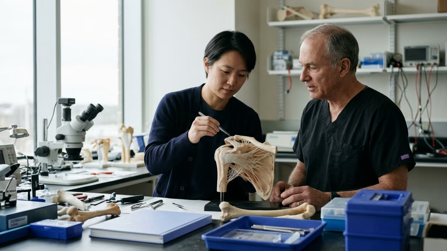

Penn’s rotator cuff scaffold targets the tendon-to-bone connection. See the early findings and the questions still ahead of human use.





On October 20, 2026, the University of Pennsylvania’s Almanac reported on a preclinical rotator-cuff repair scaffold developed by researchers at Penn’s Perelman School of Medicine. The work was described in Science Advances. Penn reported that the scaffold guided tissue formation in laboratory tests and was associated with more organized tissue formation in animal models.
The design targets a specific part of the repair: the transition from tendon to bone. It includes tendon-, fibrocartilage- and bone-like regions rather than a single uniform material.
The central idea is to give different parts of the attachment different signals. Understanding that design helps explain both the interest in the findings and the questions that come next.
The background to this work is the structure of the natural tendon-to-bone connection. According to senior author Su Chin Heo, that connection has an organized transition. Each region provides distinct signals that influence cell behavior and tissue formation.
That makes the attachment more than a place where tissues meet. In the researchers’ explanation, its regional structure matters because different areas support different kinds of tissue. Their scaffold attempts to reproduce those local conditions.
This goal also appears in the broader research context. A separate 2026 review of treatment options describes preclinical inlay scaffold research intended to resemble the native tendon-to-bone attachment. Penn’s project addresses that general research aim through a particular combination of materials and regions.
The useful background is therefore a biological question: how closely should a repair material resemble the connection it is intended to support? Penn’s researchers approached that question by giving the scaffold distinct regions instead of treating the attachment as one space with one set of signals.
Penn’s report describes both the scaffold’s construction and the early tests used to study it. These are related parts of the story, but they answer different questions.
According to Penn, the tendon- and cartilage-like regions combine nanofibers derived from cow Achilles tendons with hyaluronic acid. The porous bone region uses a citrate-based material.
The materials are arranged to represent the transition between tissues. The aim is not simply to place the same material across the whole attachment. It is to provide different cues in different areas, following the regional organization described by Heo.
The tissue labels need careful reading. Calling part of the scaffold “tendon-like” or “bone-like” describes its intended role in the design. The findings about tissue formation come from the separate cell and animal tests.
Penn also says the scaffold was designed to work with existing suture-anchor systems. That is a feature of the proposed repair system, alongside its regional materials.
In laboratory tests, Penn reported that the scaffold’s regions guided stem cells toward tendon- and cartilage-like tissues. This finding addresses the researchers’ idea that different local environments can provide different signals.
In animal models of rotator-cuff injury, the report describes more organized tissue formation at the repair site than in repairs without the scaffold. That formation included tendon, cartilage-like tissue and bone.
Read together, the findings connect the design idea with observations at different levels. The cell tests concern tissue development within the scaffold’s regions. The animal findings concern the organization of tissue at an injury repair site.
The word organized is important here. The reported result concerns how tissue formed at the connection, not merely whether material was present there. That focus matches the researchers’ goal of recreating a regional transition rather than filling a space.
The studied populations were laboratory cells and animal models of rotator-cuff injury. They were not a defined group of adults recovering from shoulder surgery.
The patients motivating the research are a separate matter. First author Zizhao (Molly) Li pointed to people with large tears, poor tissue quality or failed earlier repairs as groups who may have limited treatment options. She framed the project as an effort to improve healing quality and durability in challenging cases.
Penn’s report also identifies age-related slowing of healing as a factor that can complicate repair. These concerns explain why the researchers are interested in the attachment and its ability to support tissue formation.
For scale, Penn reports that roughly 250,000 people in the United States undergo rotator-cuff surgery each year. That figure describes surgery generally, not participation in this scaffold research.
For readers in midlife or later life, the distinction is straightforward: the clinical problems may sound familiar even though the experimental subjects are different. A person’s tear size or previous repair history may help explain their interest in the work. It does not place them within the population actually tested.
For someone recovering from a rotator-cuff injury or surgery, this development is information about a research direction. It is not a treatment currently available to patients. It does not establish a new rehabilitation protocol or a reason to change an individual care plan.
Its practical value is in making the treatment discussion more specific. Penn identifies tear size, tendon quality and previous repair history as features that can make some repairs challenging. Those are reasonable subjects for a conversation with the qualified clinician managing a person’s care.
Questions tied to the report include:
These questions focus on the person’s existing situation rather than on obtaining an experimental material. They also keep the research headline from becoming a substitute for an explanation of the current care plan.
The distinction between repair research and rehabilitation is especially useful here. A material designed for the surgical attachment and the process of returning to movement are different subjects. ReboundBody’s guide to the stages discussed after cuff repair provides related background on that recovery journey.
For readers interested in the biology behind the design, understanding how healing differs among musculoskeletal tissues offers another starting point. Neither topic requires treating an early scaffold finding as a personal recovery forecast.
The next stage identified by Penn is large-animal testing. The university says those trials are needed before the approach can be used in humans.
The report provides no sample sizes, numerical effect estimates or follow-up duration for the cell and animal tests. It also gives no detailed comparison data. Those missing details limit a reader’s ability to judge the size of the observed differences and how long they persisted.
No human outcomes are reported. Whether this scaffold reduces re-tears, improves shoulder function or speeds recovery in people remains unanswered. The researchers’ hopes for difficult repairs describe the intended future application rather than demonstrated patient benefit.
The separate review of scaffold research does not supply those missing results for Penn’s particular design. It provides context for the approach, not confirmation of its clinical value.
Likewise, compatibility with existing suture anchors does not establish approval for clinical use or predict how quickly surgeons might adopt the scaffold. A design that fits an existing system still needs evidence appropriate to its proposed use.
The findings therefore support continued investigation of a regionally designed attachment. They bring the material’s structure and its early tissue observations into the same research story. The next question is whether that approach can produce meaningful benefits beyond the experimental settings reported so far.
A more natural-looking repair site and a more dependable recovery are related hopes. They are not yet the same result.
Turning an animal scaffold result into a promise about shoulder recovery skips a vital evidence step; ReboundBody explains such research with clear limits. For active adults sorting through competing treatment claims after an orthopedic setback, the site puts rehabilitation and rebuilding physical capacity into context so personal questions stay grounded when discussed with a healthcare professional.

Follow ReboundBody for practical articles on injury recovery, rehabilitation, mobility and rebuilding strength. New posts turn research into clear ideas for the road back to activity after injury or surgery.





Read practical guidance on injury recovery, rehabilitation, mobility and rebuilding strength as you work your way back to activity.
Explore Resources