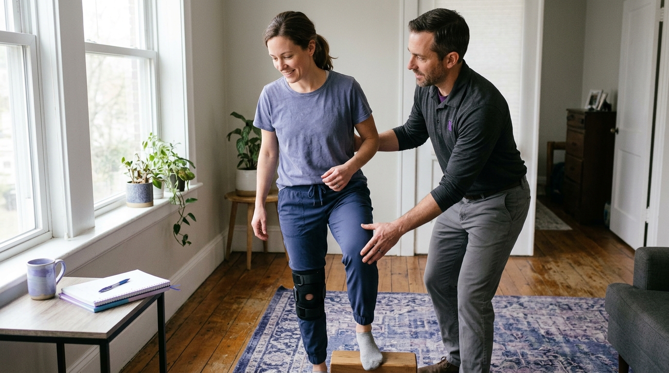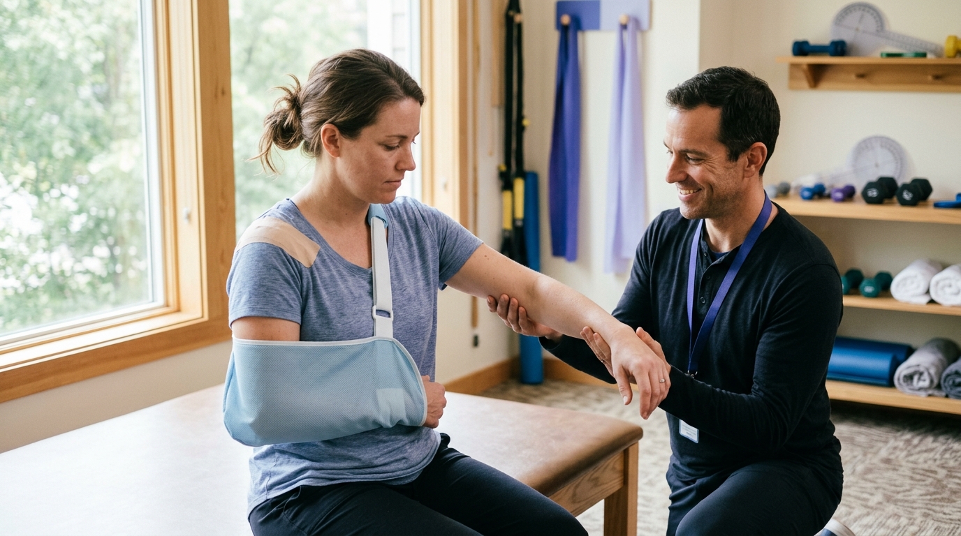

Rotator cuff recovery depends less on immediate strengthening than on phased biological healing that safely transitions your shoulder from protected rest to active mobility.





If you recently had surgery or are planning for an upcoming procedure, you have likely searched online to find out when your shoulder will feel normal again. You might want to know when you can take off the sling, drive your car, return to work, or lift weights at the gym.
The straightforward answer is that tendon-to-bone healing operates on a biological timeline that cannot be rushed. Full functional recovery typically requires six to twelve months of staged rehabilitation.
While the calendar provides a general map, your exact progression depends on your surgical details, tissue quality, and movement control. This guide outlines the standard four-phase recovery framework, summarizes the latest clinical evidence on sling use and movement timing, and explains why individual restrictions must come from your surgical team.
Understanding the language used by your surgeon and physical therapist makes following your postoperative instructions much easier. Shoulder rehabilitation separates arm movement into distinct categories based on how much muscular effort your healing tendon produces.
Passive range of motion means an outside force moves your arm while your shoulder muscles remain completely relaxed. That outside force can be a physical therapist, a family member, a pulley system, or your other arm.
Because your repaired rotator cuff muscles do not contract during passive movement, the repaired tendon is not pulled away from the bone. This protects the surgical repair site while gently moving the ball within the socket to prevent joint stiffness.
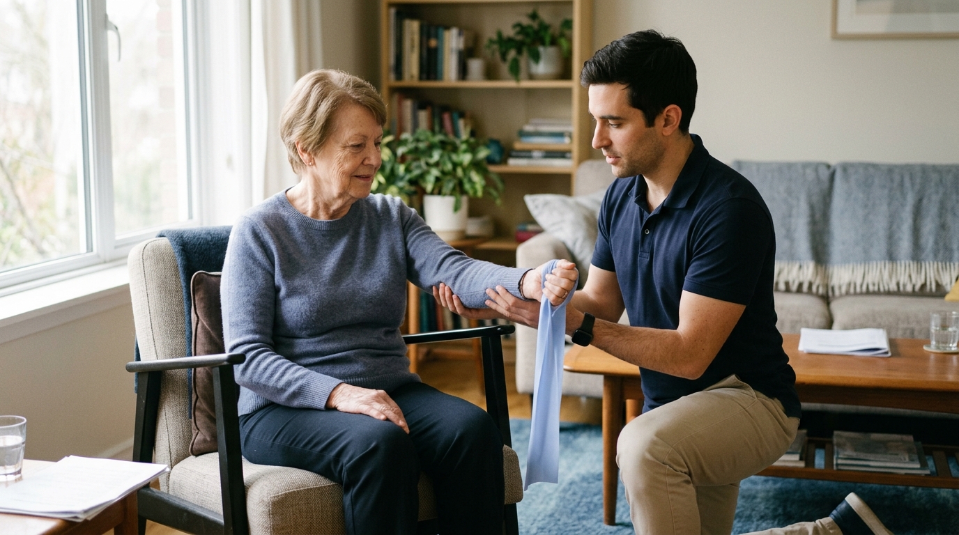
Active-assisted range of motion occurs when your shoulder muscles begin to do a portion of the work, but still receive mechanical support. You might hold a light cane, a broomstick, or use your non-operative hand to guide the surgical arm through an arc of movement.
This stage introduces mild, partial muscle activation. It helps retrain the nervous system to coordinate muscle firing without placing full gravitational loads on the healing tendon anchor.
Active range of motion means you lift and move your arm entirely under its own muscular power against gravity without any external help. At this point, the repaired rotator cuff tendon must actively contract to center the humeral head in the socket while the larger deltoid muscle lifts the arm.
Active movement requires substantial mechanical stability from the repair site. For this reason, clinical protocols delay active lifting until basic biological healing has taken root.
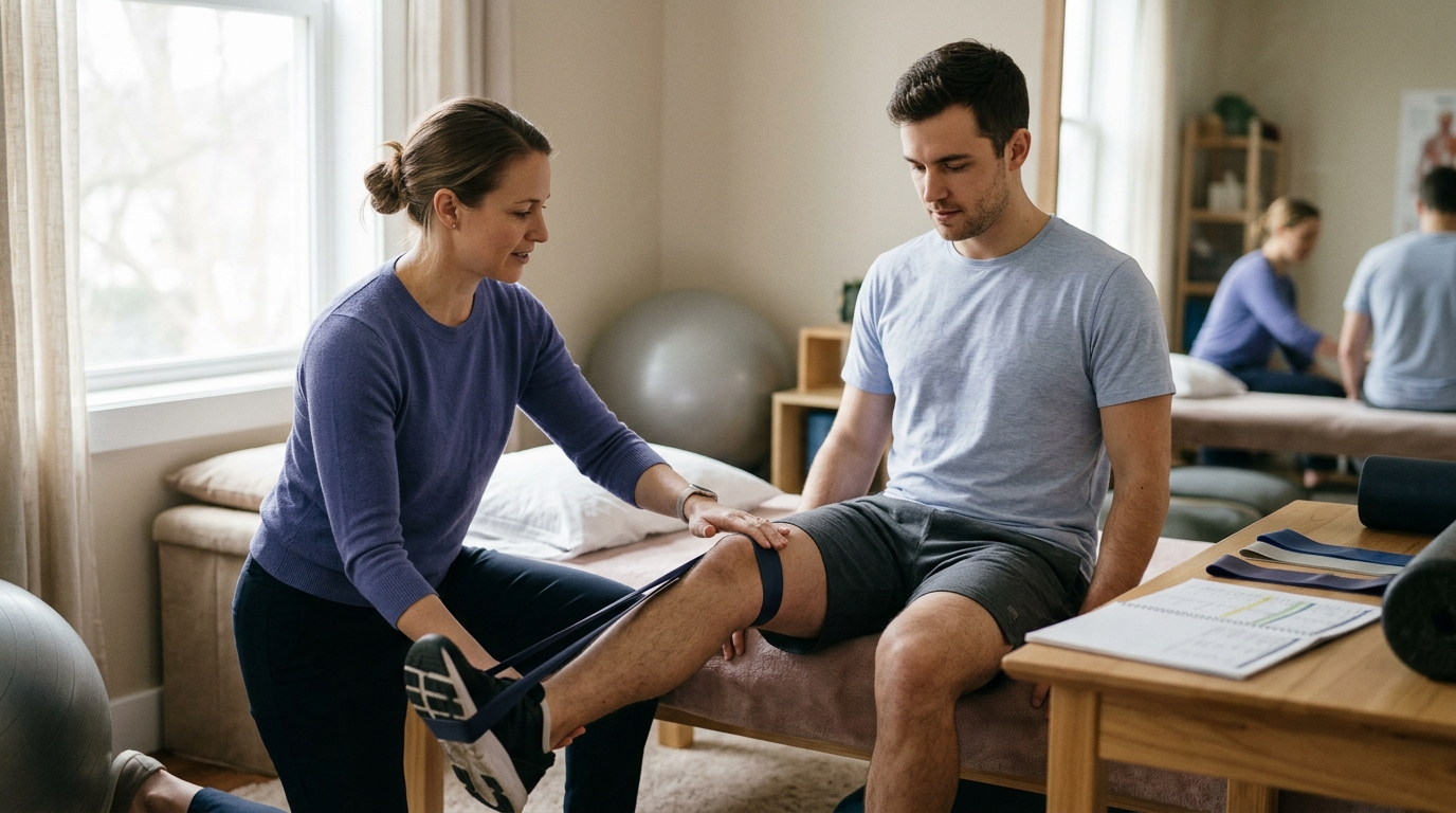
Resisted exercise adds external loads, such as elastic bands, light dumbbells, or weight machines, to active shoulder movements. These loads challenge muscle strength, tissue capacity, and muscular endurance.
Strengthening is introduced only after you demonstrate full, smooth active motion without hiking your shoulder or compensating with other muscles. Introducing resistance too early can overload the immature scar tissue at the tendon-to-bone interface.
Medical research distinguishes between structural tendon healing and functional recovery. Structural healing refers to whether the tendon physically reconnects to the bone on an ultrasound or MRI scan. Functional recovery refers to your pain levels, range of motion, and ability to complete daily tasks.
Some patients achieve excellent functional movement and return to recreation even if postoperative imaging shows incomplete structural healing. Conversely, an intact repair on an MRI does not automatically guarantee pain-free movement if mobility and strength are not properly rebuilt through structured surgical rehabilitation pathways.
A rotator cuff repair secures a torn tendon back to the humeral head using specialized anchors and sutures. The initial mechanical hold relies entirely on the strength of the suture material and the bone anchor. Over several months, your body must replace that artificial hold with a living biological bridge composed of Sharpey fibers and dense collagen.
Tendons have a limited blood supply compared to other tissues like muscle or bone. This slower metabolic rate means collagen synthesis and tissue remodeling take significant time.
During the first several weeks, the repair site consists of fragile, disorganized scar tissue. This tissue gradually reorganizes into aligned, load-bearing collagen bundles over three to six months.
Because biology sets the pace for tissue remodeling, modern orthopedic rehabilitation uses criteria-based milestones alongside broad timeframes. A time-based milestone ensures you do not load the tendon before cellular healing has occurred.
A criteria-based milestone ensures you do not advance to demanding tasks until your movement quality, joint control, and baseline strength are adequate. Combining time windows with objective movement standards provides the safest environment for long-term tendon recovery.
The initial phase begins the day of surgery and typically extends through the fourth or sixth postoperative week. The primary objective during this phase is protecting the surgical repair while managing pain, controlling swelling, and preventing severe joint stiffness.
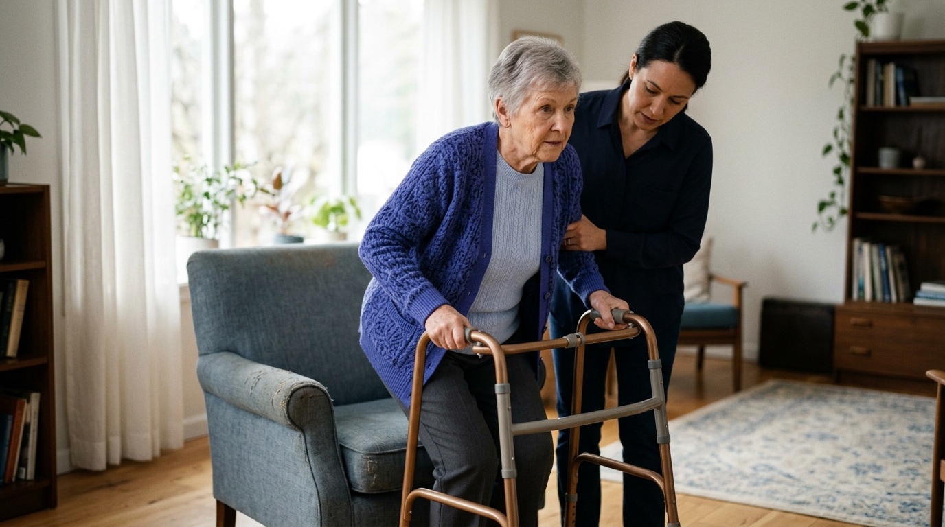
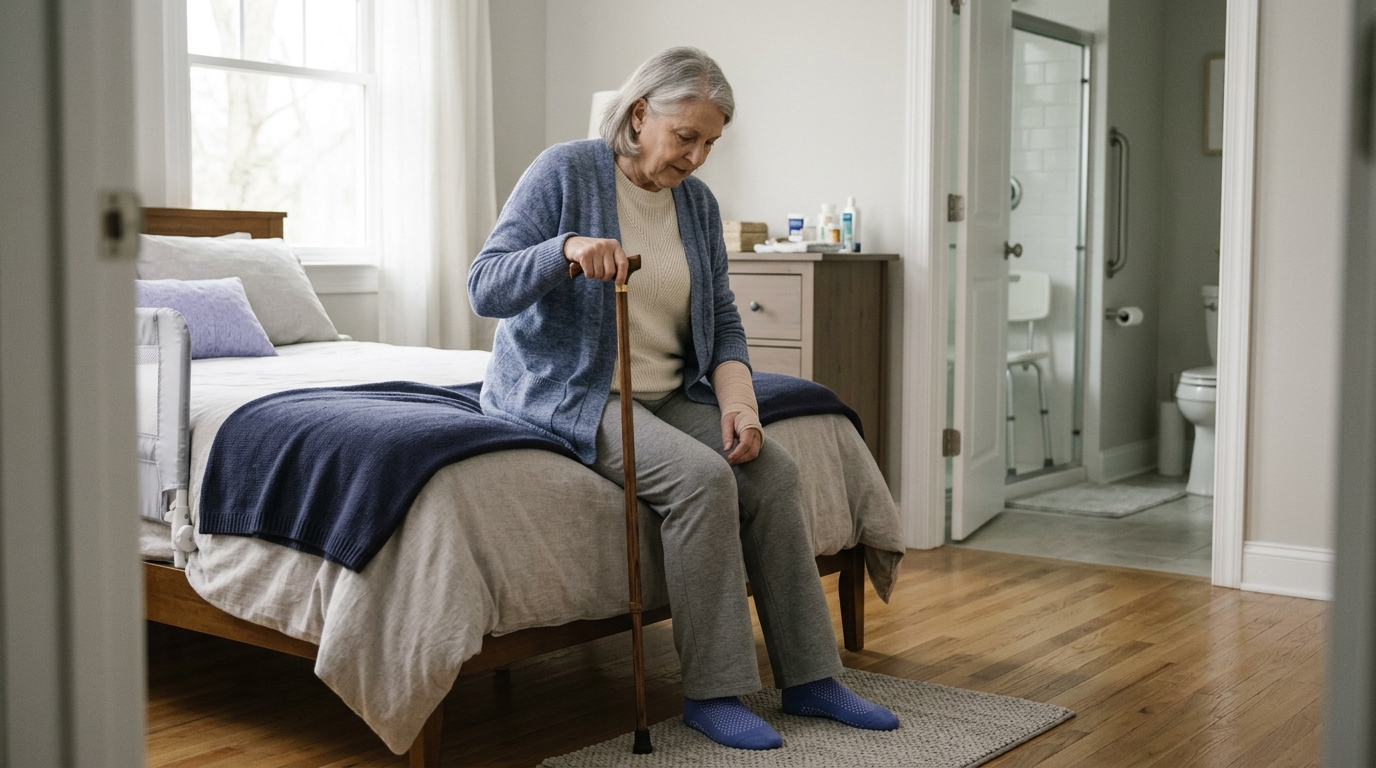
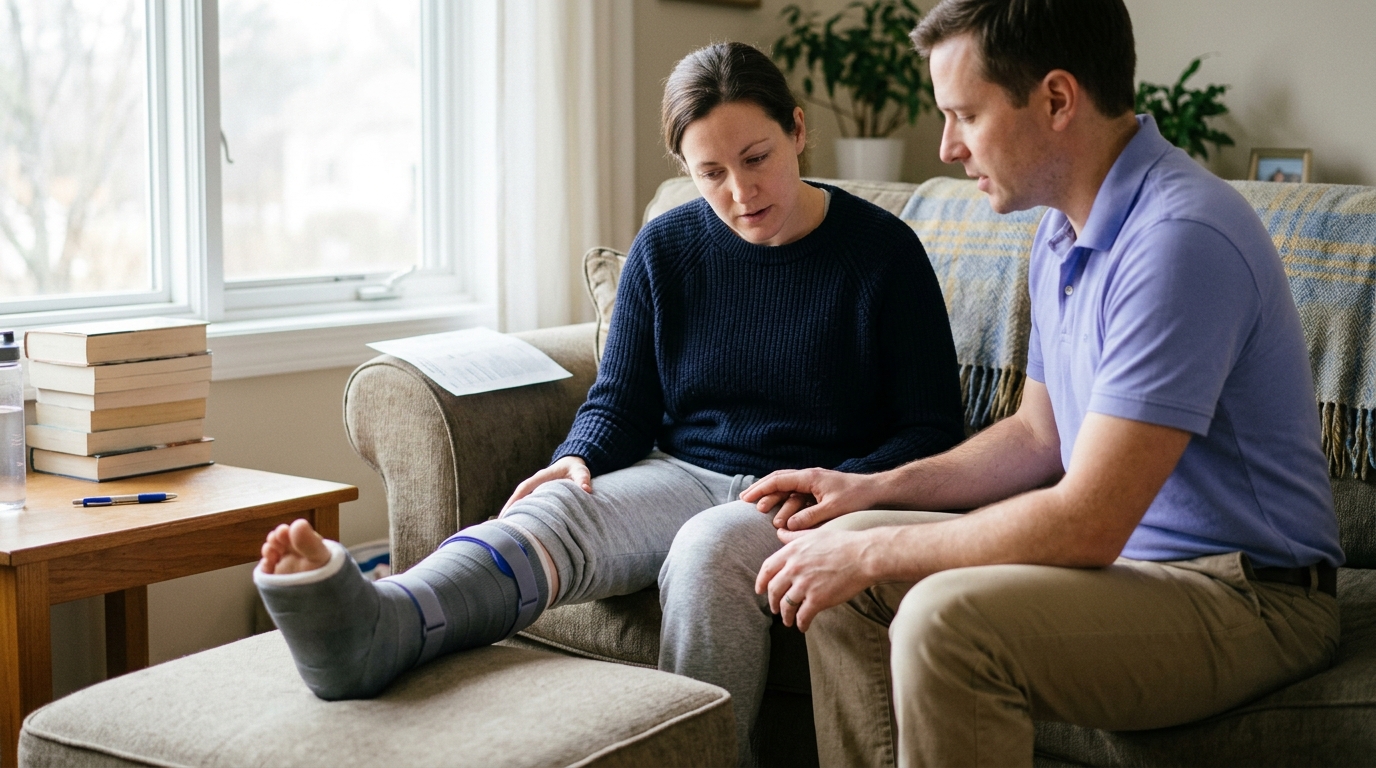
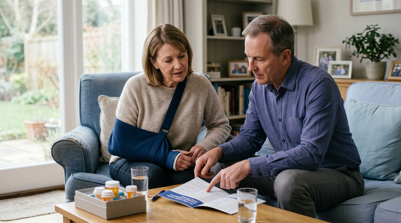
During phase one, you will wear an immobilization sling or an abduction brace as directed by your surgeon. The sling keeps your arm positioned close to your torso, taking tension off the repaired supraspinatus, infraspinatus, or subscapularis tendons.
In most standard protocols, you will wear the sling day and night, removing it only for prescribed home exercises, personal hygiene, and hand or elbow movement. You must never use your surgical arm to push up from a chair, reach for items, or support your body weight during this early window.
Sleeping in a sling can be challenging during the first few weeks. Many patients find greater comfort resting in a semi-reclined position, such as in a recliner chair or supported by a wedge pillow in bed.
Placing a small pillow or rolled towel under your operative elbow prevents the arm from dropping backward, which reduces strain on the anterior shoulder tissues.
Immobilization does not mean keeping your entire upper extremity completely still. Your care team will instruct you to perform active movements for your fingers, wrist, and forearm multiple times each day.
Opening and closing your hand, squeezing a soft stress ball, and performing gentle wrist circles maintains circulation and reduces swelling in the lower arm. Regular active elbow bending and straightening, performed while seated with your upper arm resting safely at your side, prevents elbow stiffness.
Depending on your surgeon's specific protocol, early passive shoulder exercises may begin during this phase. Common examples include:
A frequent error during early recovery is assuming that an absence of pain equals tissue healing. Modern nerve blocks and postoperative medications can reduce pain significantly within the first few weeks.
Removing your sling to carry light objects or reach into a cabinet because it does not hurt can damage the repair before biological healing has occurred.
Another mistake is performing aggressive stretching to eliminate stiffness early. Phase one aims for gentle, protected motion within specific parameters, not forcing end-range flexibility. Forcing the arm into painful ranges risks tearing sutures through fragile tendon tissue.
Before transitioning out of phase one, you should generally meet the following benchmarks:
Phase two typically spans from week six through week twelve. The primary focus shifts toward weaning from the sling, restoring full passive mobility, and gradually transitioning to active movement against gravity.
Between weeks six and eight, most patients receive clearance to begin weaning out of their sling during the day. Weaning is usually a gradual process over several days rather than an abrupt change.
You might start by leaving the sling off while sitting quietly in a safe environment, then gradually progress to wearing it only in crowded public places or while sleeping.
Once the sling is discontinued, you must continue to protect the arm from sudden, uncontrolled reaching or lifting loads.
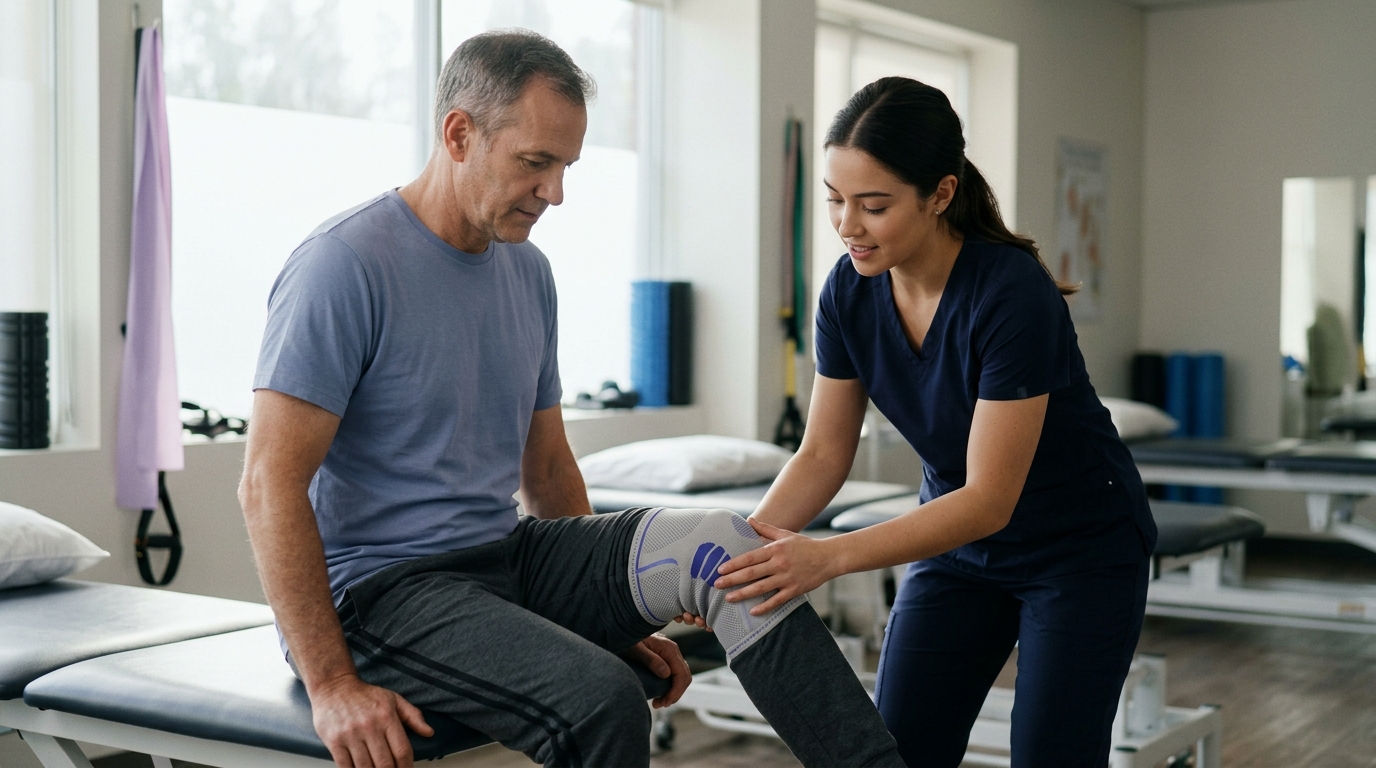
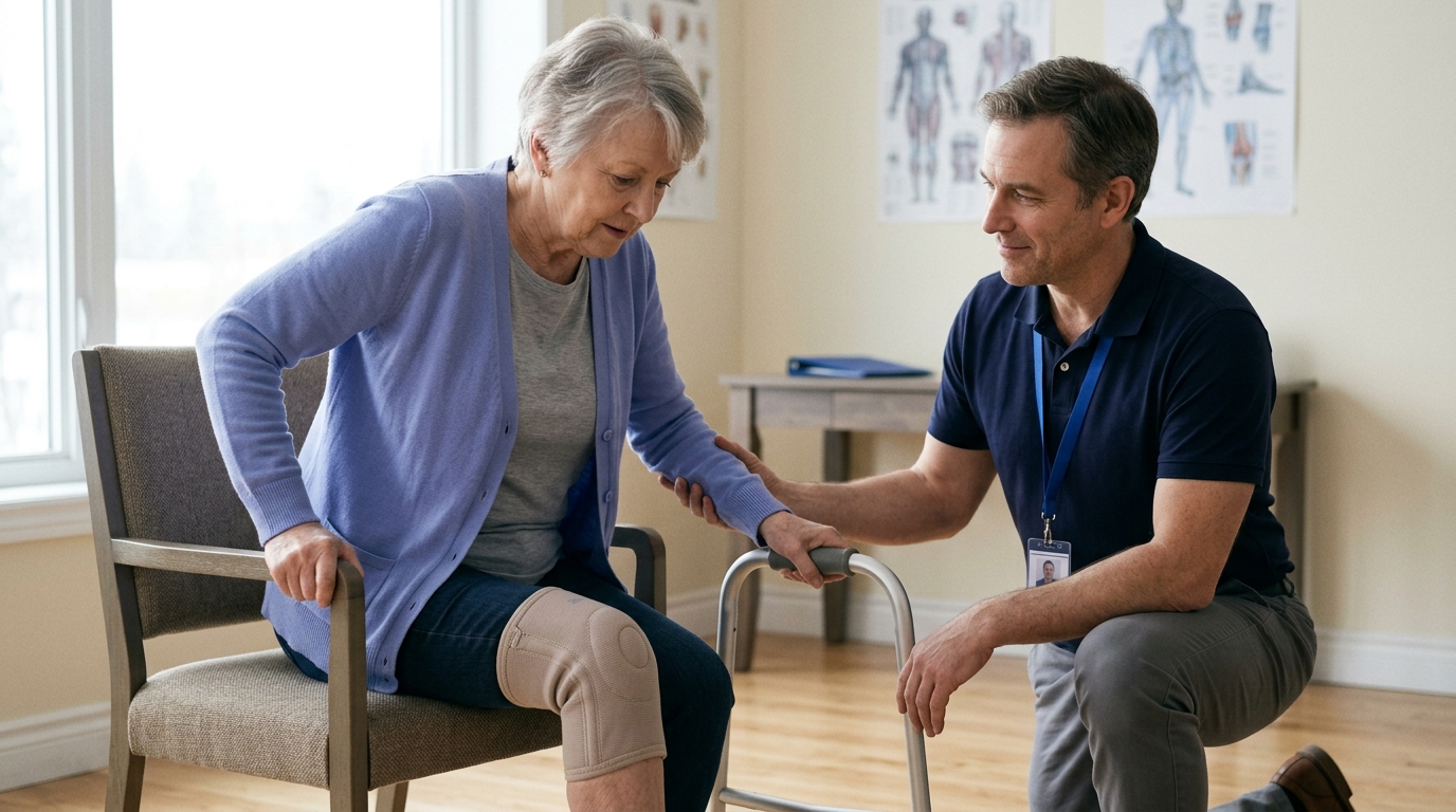
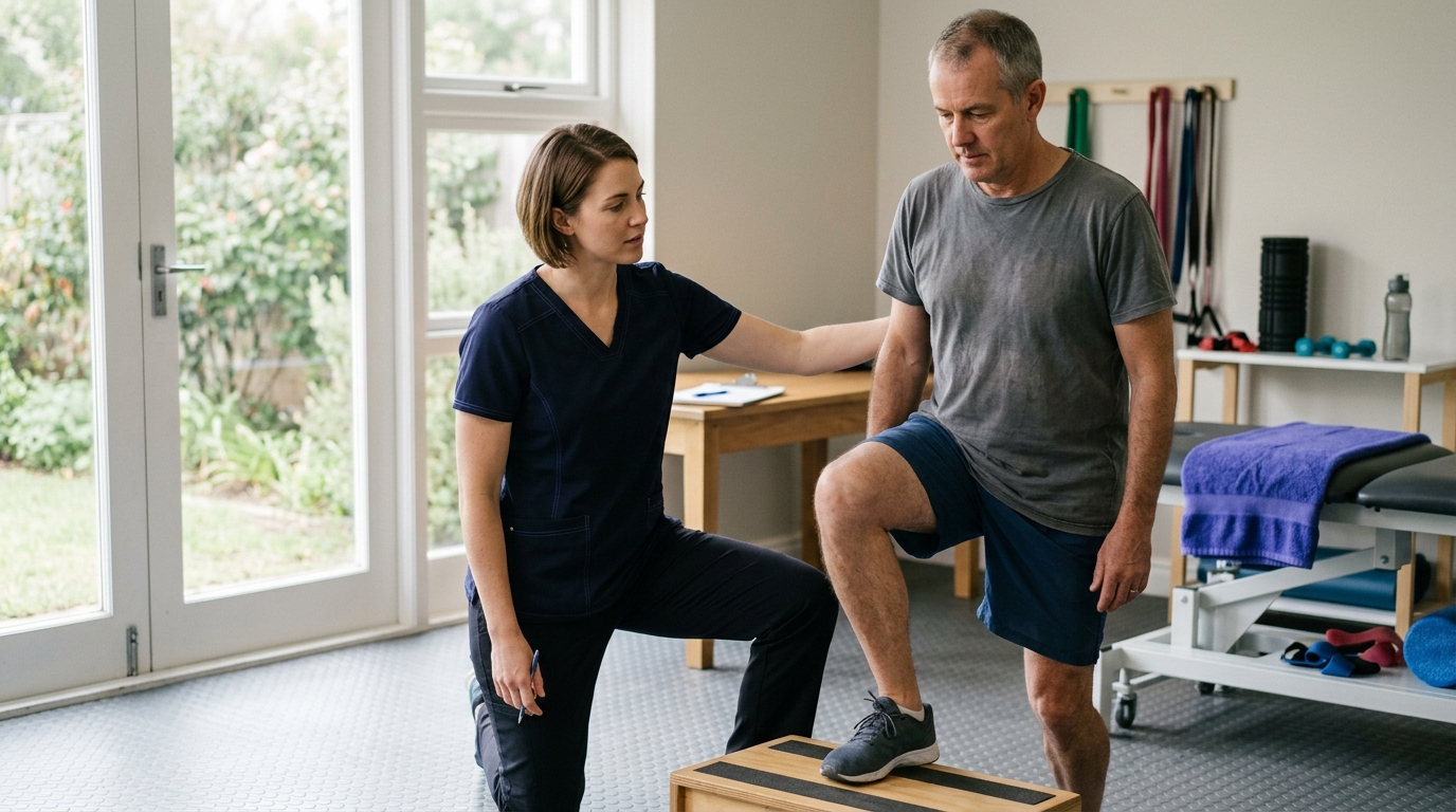
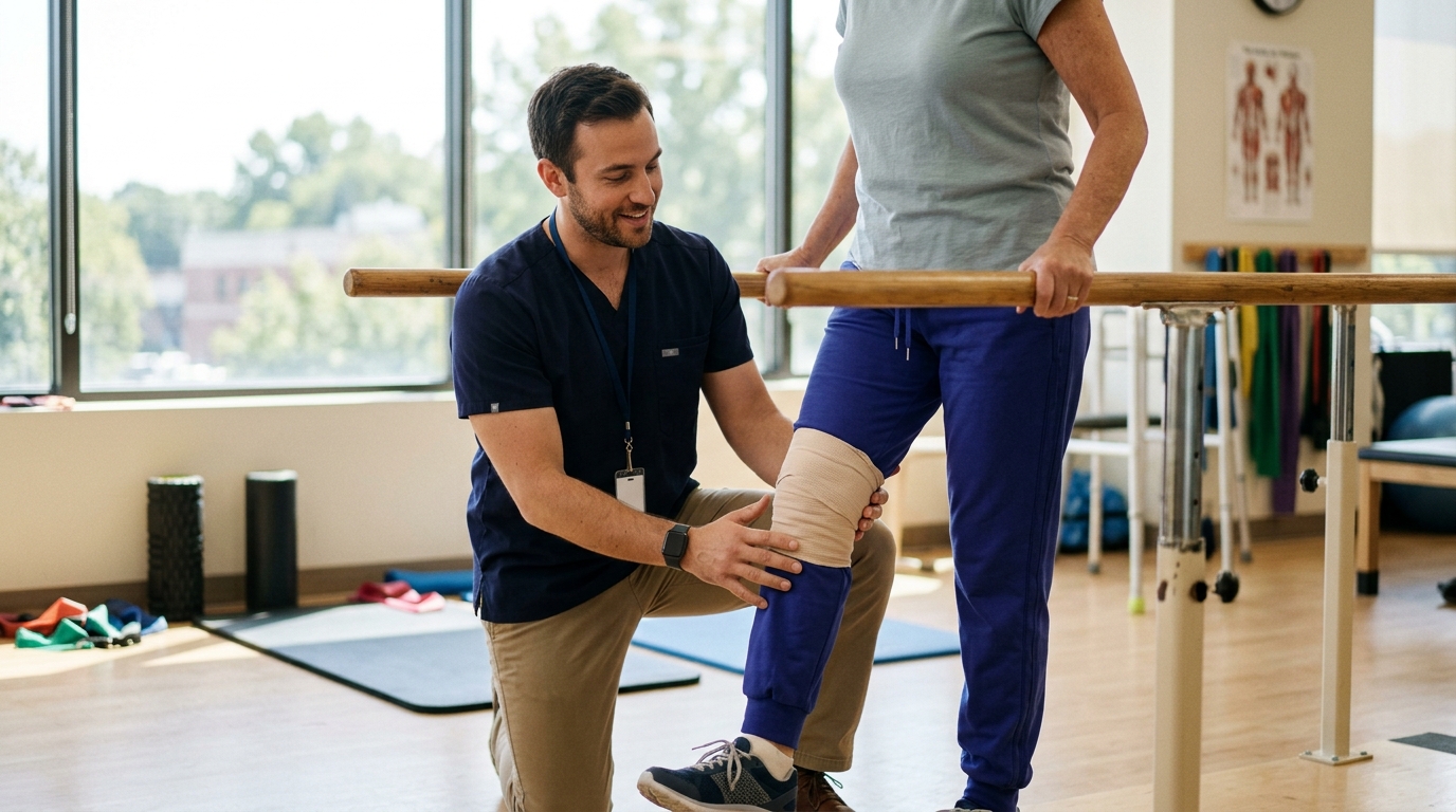
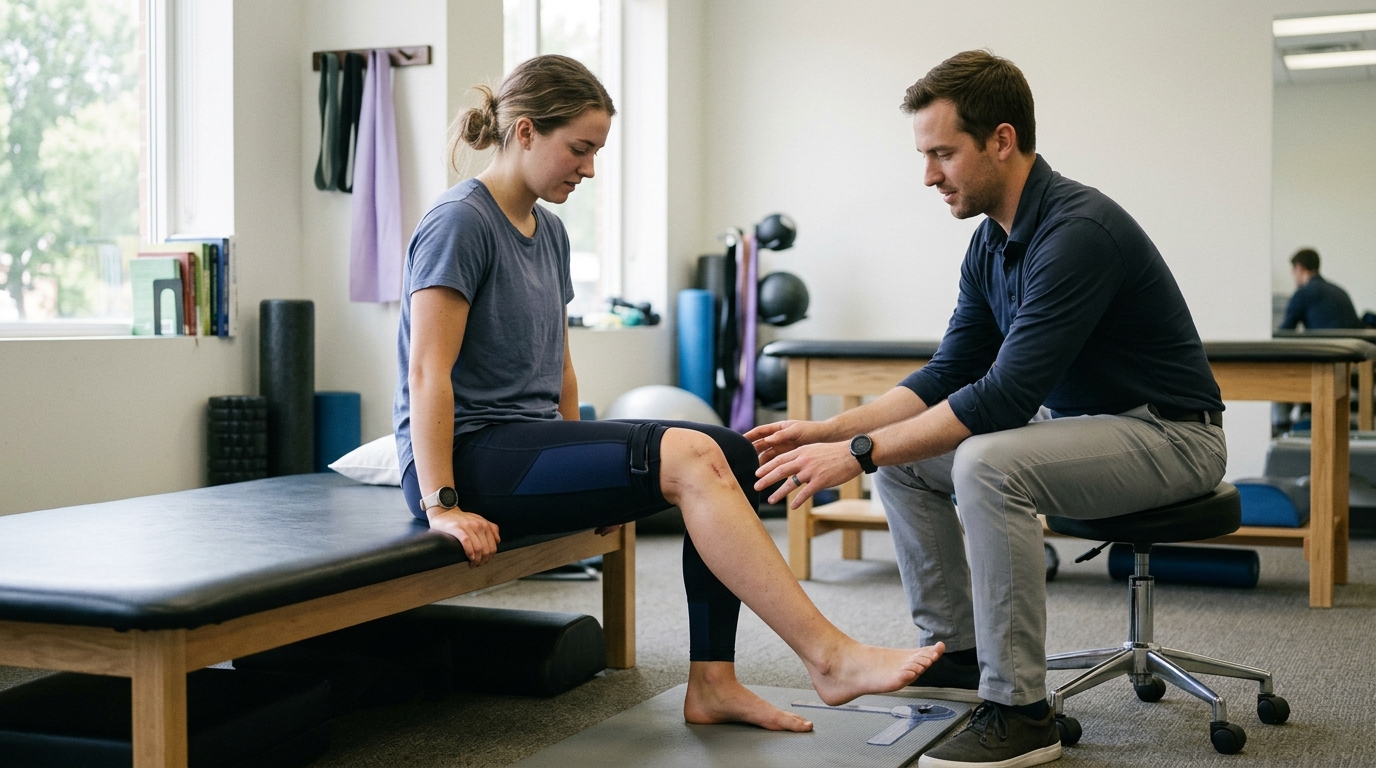
Rehabilitation in phase two builds a bridge from passive mobility to active arm control through restoring joint mobility and movement patterns. Your therapist will guide you through active-assisted exercises using tools such as canes, pulleys, or wall slides.
As your muscular control improves, you will transition to active movement against gravity. This begins in gravity-minimized positions, such as lying on your back, before advancing to sitting or standing positions.
Early phase two exercises often emphasize:
As you begin lifting your arm on your own, your brain may attempt to compensate for weak rotator cuff muscles by overusing the upper trapezius. This produces a shoulder hike, where the entire shoulder blade elevates toward the ear before the arm raises.
Repeated shoulder hiking pinches tissues against the acromion bone and creates poor movement habits.
Your physical therapist will emphasize movement quality over movement quantity. If you can only lift your arm sixty degrees before your shoulder begins to hike, you should train within that sixty-degree window until your rotator cuff can properly stabilize the joint.
Advancing to strengthening exercises requires achieving specific functional milestones:
Phase three generally encompasses months three and four of your rehabilitation program. With basic tissue healing established and active movement restored, the main objective is rebuilding muscular endurance, stability, and light functional strength.
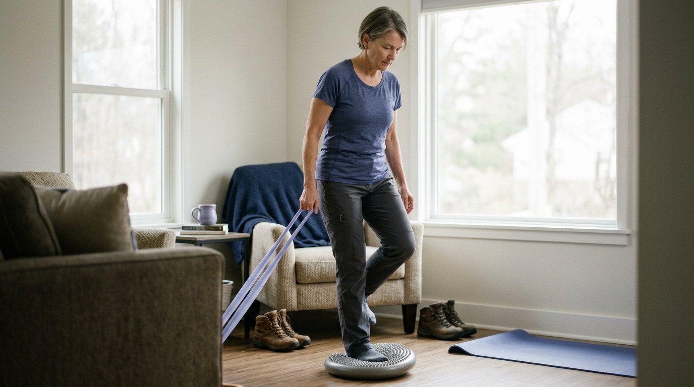
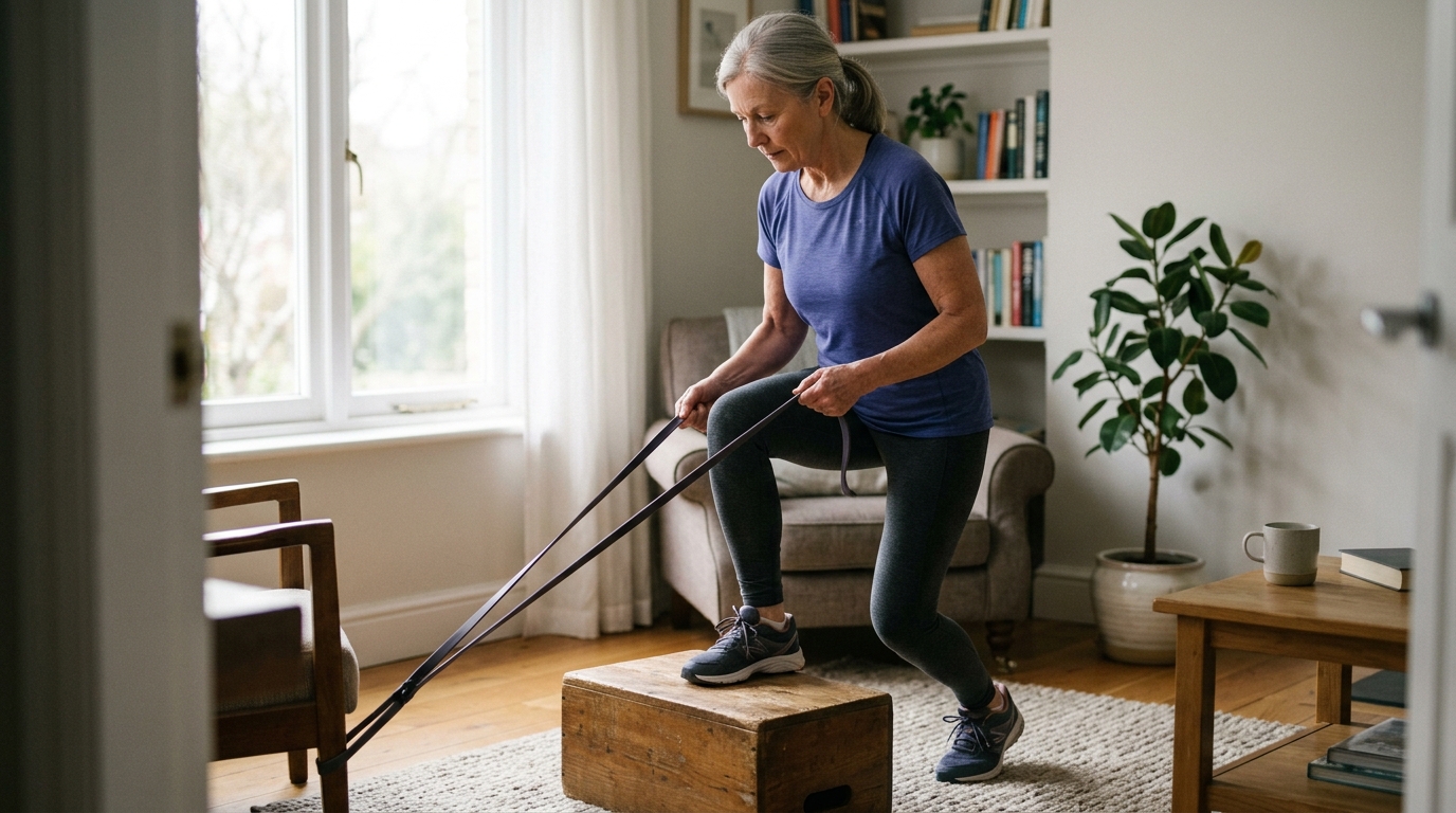
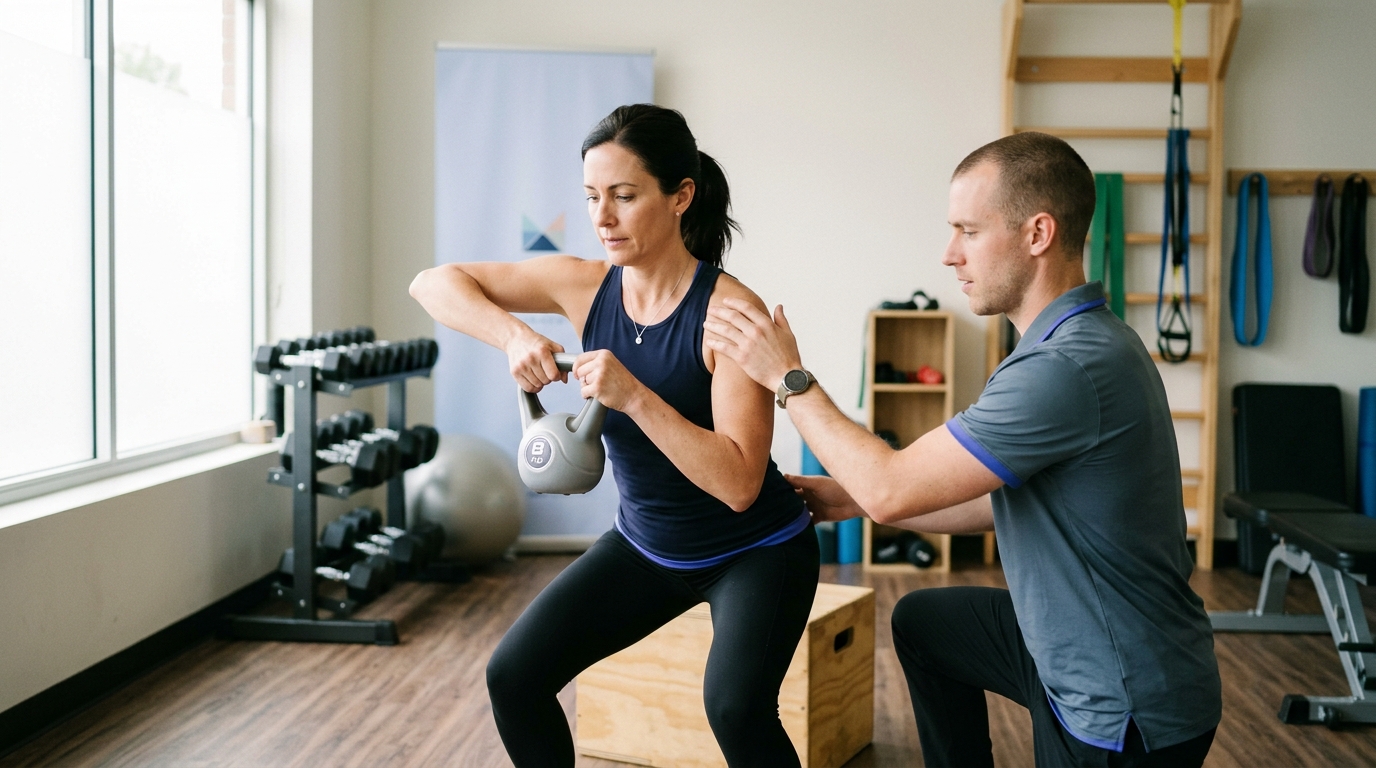
During phase three, resistance exercises are introduced systematically. Strengthening begins with light resistance bands, small hand weights, or isometric holds.
The rotator cuff consists of relatively small endurance muscles designed to stabilize the joint, not lift massive loads. High repetitions with low resistance and perfect form are far more valuable than lifting heavy weights with poor mechanics.
Phase three strengthening typically targets:
Although active range of motion is often well-developed by month three, phase three protocols generally restrict heavy overhead lifting, aggressive pushing, and ballistic pulling. Reaching overhead with an added external load multiplies the mechanical stress on the healing tendon.
Workplace tasks that involve lifting objects off high shelves or repetitive overhead tools should remain restricted during this stage.
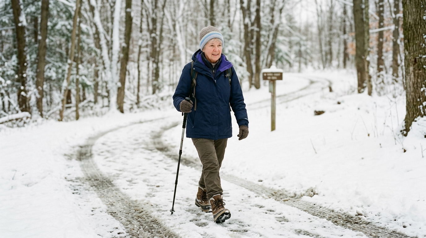
Daily functional tasks, such as light meal preparation, typing, driving, and carrying light groceries under five pounds close to the body, are usually well-tolerated.
You should continue to avoid sudden, jerky movements, such as catching a falling object or walking a large, pulling dog on a leash.
Moving into advanced functional training requires clearing several intermediate benchmarks:
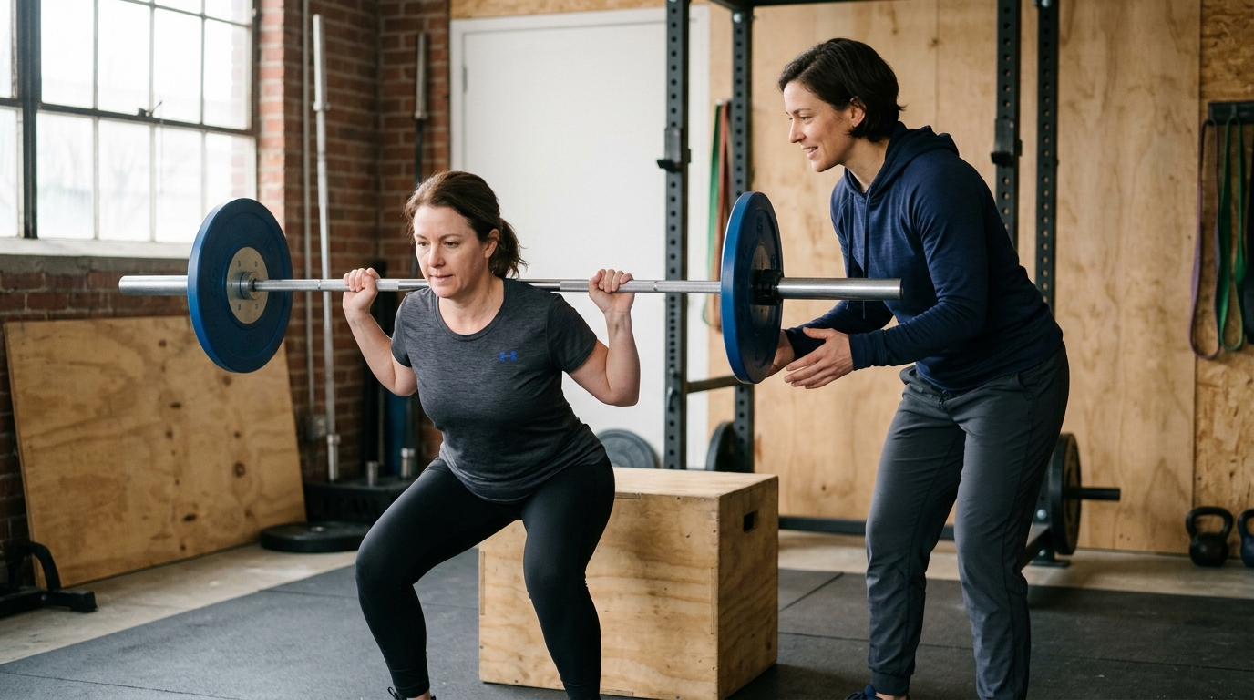
Phase four typically begins around month four and extends through month six, nine, or twelve depending on your physical goals. This phase prepares the shoulder for higher physical demands, including manual labor, recreational athletics, heavy lifting, and overhead sports.
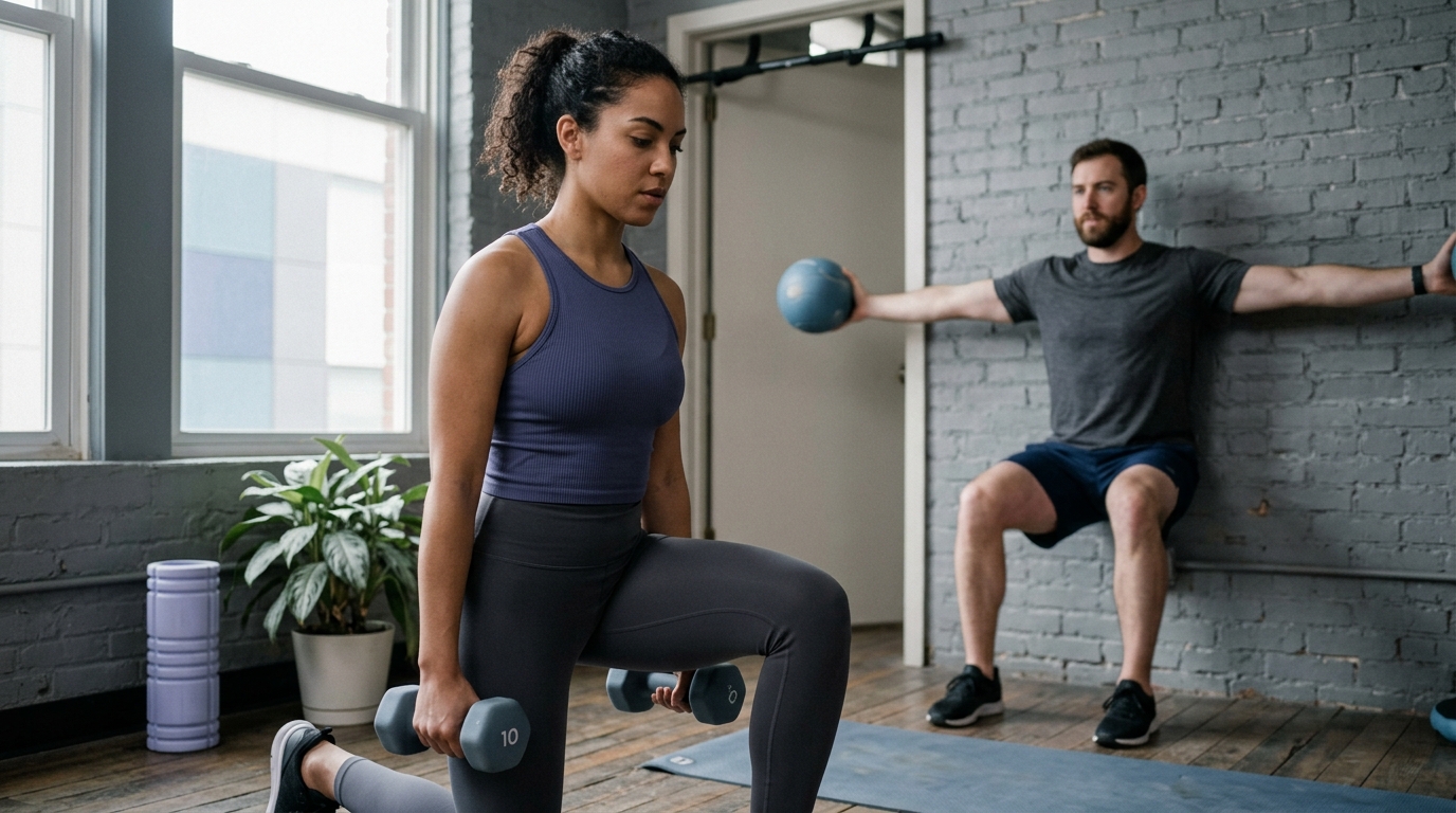
Rehabilitation in phase four focuses on dynamic joint stability, multi-planar movements, and progressive overload through strength rebuilding resources. Exercises become more complex and functional, challenging the shoulder across different angles and speeds.
Standard phase four training includes:
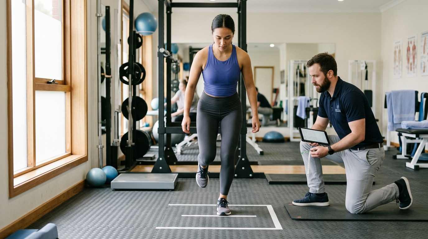
Clearing a patient to return to recreational sports or demanding occupations should never rely solely on a calendar date. Biological healing timelines must intersect with objective physical performance testing.
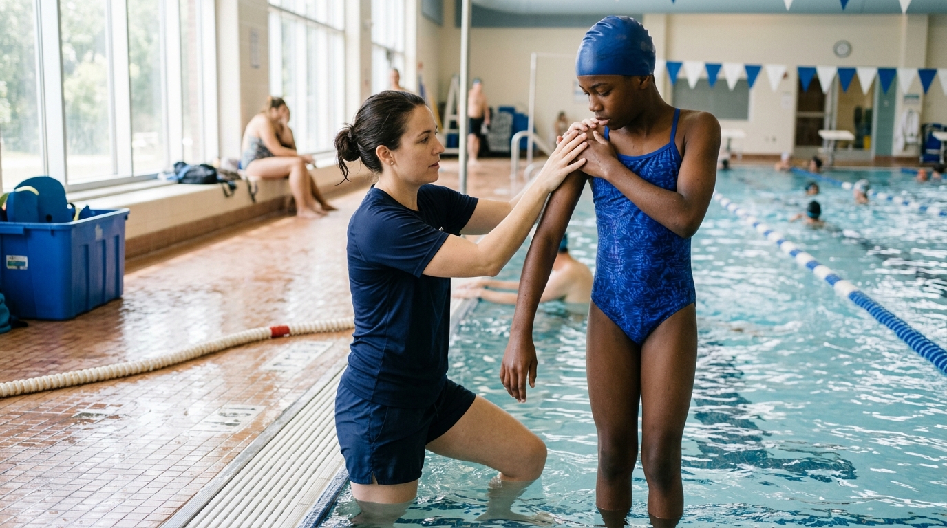
Returning to overhead athletics, such as tennis, swimming, or baseball, requires passing a structured return to sport progression under clinical supervision.
Clearance criteria typically require:
Surgeons and physical therapists continue to study the ideal balance between early joint movement and structural protection. Reviewing the published medical evidence clarifies why protocols vary across clinical practices.
A central question in shoulder rehabilitation is whether early passive motion increases the risk of a tendon retear compared to keeping the arm immobilized longer.
A systematic review and meta-analysis evaluated this question and found no statistically significant difference in retear risk between early and delayed motion. The calculated risk ratio was 1.01 with a 95 percent confidence interval of 0.63 to 1.64 (P = 0.95).
The review also noted a small difference in forward elevation of only 1 degree in favor of early passive motion, which researchers characterized as clinically unimportant.
In a randomized clinical trial of 124 patients analyzed within that review, 92 percent of tears healed successfully. Structural healing rates did not differ significantly between early and delayed rehabilitation groups (P = 0.46).
Active elevation and external rotation favored the traditional delayed group at three months. By the final follow-up, functional scores, range of motion, and strength were identical between both groups.
Another meta-analysis on early versus delayed mobilization similarly found no apparent increase in retear risk with early movement, reporting an odds ratio of 1.05 with a 95 percent confidence interval of 0.64 to 1.75.
However, findings across the wider literature are not completely uniform. A separate systematic review of retear rates suggested that early motion might provide slightly faster early mobility gains but could carry a higher retear risk in certain patient subsets.
The authors of that review suggested that early motion might be best suited for small tears, while delayed motion may be safer for larger tears.
The American Academy of Orthopaedic Surgeons (AAOS) addressed postoperative mobilization in its clinical practice guideline updates. The guideline notes high-level evidence showing that after arthroscopic repair of small-to-medium full-thickness tears, outcomes are comparable whether sling immobilization begins early or is maintained for up to eight weeks.
The AAOS guideline also reports that selected patients with small-to-medium repairs may begin weaning from their sling as early as two weeks after surgery to allow light active range of motion for basic daily activities.
These patients demonstrated similar tendon healing and functional scores compared to patients who maintained prolonged sling use for four to six weeks.
This finding is an option for specific patients under medical supervision, not a universal rule to discard the sling without physician clearance.
The medical literature confirms that there is no single rehabilitation timetable suitable for every patient. Early passive motion can safely reduce early stiffness in selected repairs, but it does not justify aggressive active loading.
Results from small-to-medium repairs cannot be automatically applied to massive, multi-tendon tears.
If you compare your rehabilitation timeline with another person who had rotator cuff surgery, you may discover substantial differences. These variations reflect critical anatomical and surgical factors documented in clinical protocols, including those from Mass General Brigham and Sanford Health.
Rotator cuff tears are categorized by size: small (less than 1 centimeter), medium (1 to 3 centimeters), large (3 to 5 centimeters), and massive (greater than 5 centimeters or involving two or more tendons).
Small tears with healthy tissue edges have a high healing capacity and can tolerate earlier passive movement.
Large or massive tears involve substantial tissue loss, higher suture tension, and a greater risk of structural failure. Rehabilitation for complex or multi-tendon tears follows a conservative protocol, often extending phase one protection for six to eight weeks before beginning active motion.
The rotator cuff consists of four distinct muscles: the supraspinatus, infraspinatus, teres minor, and subscapularis. The location of your tear determines which movement directions place tension on the repair.
A repair involving the subscapularis tendon on the front of the shoulder requires strict movement restrictions. Subscapularis protocols strictly limit passive external rotation and prohibit resisted internal rotation for several weeks.
Performing generic rotator cuff exercises without knowing which tendon was repaired can tear a subscapularis repair.
Tendon tissue quality varies significantly between individuals. Healthy, thick tendon tissue holds sutures securely, allowing standard progression.
Frayed, thin, or fatty-infiltrated tendon tissue provides a weaker hold for surgical anchors.
When tissue quality is compromised, the surgeon will deliberately slow down the rehabilitation timeline to give the repair site extended time to form a biological bond.
Overall health status directly influences the speed and quality of collagen synthesis:
Your daily physical demands dictate the length of your later rehabilitation phases. A desk worker may return to typing and modified office duties within two to four weeks while wearing a sling.
A carpenter, electrician, or heavy laborer who lifts tools overhead may require six to nine months of structured conditioning before safely resuming unrestricted manual work.
Rehabilitation timelines must be customized to bridge the gap between basic joint motion and specific daily loads, as outlined in injury recovery and healing guides.
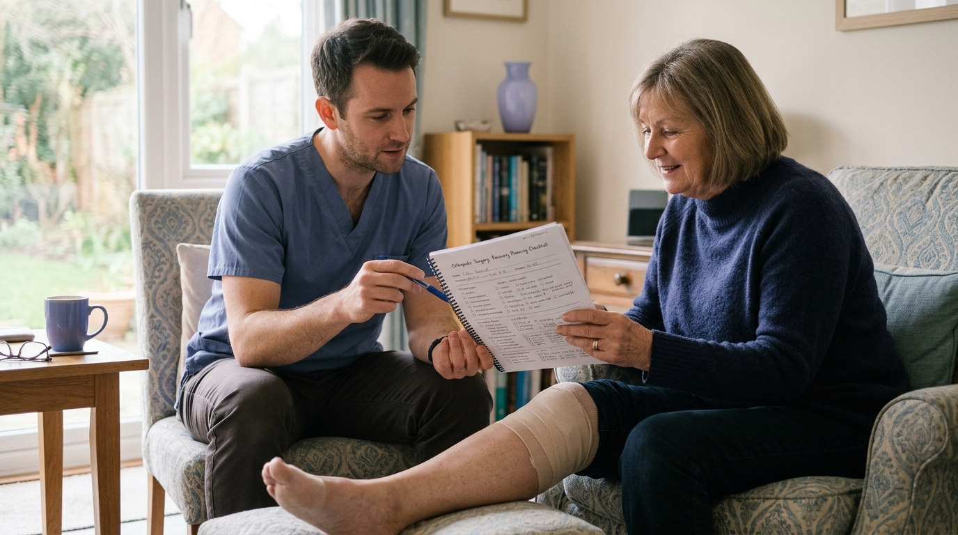
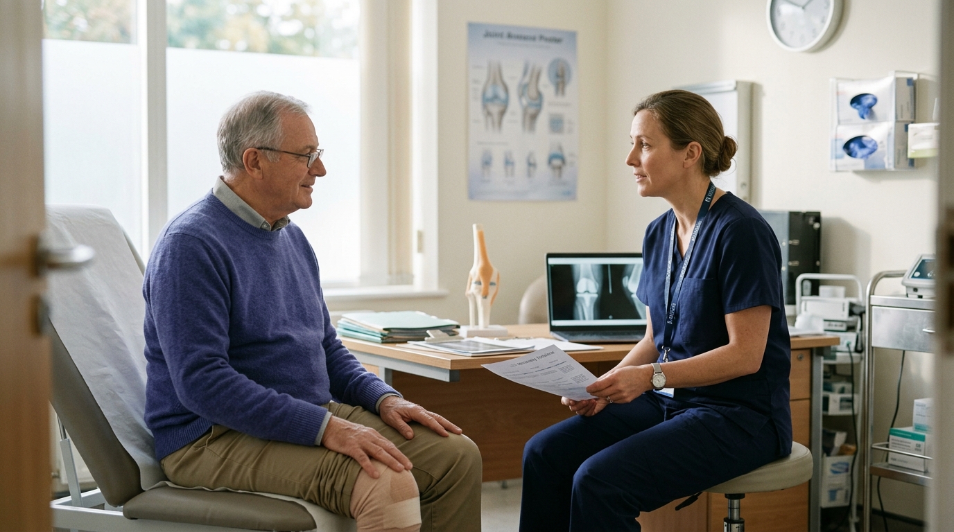
Because every shoulder repair is unique, you should discuss your specific surgical details and restrictions directly with your care team. Consider bringing these questions to your follow-up appointments:
Recovering from rotator cuff repair surgery requires patience, consistency, and respect for biological healing timelines. Regaining active arm elevation is a major milestone, but functional motion does not mean the tendon is ready for heavy resistance.
Protecting the surgical repair during early phases establishes the structural foundation needed for advanced strength and endurance later.
Use published recovery phases as a helpful orientation for what lies ahead, but always follow the specific restrictions established by your surgeon and physical therapist.
To keep your recovery on track this week, focus on these practical actions:

Use ReboundBody resources to understand common recovery stages, rehab terms, movement limits and strength rebuilding. Each guide is designed to make a complex comeback easier to understand.


Read practical guidance on injury recovery, rehabilitation, mobility and rebuilding strength as you work your way back to activity.
Explore Resources