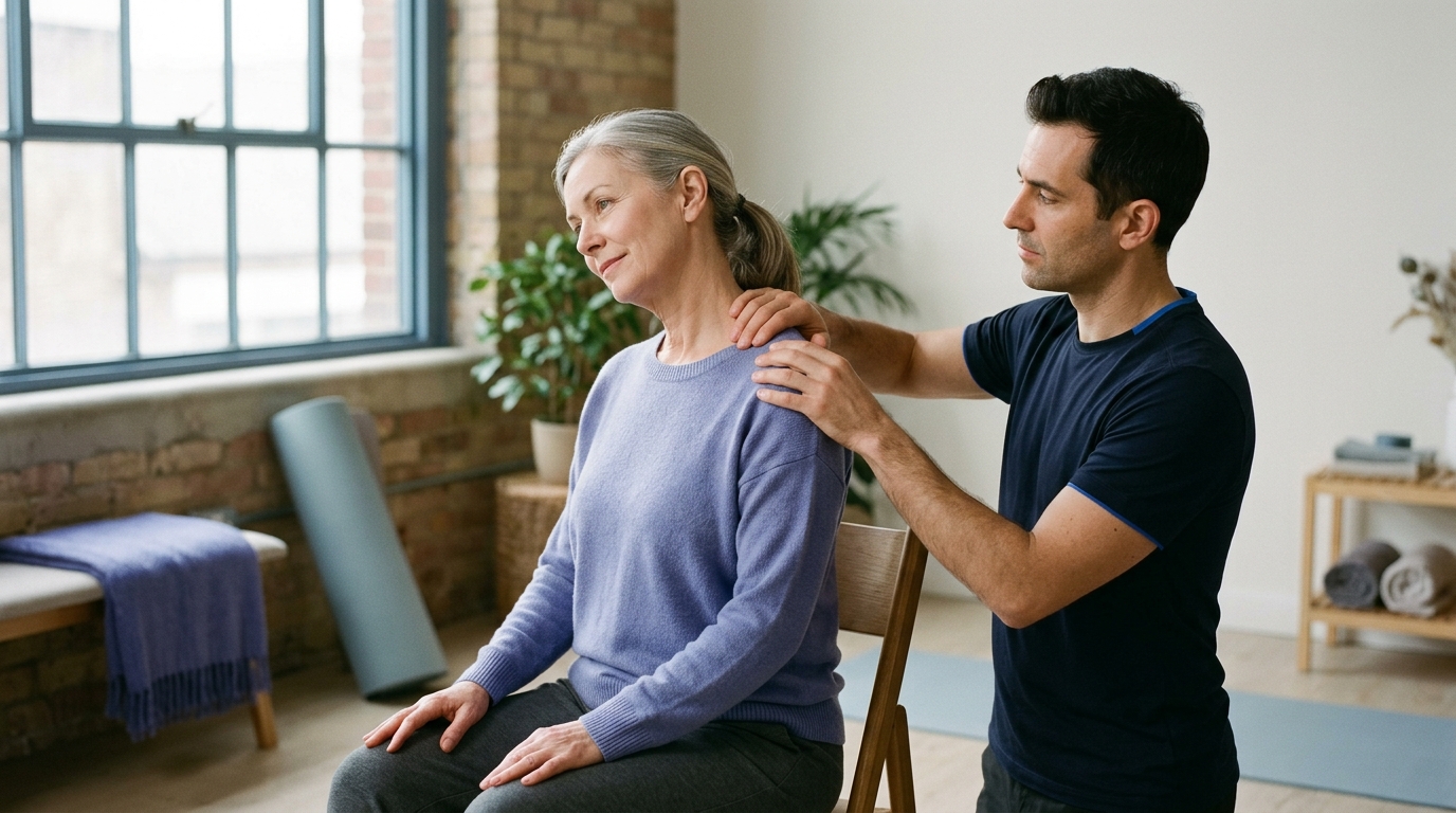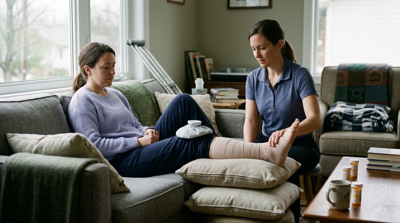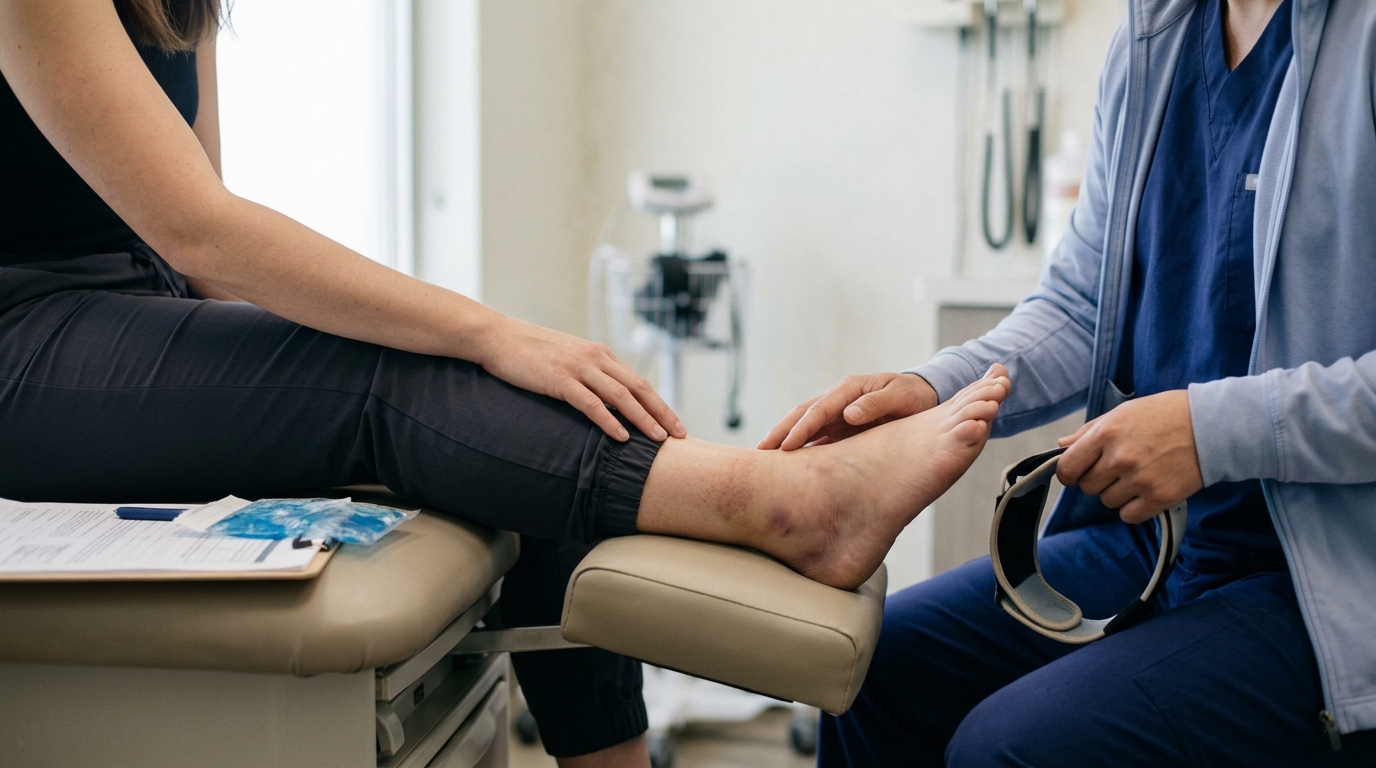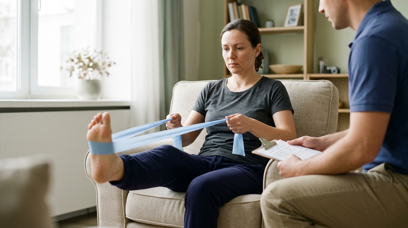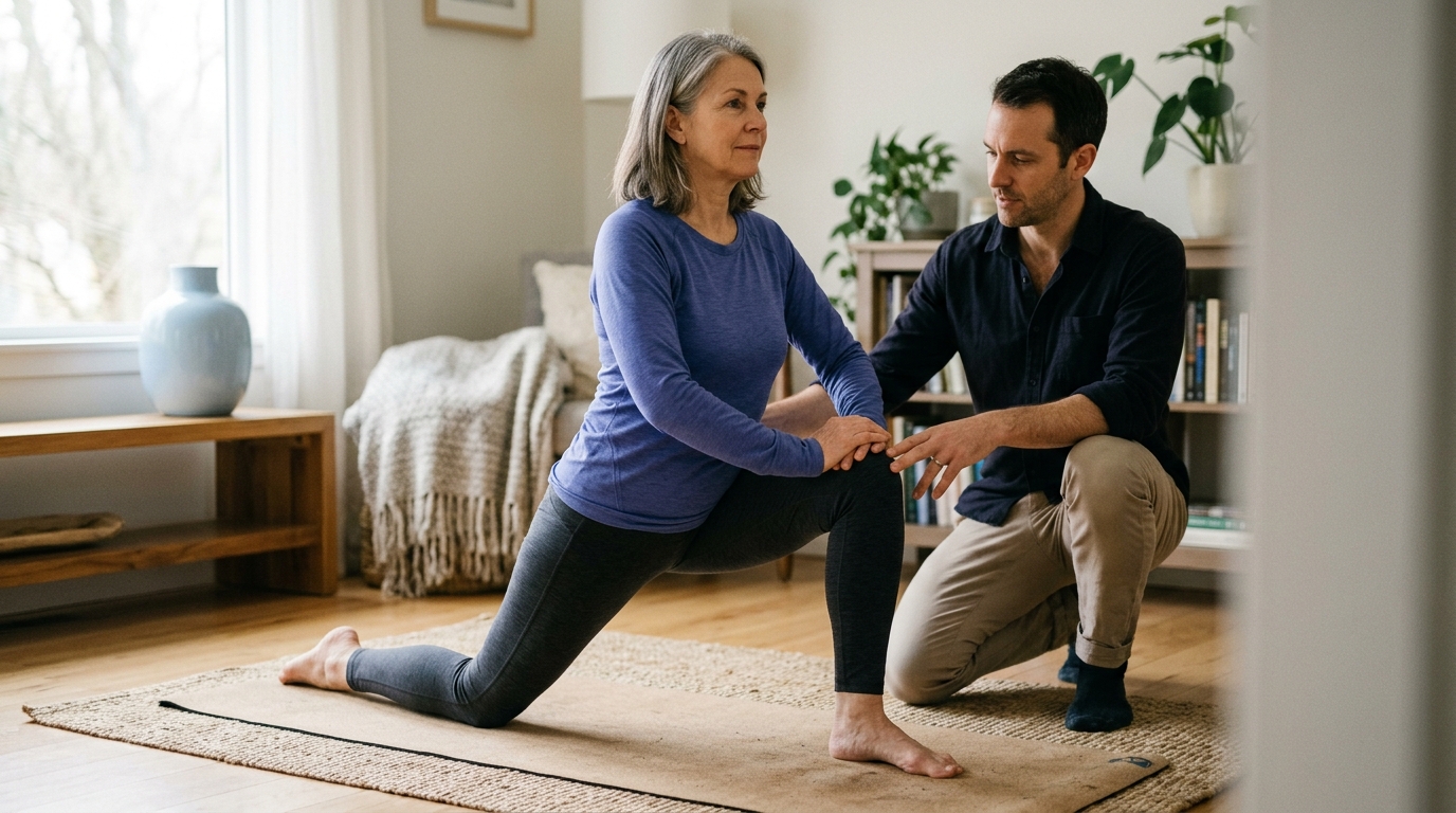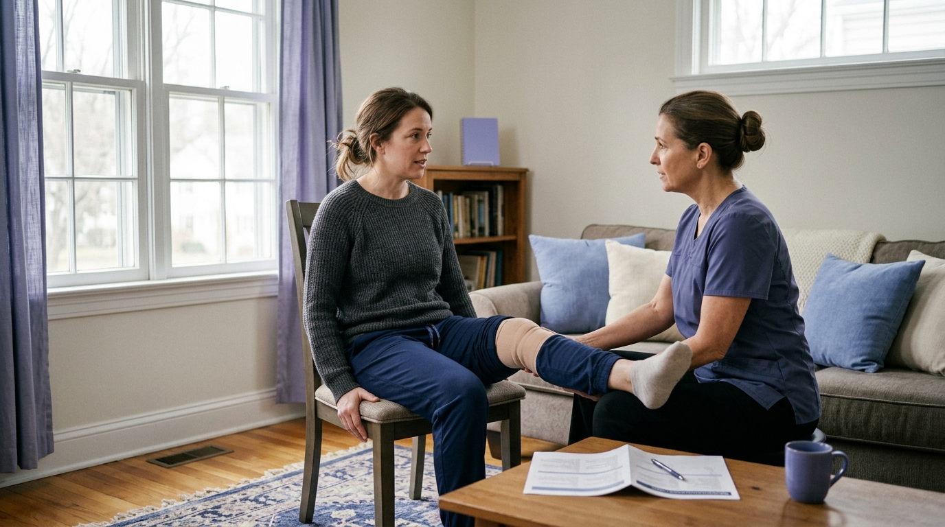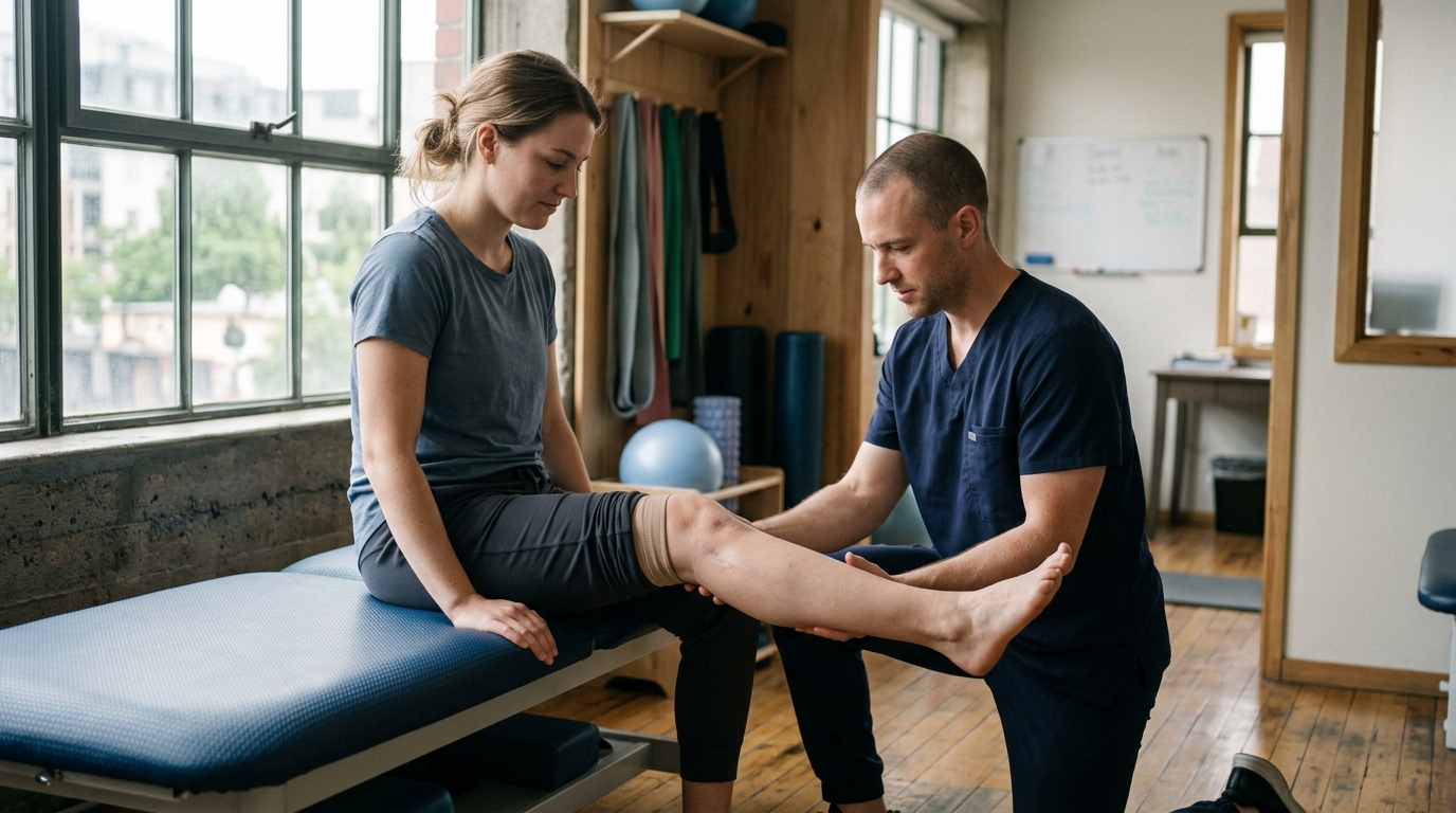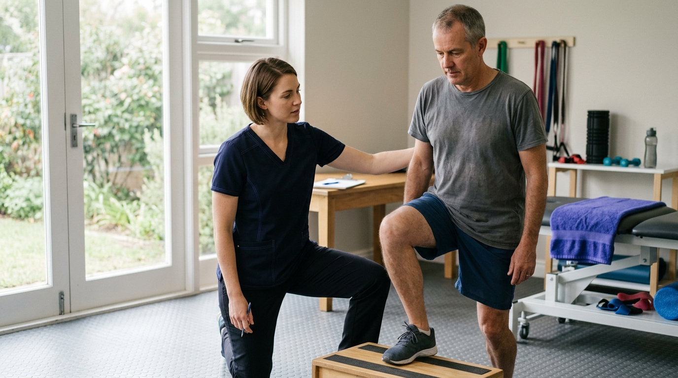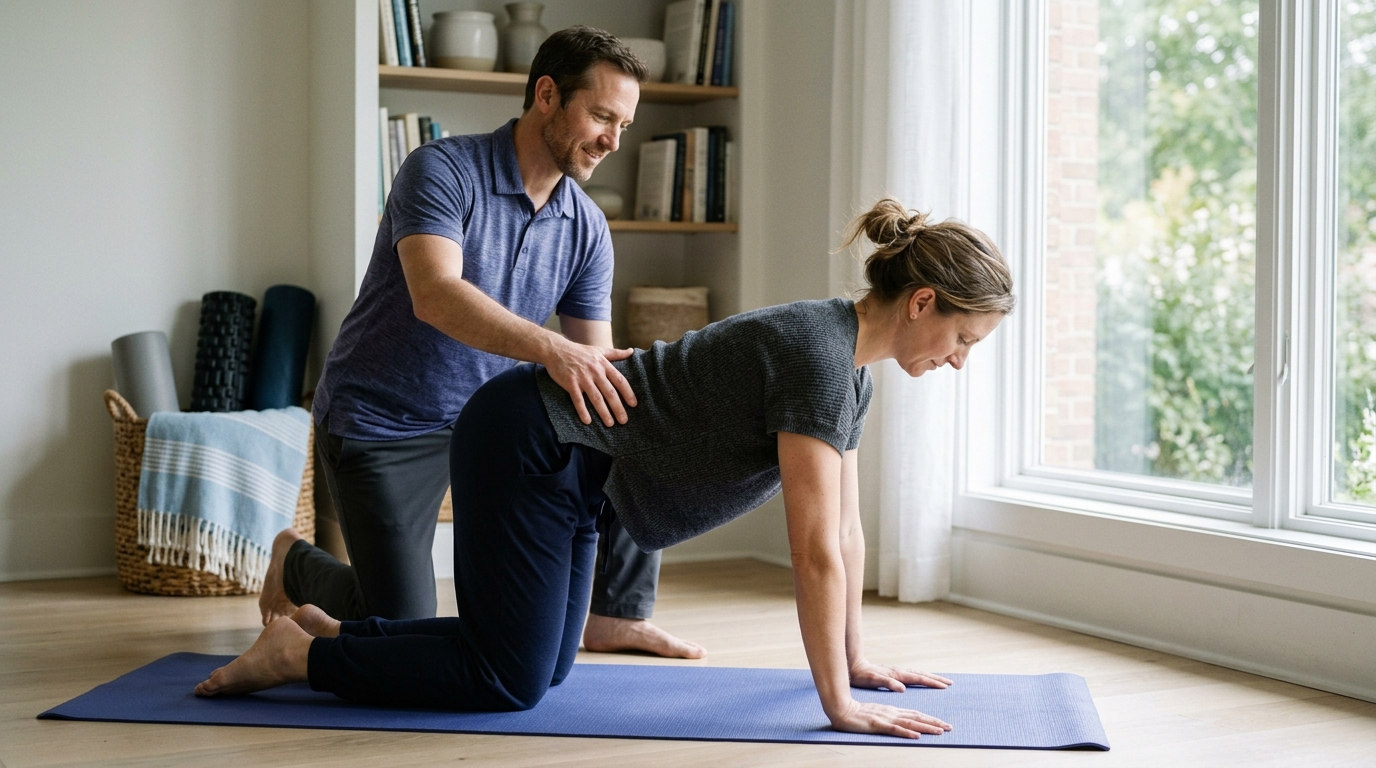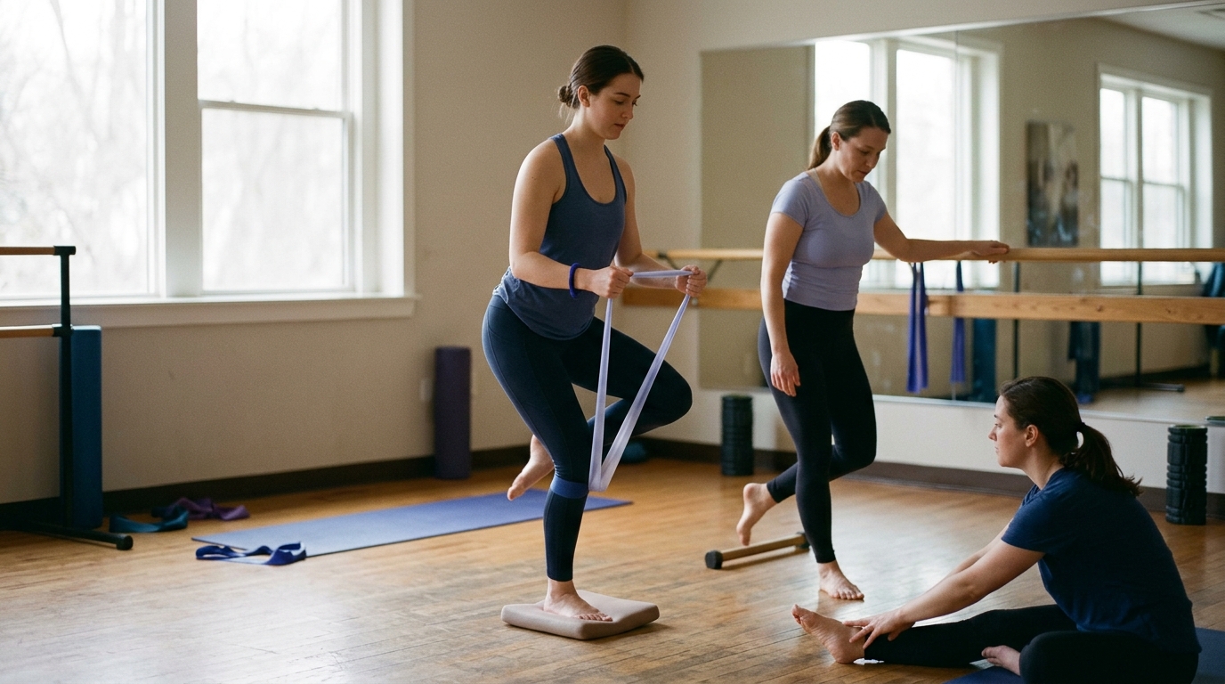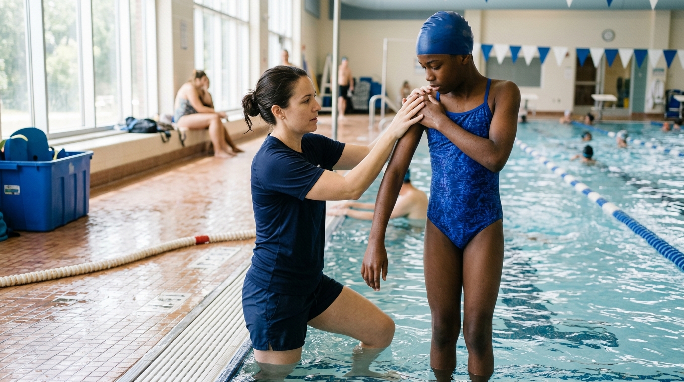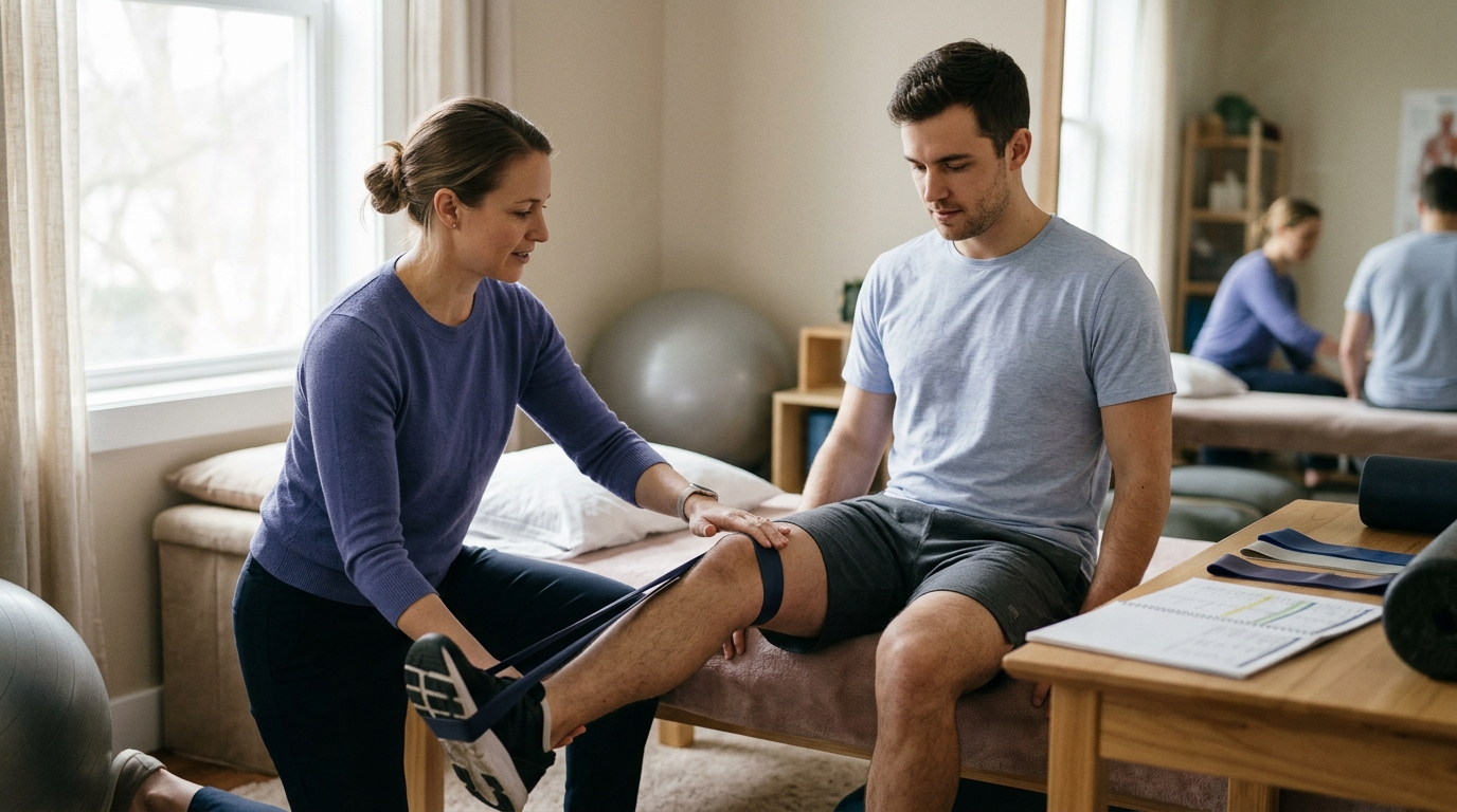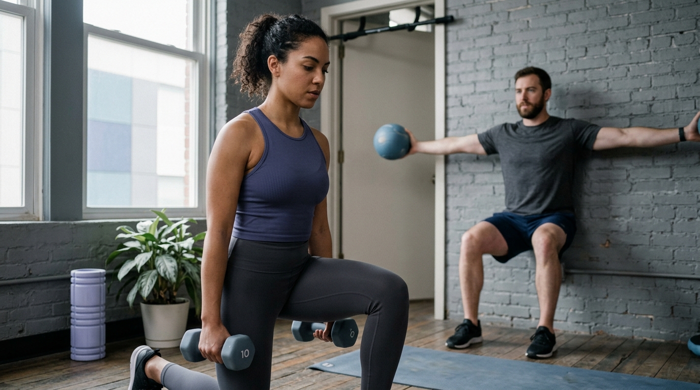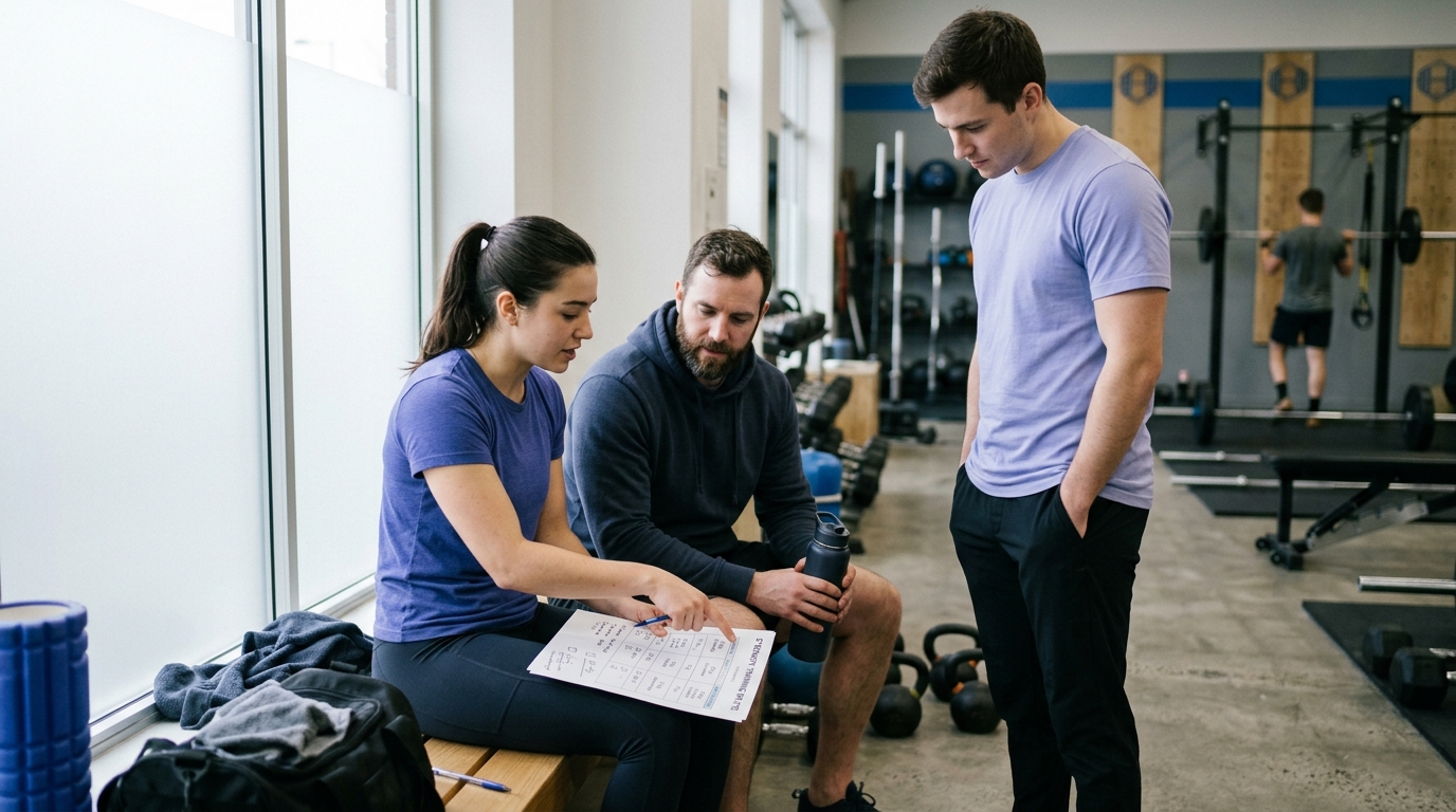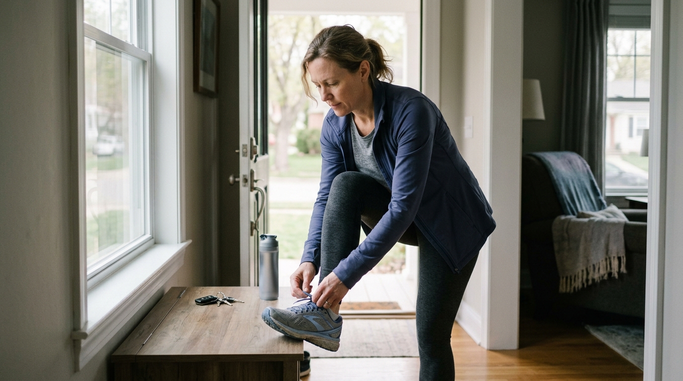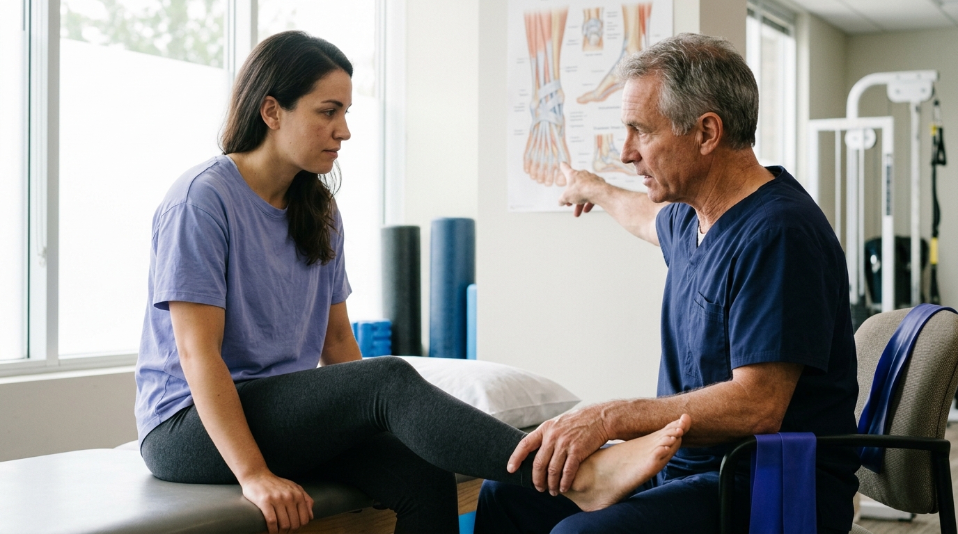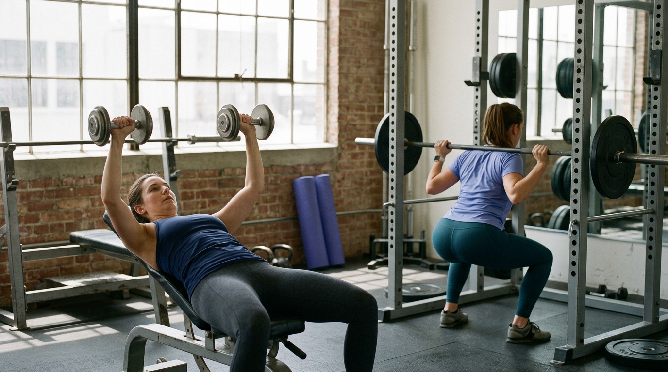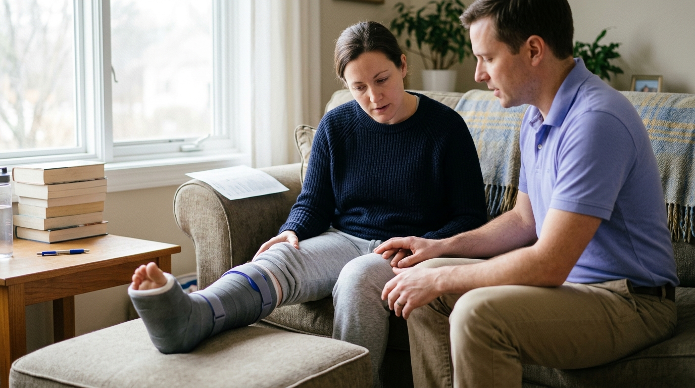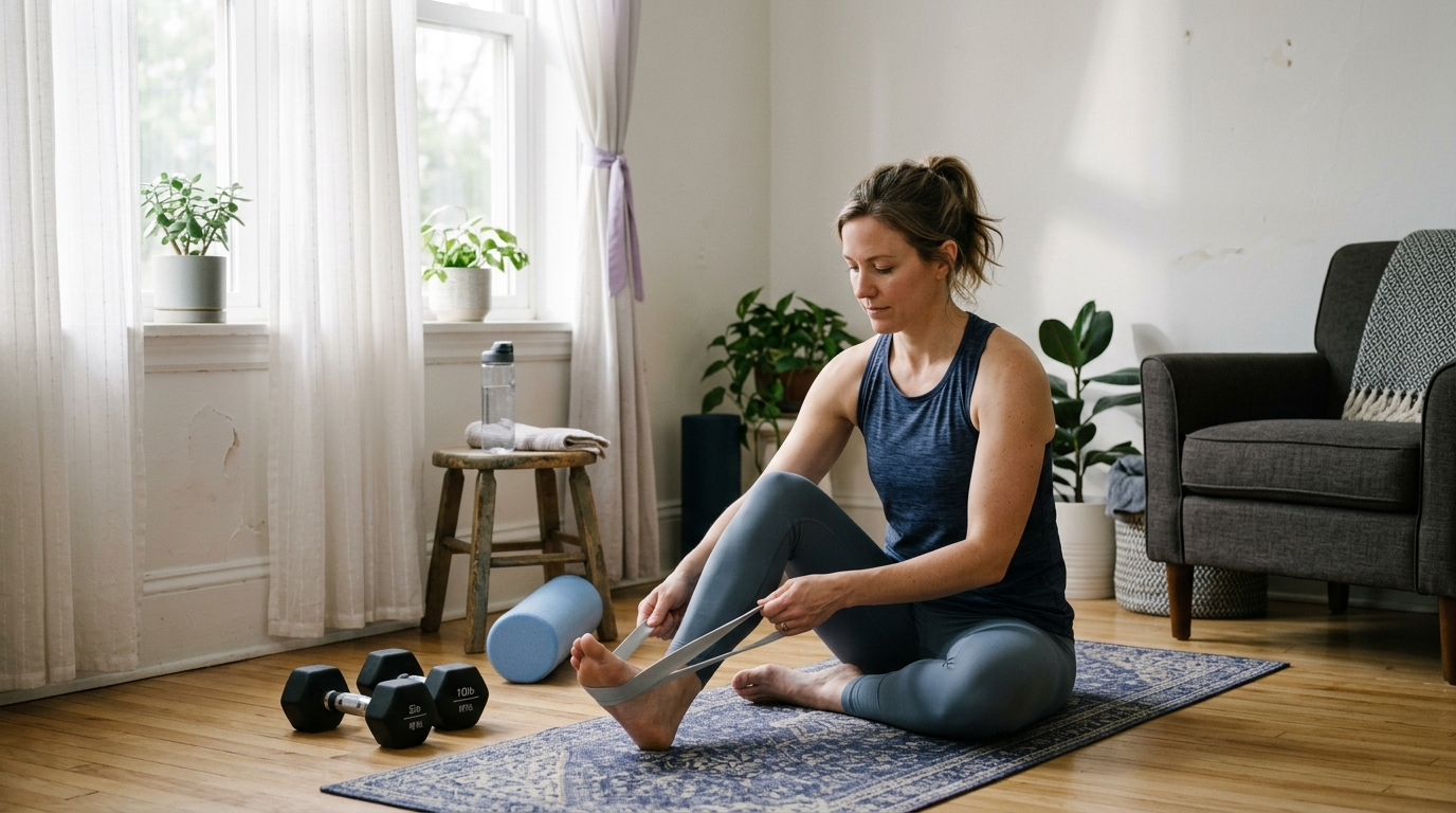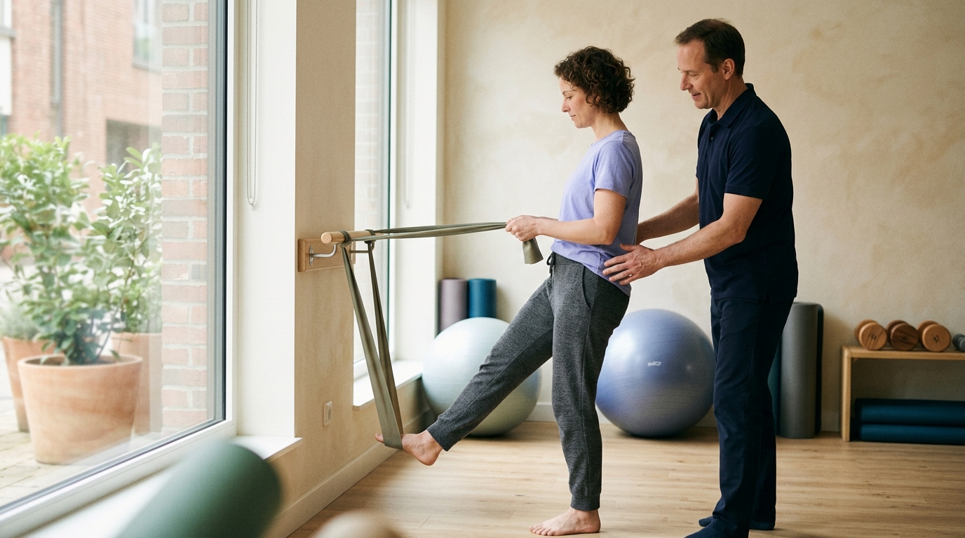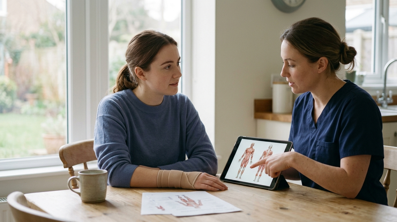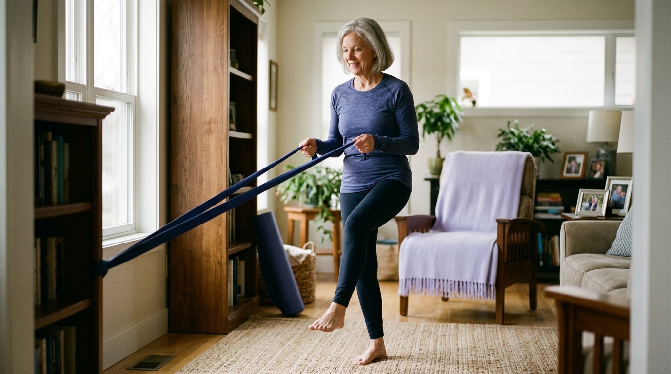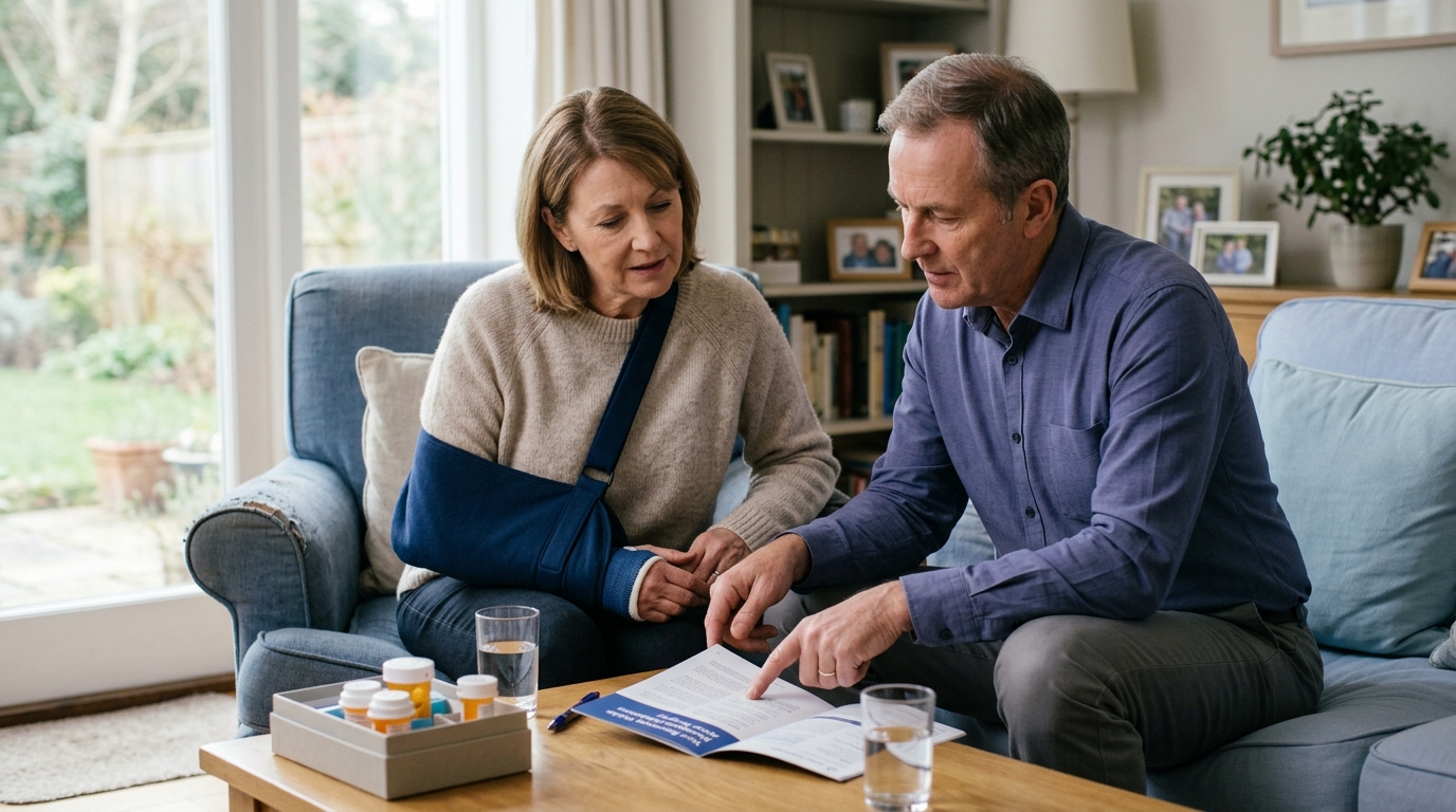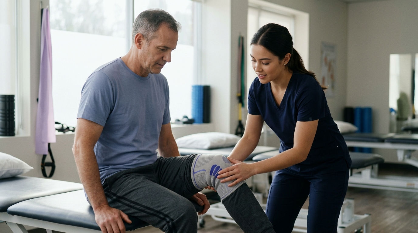

Descending stairs or rising from a chair feels manageable again with evidence-based rehabilitation strategies that rebuild knee strength, mobility, and coordination.





Knee rehabilitation is a structured process of restoring how the joint moves, bears weight, and manages daily activity. It is not a passive waiting period, nor is it simply a race to bend the joint to a certain angle.
True joint recovery requires restoring four distinct pillars of physical function. These pillars are joint mobility, muscular strength, movement coordination, and activity tolerance. Measuring only how far the knee bends leaves out critical information about how the leg handles real life.
Everyday movements such as walking, climbing stairs, and standing up from a low chair require these four elements to work together. When one element is missing, other joints and muscles must compensate.
Your specific recovery path depends heavily on whether you are managing long-term cartilage changes, recovering from a total joint replacement, or rehabilitating a reconstructed ligament. Protocols vary significantly between procedures, meaning that general timelines cannot replace personalized clinical guidance.
This guide provides a comprehensive overview of how knee function is evaluated and rebuilt. You will learn how basic daily tasks serve as functional tests, what scientific research says about exercise therapy, and how criterion-based milestones help protect healing tissues.
Restoring knee function requires looking past single measurements like degrees of flexion. A knee that bends fully while lying on an examination table may still struggle to support body weight on a flight of stairs.
Clinicians evaluate function using a four-part framework that examines how the joint performs under load. These four dimensions are mobility, strength, coordination, and activity tolerance.
Each dimension addresses a different aspect of joint mechanics and muscular control. Working on all four areas creates a balanced foundation for everyday tasks and recreational pursuits.
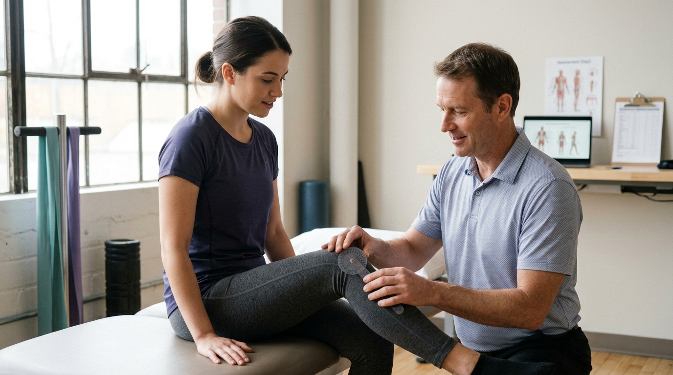
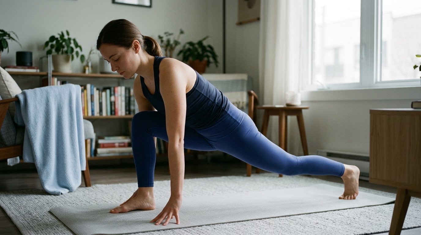
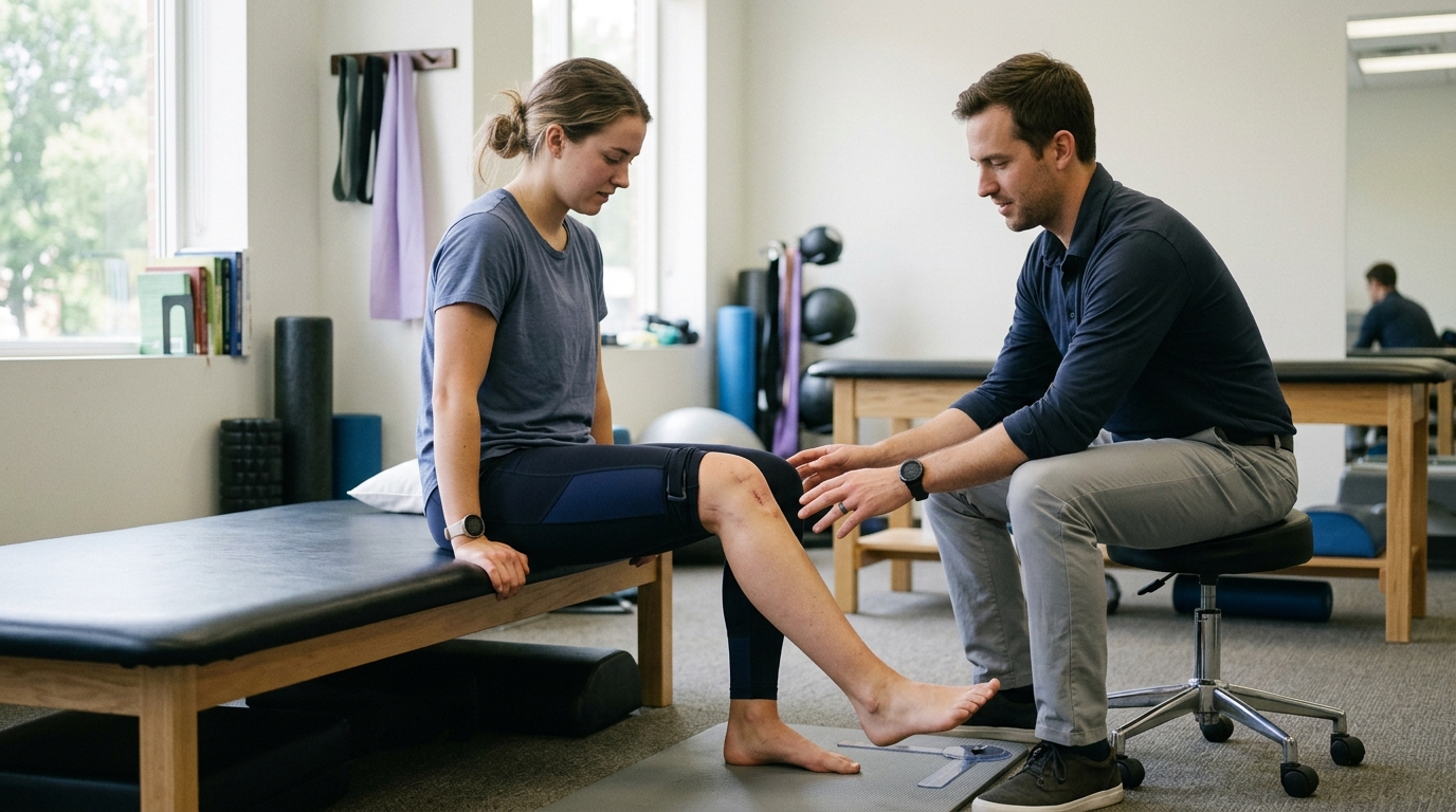
Mobility refers to the available range of motion in the knee joint during active and passive movement. It includes the ability to straighten the leg fully, known as extension, and the ability to bend the knee, known as flexion.
Full knee extension is vital for a normal walking pattern and efficient standing posture. Even a minor loss of extension increases the muscular effort required by the quadriceps during normal standing.
Flexion is equally necessary for common daily tasks. Sitting comfortably in a standard chair requires roughly 90 degrees of knee bend, while descending stairs and squatting demand even greater range.
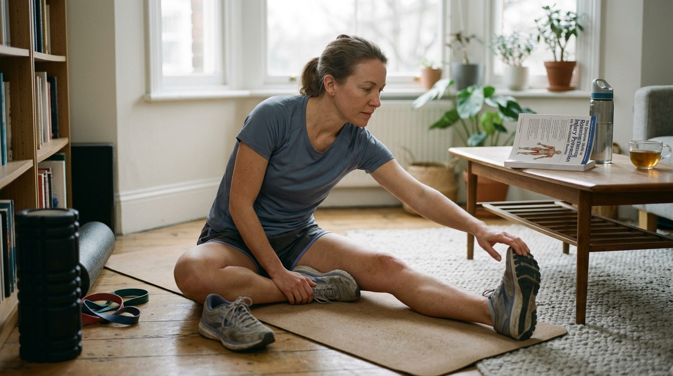
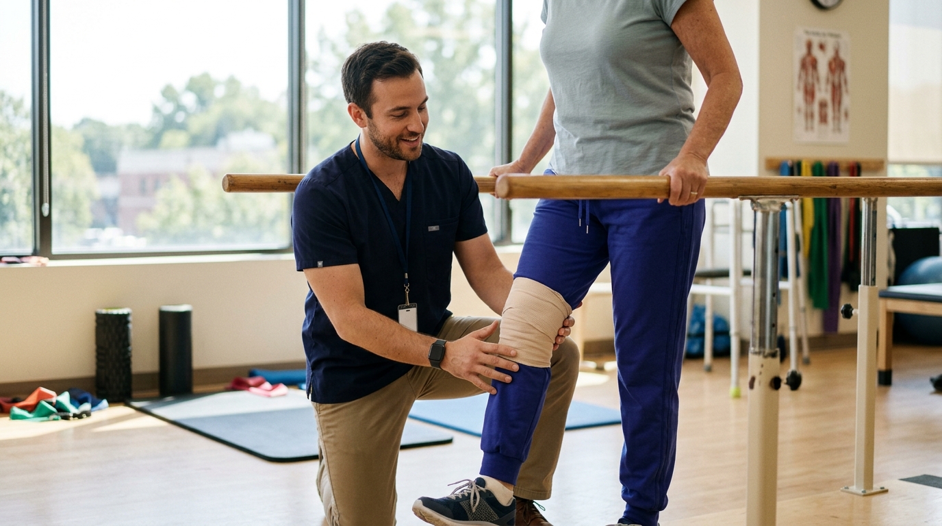
Rehabilitation exercises work to regain this usable range without irritating healing tissues. However, mobility must always be matched with muscular control to ensure the joint remains stable throughout its entire arc.
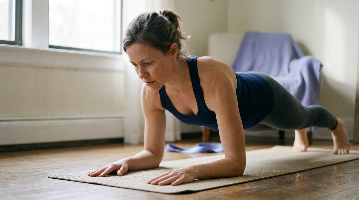
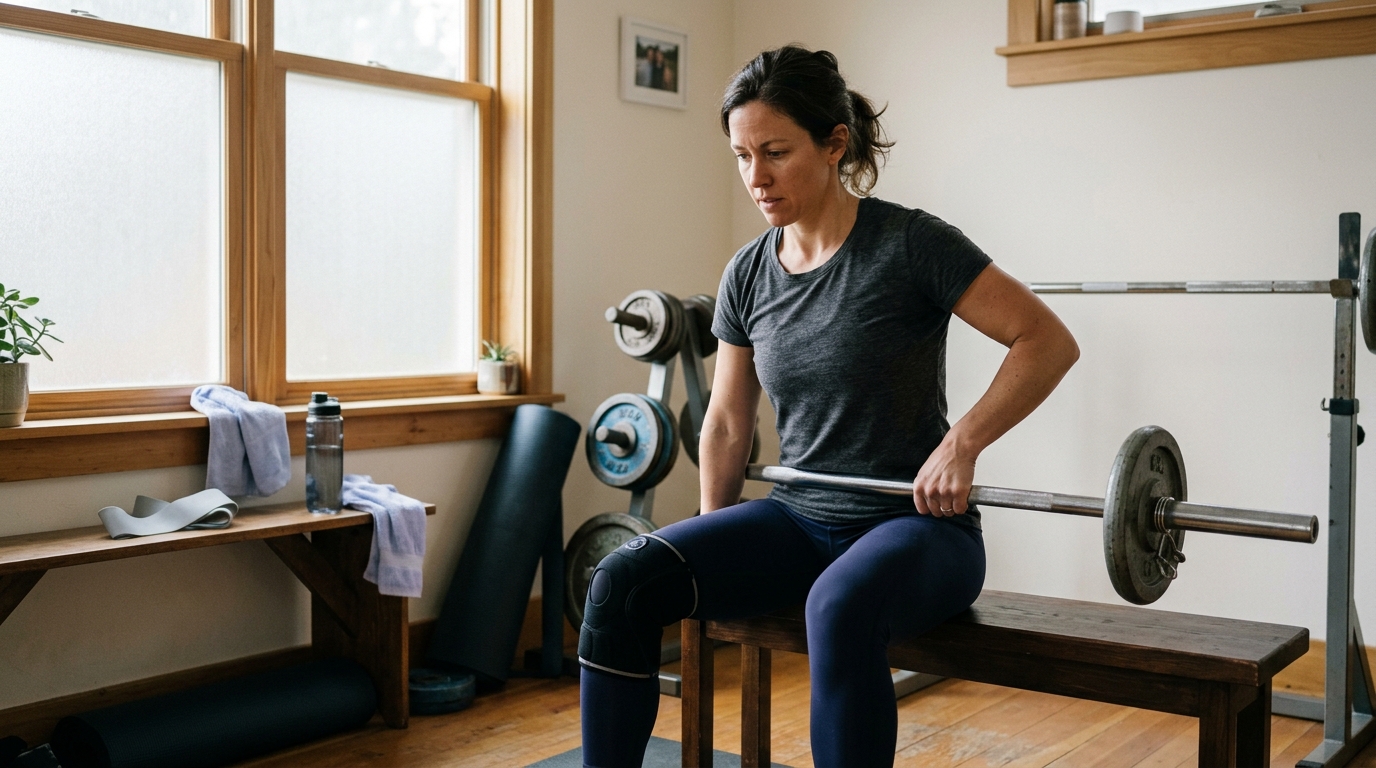
Strength is the capacity of surrounding muscles to produce force, absorb impact, and stabilize the knee joint. The quadriceps muscles on the front of the thigh play a central role in controlling knee flexion when you sit down or walk downstairs.
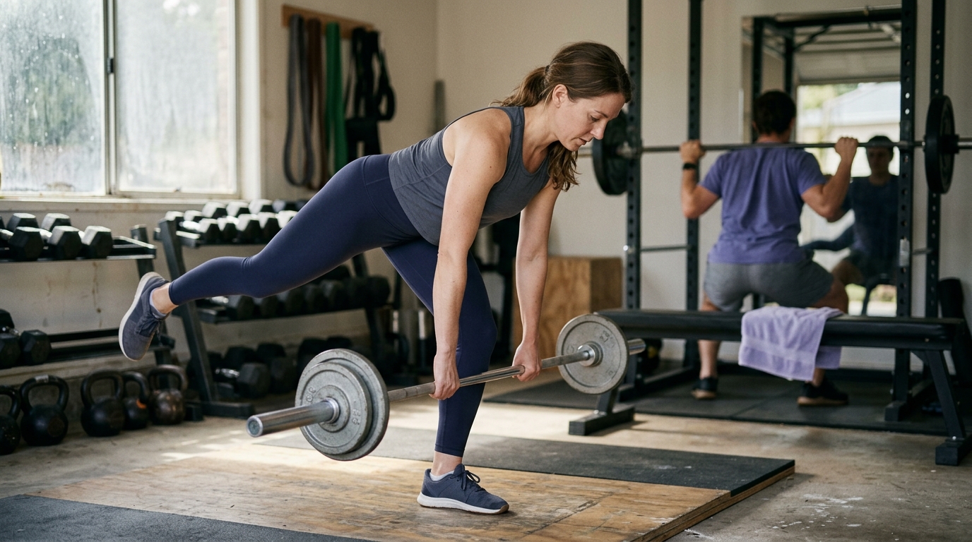
The hamstrings on the back of the thigh and the gluteal muscles in the hip provide essential support by controlling rotation and stabilizing the pelvis. Calf muscles also contribute significantly to forward propulsion and ankle stability during walking.
Strength rehabilitation focuses on both concentric actions, where the muscle shortens to create movement, and eccentric actions, where the muscle lengthens under tension to brake motion. Eccentric quadriceps strength is especially critical for safe stair descent and shock absorption.
Without adequate strength, daily mechanical forces are transferred directly to joint surfaces and passive ligaments. Building balanced muscular support protects internal structures from excessive physical stress.
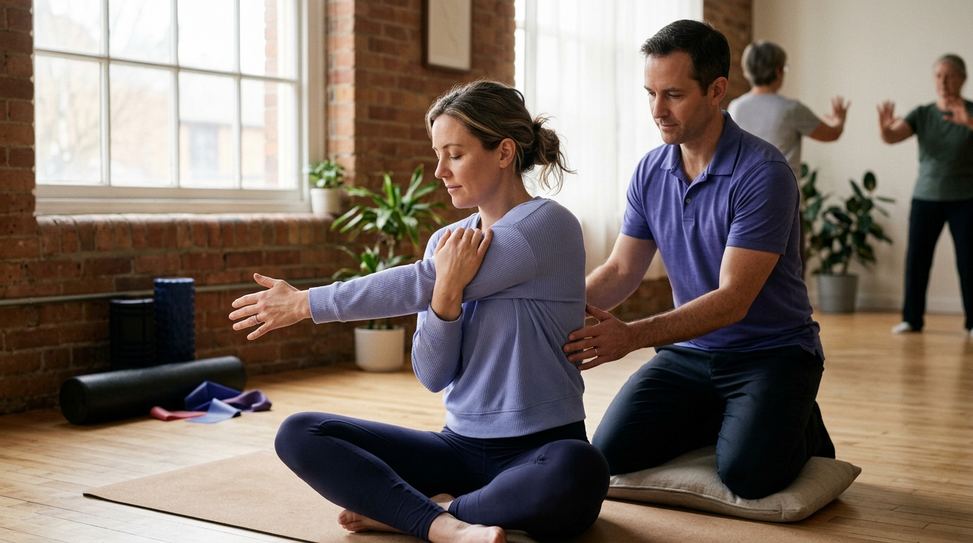
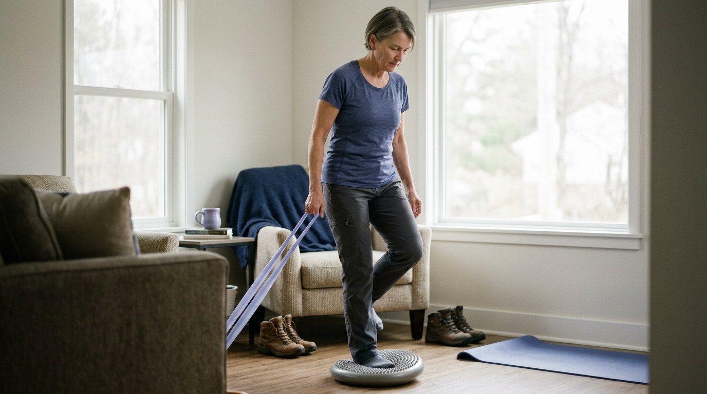
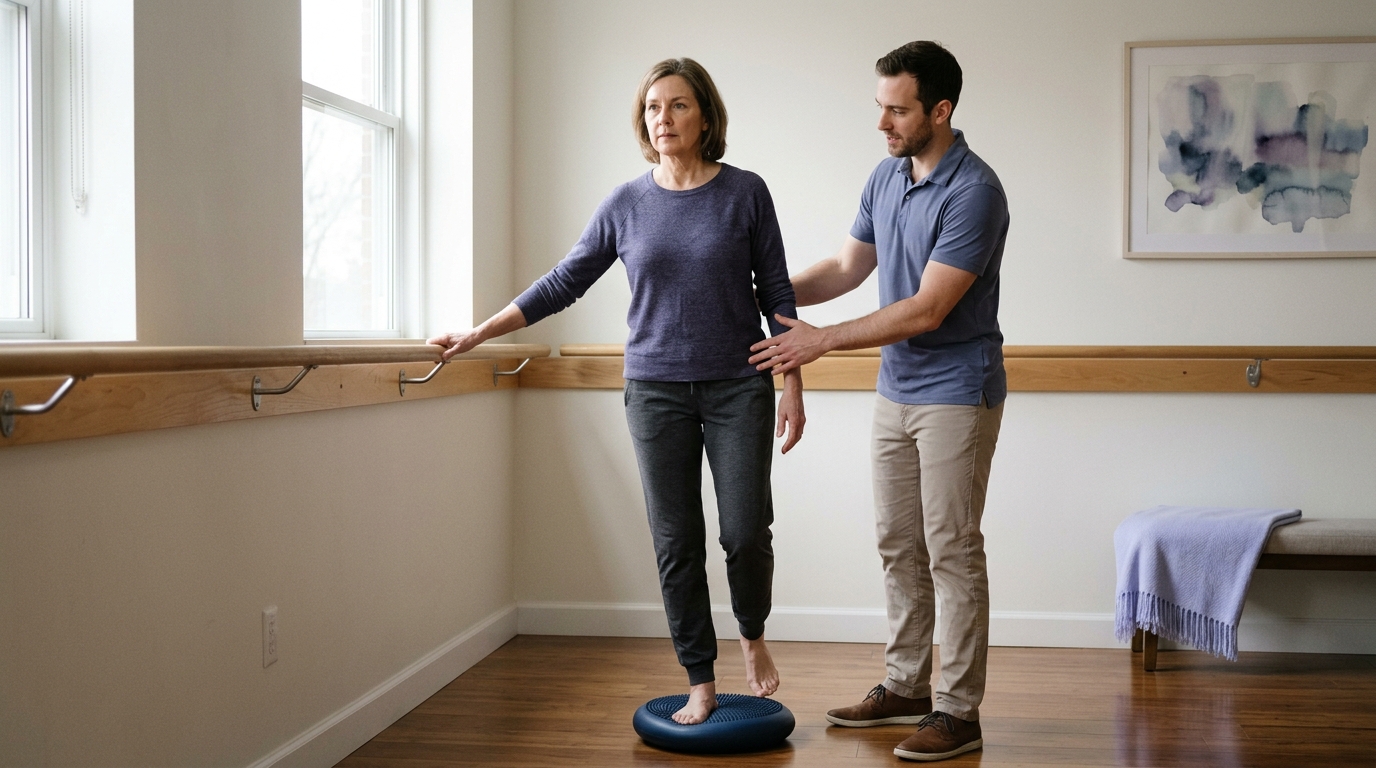
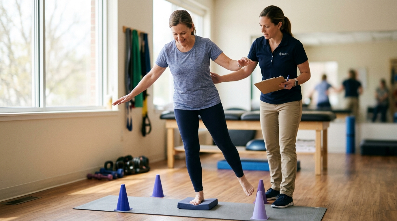
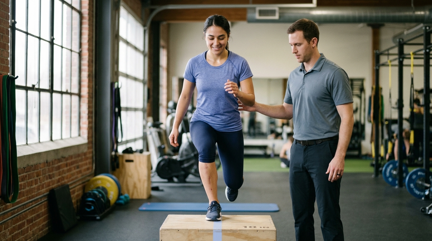
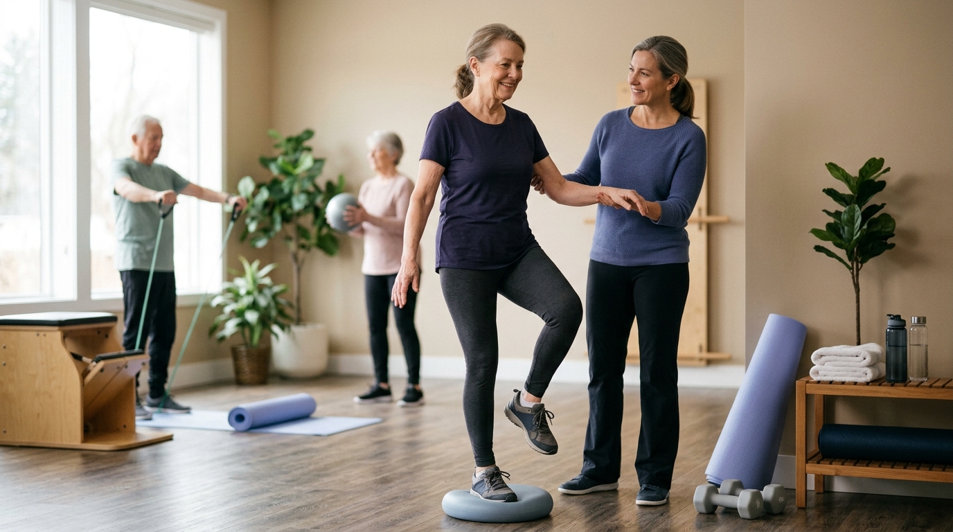
Coordination describes how the nervous system organizes muscles to control the alignment and motion of the limb. It is often referred to as movement quality or neuromuscular control.
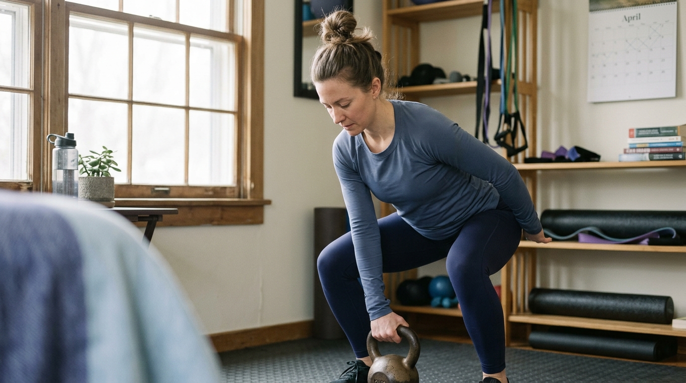
A person may possess strong leg muscles but still allow the knee to collapse inward during a step or a squat. This inward collapse, termed dynamic valgus, places uneven mechanical stress across the joint cartilage and ligaments.
Coordination training helps re-establish communication between joint position sensors, called mechanoreceptors, and the brain. This sensory feedback loop is frequently disrupted after joint injury, prolonged swelling, or surgical intervention.
Practicing stable movement patterns ensures that strength is applied effectively and safely. Better coordination helps keep the knee properly aligned over the middle toes during functional tasks.
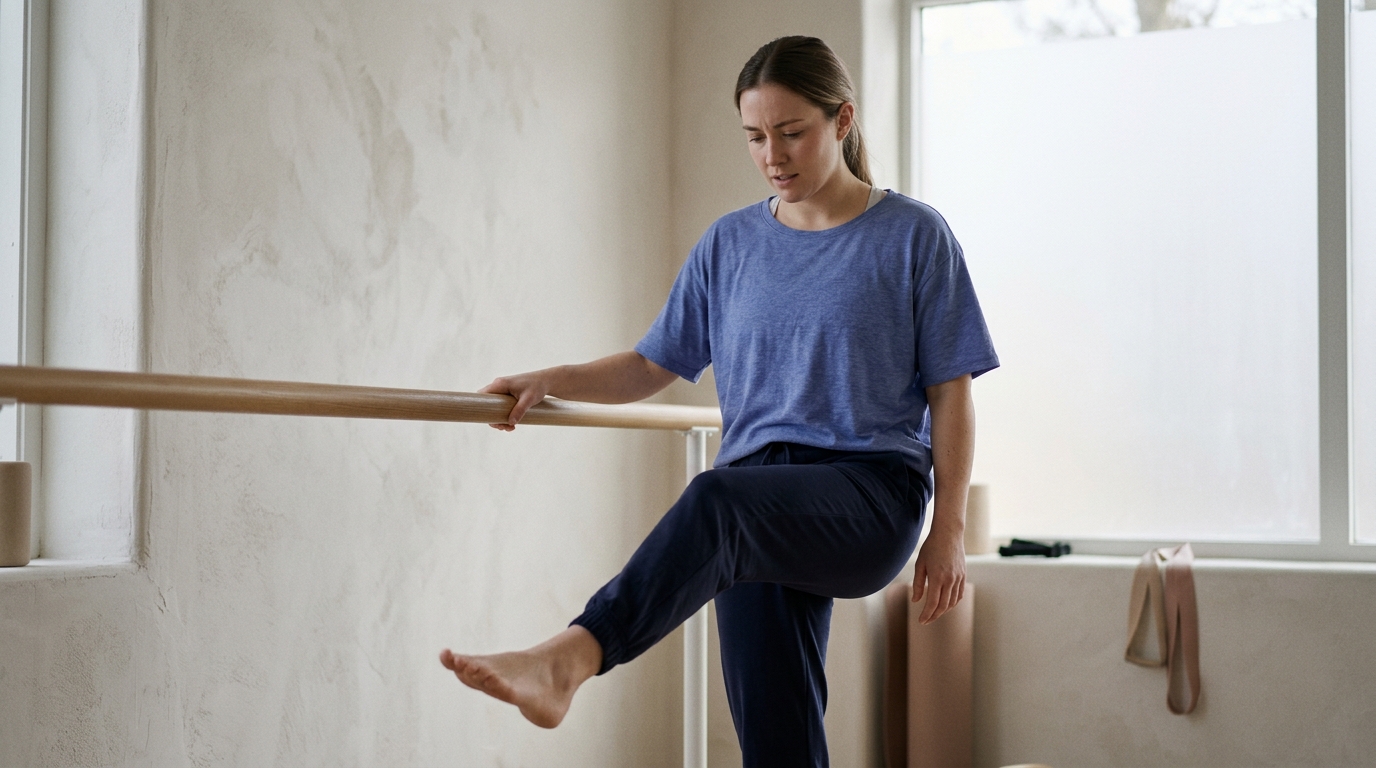
Activity tolerance is the capacity of the joint and surrounding tissues to handle repeated loading over time without excessive pain or swelling. A person might complete a single chair transfer or walk a short hallway with good form, yet experience significant stiffness and warmth several hours later.
Tolerance reflects tissue capacity, which builds gradually in response to progressive physical demands. If training loads increase too quickly, cartilage, bone, and tendons can become irritated.
Rehabilitation programs monitor the joint's 24-hour response to identify whether an activity level is appropriate. Stable joint symptoms indicate that current tissue capacity matches the applied physical demands.
Building activity tolerance allows you to transition from performing isolated exercises to managing full days of work, household chores, and recreation.
Clinical guidelines strongly support exercise therapy as a primary tool for restoring knee function and managing joint symptoms. Major orthopedic and rheumatology organizations consistently recommend structured movement for both degenerative conditions and surgical recovery.
Evidence confirms that tailored physical activity improves joint lubrication, stimulates muscle growth, and enhances functional independence. However, research also shows that outcomes vary, and exercise should be viewed as an ongoing management strategy rather than an overnight solution.
Understanding the quality and scope of this research helps set realistic expectations for your rehabilitation journey.
Clinical guidelines from the American Academy of Orthopaedic Surgeons recommend self-management programs, strengthening exercises, low-impact aerobic activity, and neuromuscular education for individuals with symptomatic knee osteoarthritis. These interventions help distribute mechanical loads more evenly across the joint.
Guidelines published by the National Institute for Health and Care Excellence similarly recommend tailored therapeutic exercise for adults with osteoarthritis. These recommendations emphasize local muscle strengthening combined with general aerobic fitness to improve daily capacity.
A systematic review published in the Cochrane Database evaluated exercise therapy for knee osteoarthritis across 28 randomized trials with 2,873 participants. The authors reported that exercise produced an average pain reduction of 8.70 points on a 0 to 100 scale compared to control groups, although this pain evidence was rated as low certainty.
The same Cochrane summary evaluated 24 studies with 2,536 participants and found an average physical function improvement of 11.27 points on a 0 to 100 scale. This physical function evidence was graded as moderate certainty, indicating clear benefits for everyday tasks.
An earlier 2015 Cochrane review analyzed land-based exercise and found standardized mean differences of 0.49 for pain reduction and 0.52 for physical function improvement immediately following treatment. The review noted that functional benefits were sustained for two to six months after formal treatment concluded.
Rehabilitation following knee surgery relies on phased, criteria-driven protocols rather than fixed calendar timelines. A consensus guideline published in the British Journal of Sports Medicine outlines a structured pathway for anterior cruciate ligament reconstruction.
This consensus framework begins with a prehabilitation phase before surgery to optimize range of motion and muscle strength. Postoperative care is then divided into three distinct phases: impairment-based recovery, sport-specific training, and return to full activity.
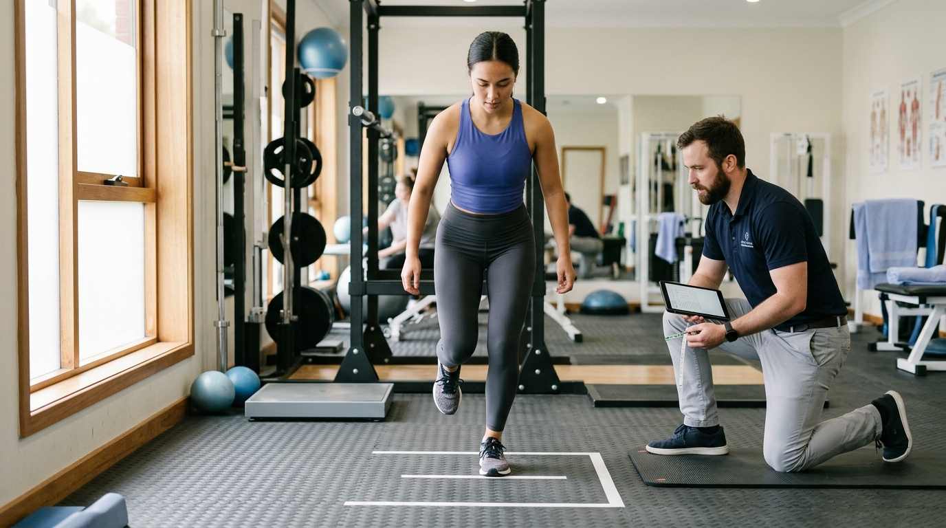
Progression between these stages depends on objective testing, including quadriceps strength symmetry, functional hop tests, movement quality assessments, and psychological readiness measures. Research shows that passing objective physical criteria significantly reduces the risk of secondary joint injury.
For joint replacement procedures, hospital clinical guidelines emphasize early mobilization and functional training. Patients practice safe bed mobility, walking aid progression, and stair climbing within the initial postoperative days to support safe discharge home.
You can learn more about structured protocols by reviewing our surgical recovery comeback resources.
Scientific literature demonstrates clear, measurable improvements in physical function and mobility from exercise therapy. However, the evidence does not support claims of immediate symptom elimination or complete cartilage regeneration.
Exercise alters how loads are distributed through the joint and improves the shock-absorbing capacity of surrounding muscles. It helps keep joint capsule tissues pliable and reduces systemic inflammatory markers through regular movement.
Yet, individual responses to exercise vary based on joint anatomy, sleep habits, overall health, and program adherence. Recognizing that exercise provides moderate, lasting improvements helps patients stay consistent without becoming discouraged by short-term fluctuations.
Rehabilitation exercises are most effective when they translate directly into everyday tasks. Basic movements such as walking across a room, navigating stairs, and standing up from a chair serve as practical benchmarks of functional recovery.
These tasks require complex interactions between joint range of motion, muscle strength, and balance. Evaluating how you perform these activities provides real-world insight into joint readiness.
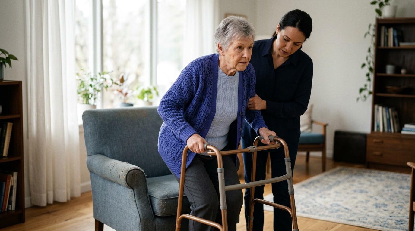
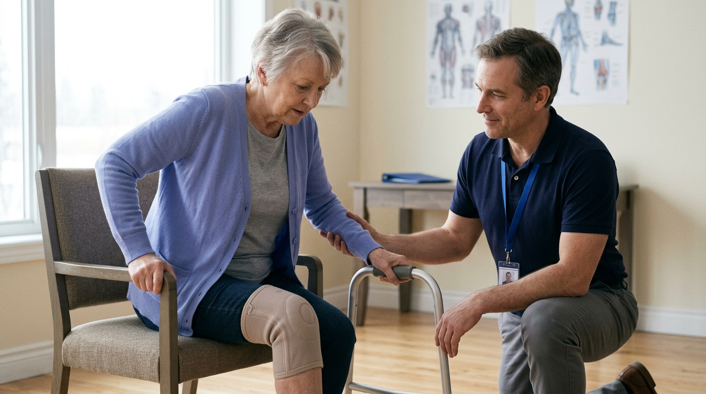
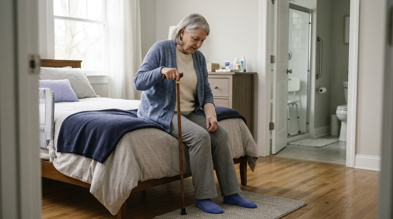
Walking is both a primary daily activity and a fundamental way to assess lower-body function. Following joint replacement or reconstructive surgery, early walking is typically restricted to short household distances using an appropriate walking aid.
A walking aid, such as a walker or crutches, reduces joint contact forces and provides stability while muscle activation returns. Progressing from two crutches to a single stick or unaided walking should be guided by movement quality rather than speed.
Clinicians look for a non-antalgic gait, which means walking smoothly without a limp. Walking with a limp reinforces poor movement habits and overloads other joints such as the lower back and hips.
If swelling increases or walking mechanics deteriorate after a walk, the distance should be reduced temporarily. Exploring our detailed rehabilitation and mobility movement resources can help you understand how to structure your daily walking progression.
Climbing and descending stairs require significantly more joint flexion and muscle strength than level walking. Ascending a step requires concentric power from the quadriceps and gluteals to lift body weight upward.
Descending stairs requires controlled eccentric lowering, demanding high quadriceps strength and ankle flexibility. Because of these physical demands, early postoperative protocols teach specific compensation strategies to ensure safety.
A standard pattern taught in postoperative hospital guidance uses a step-by-step approach:
This familiar pattern, often summarized as up with the good leg and down with the operated leg, is a temporary safety strategy. It protects healing tissues during early recovery but is not a permanent movement pattern for healthy joints.
As strength, balance, and range of motion return, patients work toward a reciprocal stair pattern, taking one step per foot. Reciprocal stair climbing requires at least 90 to 105 degrees of knee flexion and solid eccentric quadriceps control.
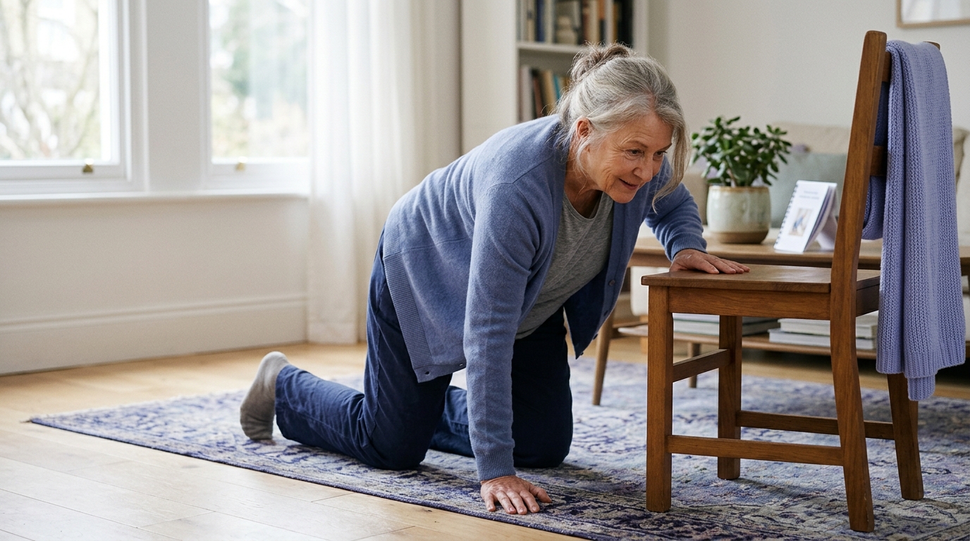
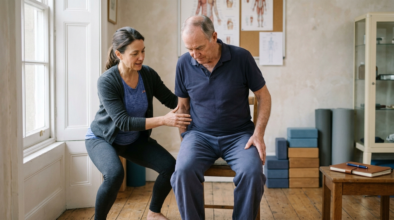
Standing up from a seated position, known as a chair transfer, requires adequate knee flexion, quadriceps force, and forward trunk lean. Low or soft chairs increase the difficulty by requiring deeper joint angles and greater muscular effort.
Hospital patient guidance for joint replacement recovery outlines practical transfer techniques. Patients are instructed to slide forward toward the edge of the seat, place both feet firmly on the floor, and push upward using the chair armrests.
Pushing through armrests reduces initial knee contact forces while quadriceps activation is recovering. When sitting down, patients step backward until both legs touch the chair seat, reach back for the armrests, and lower themselves slowly.
Dropping quickly into a chair indicates insufficient eccentric strength and control. Practicing chair rises from a firm, elevated surface helps rebuild strength safely before progressing to lower seating.
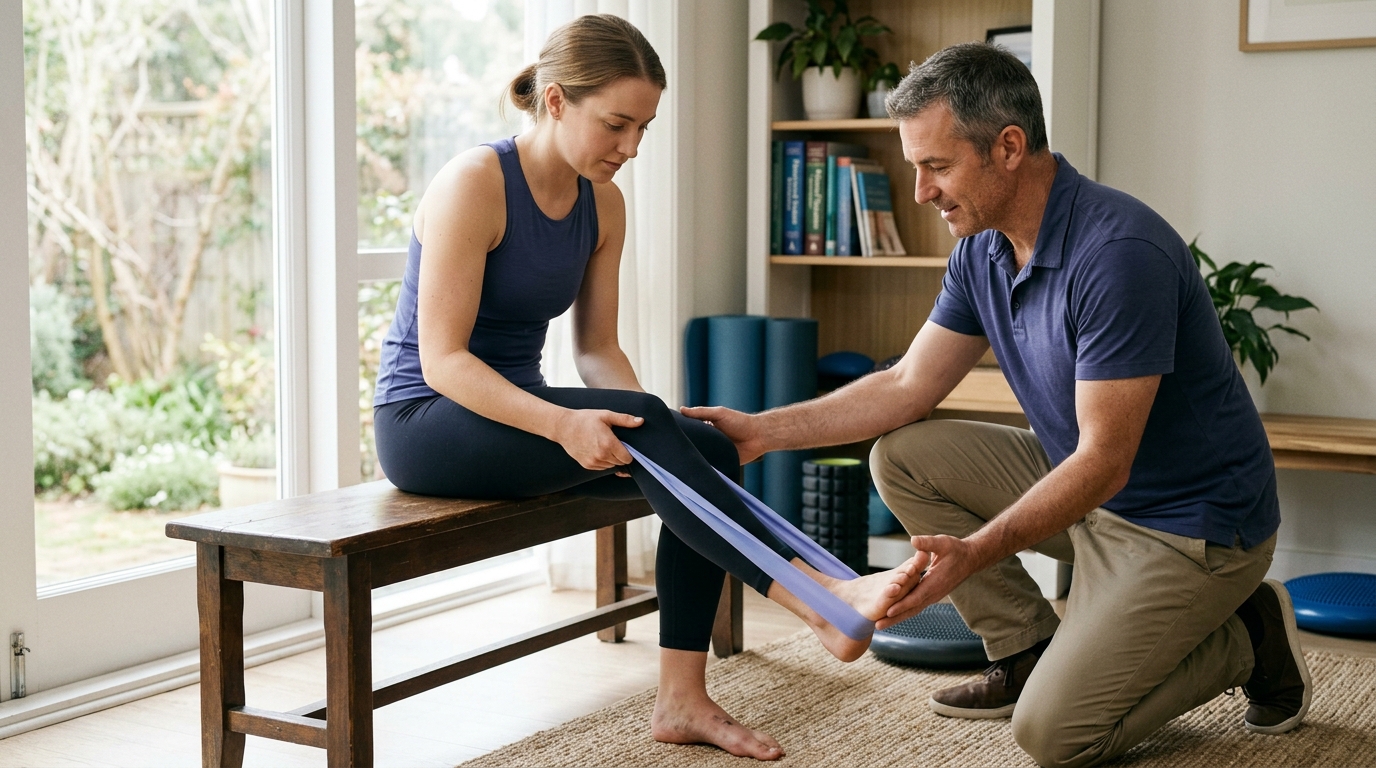
Chair-based exercises are commonly included in early home programs. Standard exercises described in hospital handouts include seated knee extensions to activate the quadriceps and seated heel slides to improve flexion range.
Rehabilitation is not a one-size-fits-all process. The underlying biology of your knee condition dictates how quickly you can safely apply load, bend the joint, and return to demanding activities.
Treating a degenerative joint condition involves different biological timelines than protecting a surgically repaired tendon or ligament. Understanding these differences prevents the misapplication of protocols designed for other conditions.
Total knee replacement involves resurfacing damaged bone and cartilage with metal and polyethylene components. Early rehabilitation focuses on pain management, rapid restoration of extension and flexion, and safe household mobility.
Postoperative home exercise programs provided by hospitals frequently recommend performing specific movements three times per day. For example, a standard NHS handout advises 10 repetitions of ankle pumps, static quadriceps sets, and heel slides, holding each contraction for five seconds.
These early exercises are designed to restore circulation, prevent muscle shutdown, and gradually stretch joint tissues. Weight-bearing is typically permitted immediately as tolerated, supported by crutches or a walker.
Progression focuses on achieving independent walking, mastering stairs, and regaining functional independence for daily living. You can find comprehensive articles on joint replacement recovery in our surgical recovery articles section.
Reconstruction of the anterior cruciate ligament involves replacing a torn ligament with a tissue graft. Because the graft must undergo a biological healing process called ligamentization, mechanical stress must be carefully managed.
Anterior cruciate ligament protocols use strict criteria before allowing patients to progress to running, jumping, or cutting movements. For instance, clinical guidelines specify that impact activities should not begin until the patient demonstrates full range of motion, no joint swelling, and adequate baseline strength.
Progression guidelines often require a limb symmetry index of at least 90 percent on quadriceps and hamstring strength tests and functional hop testing before approving return-to-sport activities. Psychological readiness tests are also used to assess patient confidence and reduce fear of reinjury.
Applying knee replacement guidelines to an athlete recovering from ligament reconstruction would result in premature loading and potential graft failure. Detailed guidance on athletic transitions can be explored in our strength rebuilding and sport progression articles.
Rehabilitation for knee osteoarthritis does not involve protecting healing surgical incisions or biological grafts. Instead, the focus is on improving load tolerance, building periarticular muscle mass, and maintaining cardiovascular health.
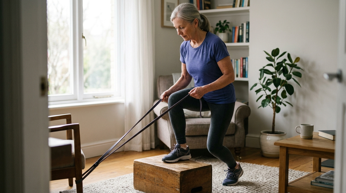
Because joint degeneration is an ongoing condition, exercise programs are designed for long-term consistency. Low-impact aerobic activities, such as stationary cycling, aquatic exercise, or brisk walking, are combined with resistance training.
Programs are tailored to the individual's baseline capacity, gradually increasing volume as symptoms allow. For additional insights on maintaining joint function over time, review our active aging and long-term joint health resources.
Beyond the specific diagnosis, several personal and biological factors influence how quickly knee function returns:
Recognizing these variables helps you focus on personal functional progress rather than comparing your recovery to others.
Relying solely on a calendar to determine when to advance rehabilitation can lead to setbacks. Biological healing times provide general guardrails, but tissue capacity and functional ability vary widely between individuals.
Criterion-based progression requires meeting objective physical benchmarks before advancing to higher-demand movements. This approach ensures that the joint possesses the strength, mobility, and stability required for new physical tasks.
A calendar-only approach assumes that tissue healing automatically equals functional readiness. For example, assuming a patient is ready to run at twelve weeks post-surgery simply because twelve weeks have passed ignores underlying muscular deficits.
If the quadriceps have not regained sufficient strength, running places excessive impact shock directly on articular cartilage and healing repairs. This premature loading often results in joint effusion, pain flares, and compensatory movement habits.
Using objective criteria protects recovering joints from being exposed to forces they cannot yet absorb safely. It provides clear goals for both the patient and the physical therapist.
Clinicians use a variety of validated tests to evaluate readiness for activity progression:
Passing these objective tests demonstrates that the muscular and nervous systems can handle dynamic forces.
A key component of criterion-based progression is monitoring how the knee responds over the full 24 hours following an activity. Tissues often feel acceptable during exercise, but react several hours later with increased swelling, warmth, or morning stiffness.
A mild ache that resolves within two hours of exercise is generally considered an acceptable training response. However, sharp pain during movement or swelling that lasts into the next day signals that tissue capacity was exceeded.
Tracking this 24-hour response helps calibrate exercise dosage, ensuring that progression stimulates adaptation without causing inflammatory setbacks.
Navigating knee rehabilitation requires balancing active exercise with joint protection. Avoiding common recovery mistakes helps maintain steady progress and prevents unnecessary flare-ups.
A frequent error is focusing entirely on bending the knee while neglecting full knee extension and muscular strengthening. While regaining flexion is necessary, full extension is what allows normal walking mechanics and efficient standing.
Furthermore, achieving joint range without developing strength in that new range leaves the joint vulnerable. Mobility exercises must always be paired with active strengthening to ensure functional stability.
Being able to perform a single squat, step-up, or transfer does not mean the knee is ready for sustained activity. Everyday tasks and recreational sports require muscular endurance and repeated force absorption.
When stabilizing muscles fatigue, movement quality deteriorates, and mechanical loads shift directly onto passive joint structures. Rehabilitation must build local muscular endurance through appropriate volume before advancing to high-demand tasks.
More exercise is not always better. Healing cartilage, bone, and tendons require adequate rest periods to synthesize new structural proteins and adapt to applied loads.
Performing aggressive home exercises multiple times a day without adequate recovery can lead to chronic joint inflammation and persistent quadriceps inhibition. Following prescribed exercise dosages and allowing rest days supports consistent, long-term tissue adaptation.
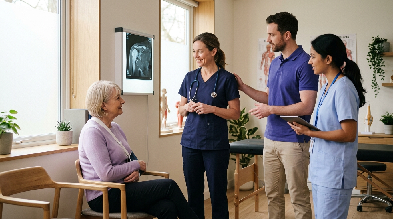
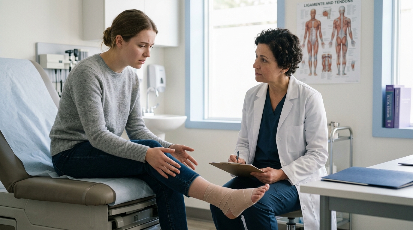
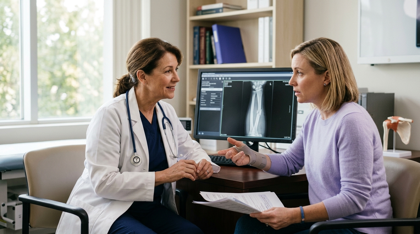
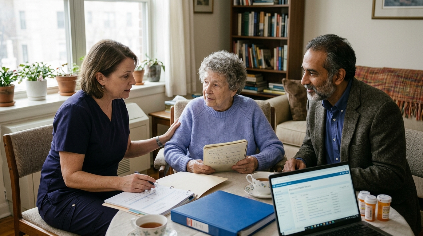
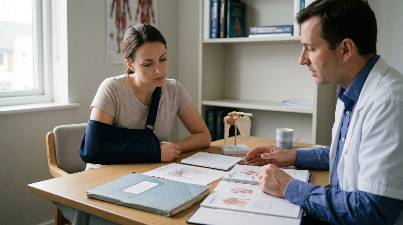
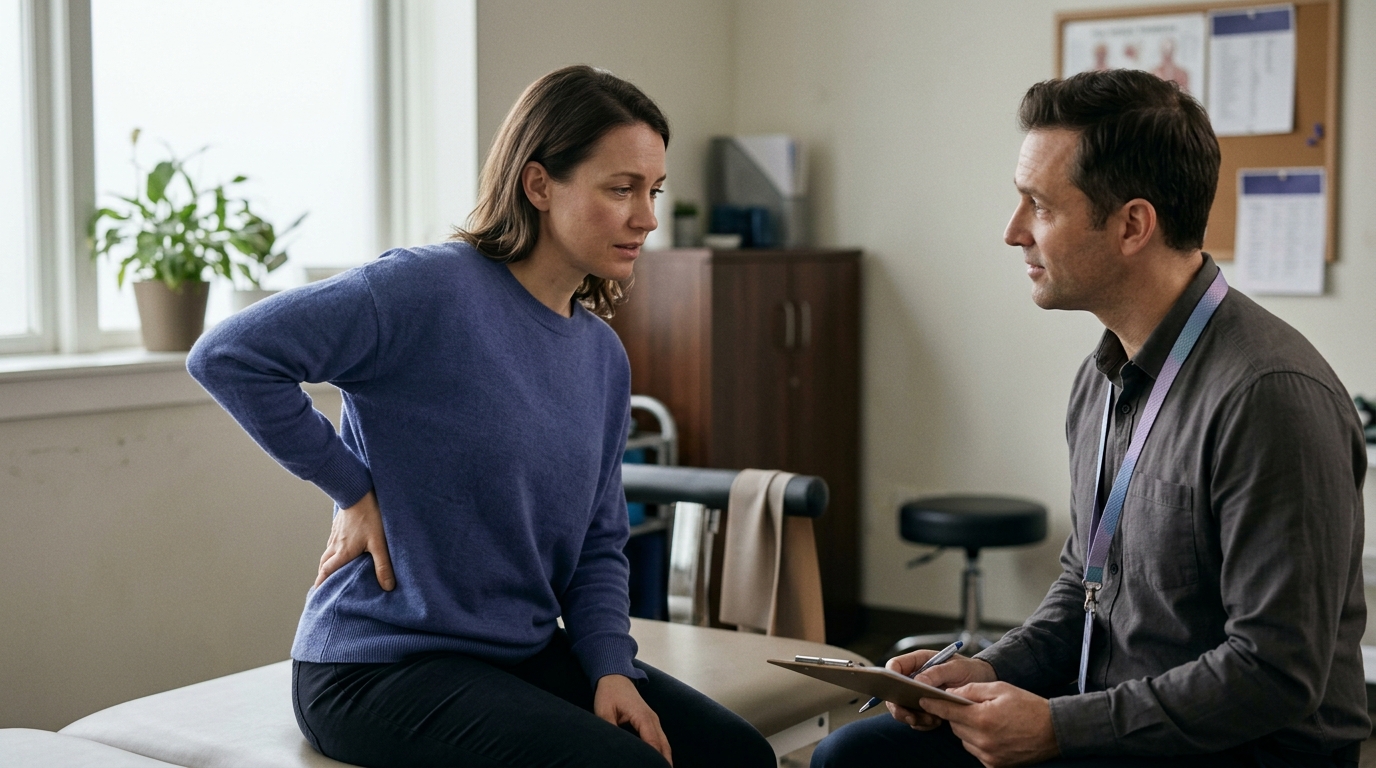
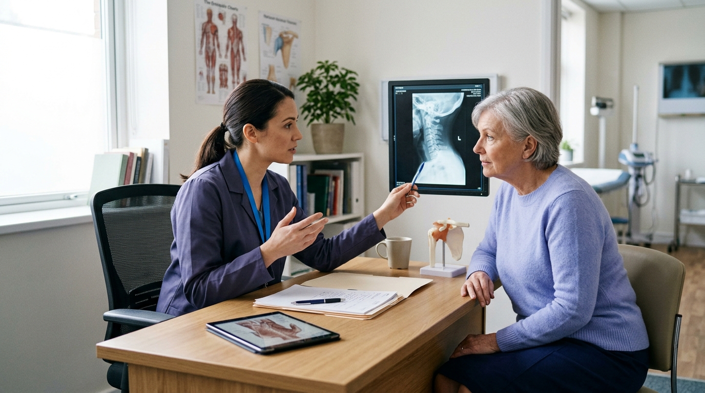
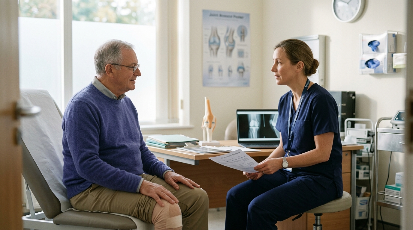
Clear communication with your orthopedic care team ensures that your rehabilitation aligns with your specific surgical procedure or clinical diagnosis. Bringing targeted questions to your appointments helps clarify expectations and safety boundaries.
Consider discussing the following practical questions during your clinical visits:
Having these clear boundaries prevents guesswork and gives you confidence during daily practice.
Restoring knee function requires a balanced approach that addresses joint mobility, muscular strength, movement coordination, and activity tolerance. Relying on range of motion alone or following a rigid calendar timeline does not account for the biological reality of tissue healing.
Everyday tasks such as walking, managing stairs, and rising from a chair provide practical opportunities to build strength and assess movement control. Tailored exercise is backed by solid clinical evidence for improving function and reducing discomfort in both degenerative conditions and surgical recovery.
Progressing through rehabilitation requires patience, objective testing, and consistent communication with your treating healthcare team. Respecting your knee's current tissue capacity while systematically challenging it leads to safe, lasting functional movement.
If you must navigate stairs without a handrail during early recovery, use two crutches or canes to provide bilateral support. Place both crutches down on the step below before stepping down with the operated leg, or push through both crutches while stepping up with the unoperated leg. If balance remains uncertain, having an assistant stand below you or considering temporary sleeping arrangements on the ground floor is recommended.
Post-activity stiffness and warmth represent an inflammatory response, indicating that the cumulative physical load exceeded current tissue capacity. While joint tissues can often tolerate the mechanical stress during the walk itself, delayed swelling occurs as fluid accumulates in the joint capsule. Reducing walking distance temporarily and icing or elevating the limb can help manage symptoms until tissue tolerance improves.
You should only transition to a single walking aid when you can bear full weight on the operated leg without significant pain or compensatory limping. When using a single cane or crutch, hold it in the hand opposite the recovering leg to encourage a normal reciprocal arm swing and maintain balanced pelvis alignment. Always confirm this transition with your physical therapist to ensure your movement mechanics are stable.
Knee replacement protocols focus on early mobilization, functional household transfers, and managing stable prosthetic components that allow immediate weight-bearing. In contrast, ACL reconstruction involves a biological tissue graft that requires months to incorporate, remodel, and mature. Consequently, ACL protocols enforce strict restrictions on impact and cutting activities until rigorous strength and functional criteria are met.

Use ReboundBody resources to understand common recovery stages, rehab terms, movement limits and strength rebuilding. Each guide is designed to make a complex comeback easier to understand.


Read practical guidance on injury recovery, rehabilitation, mobility and rebuilding strength as you work your way back to activity.
Explore Resources