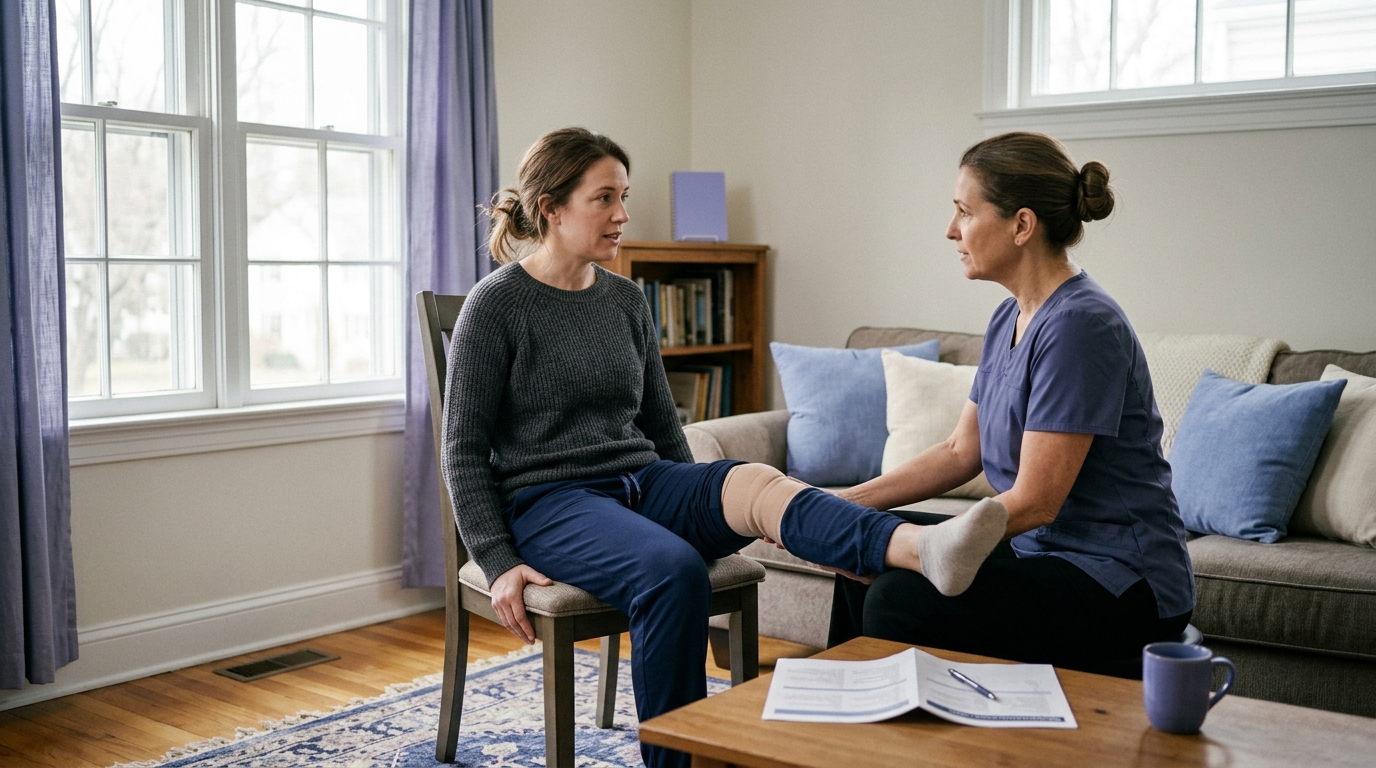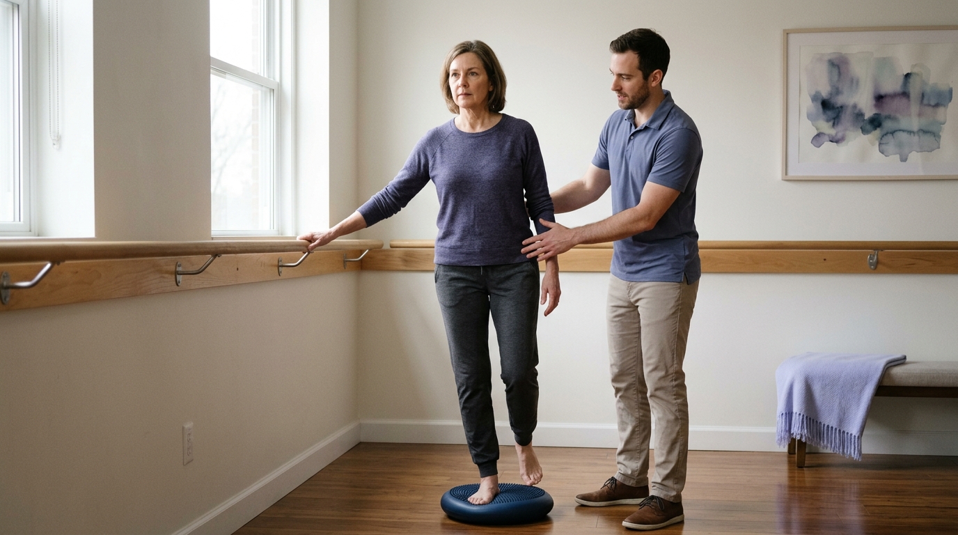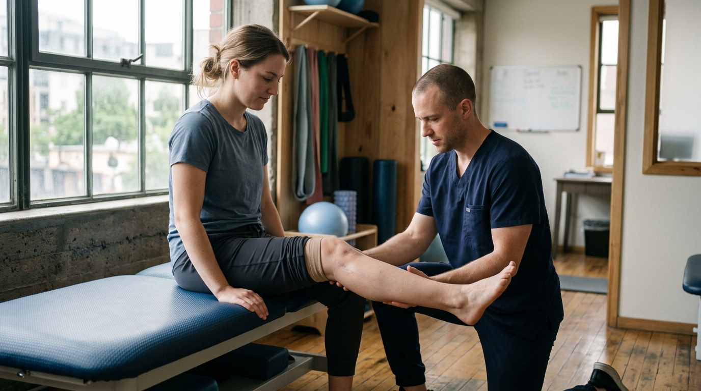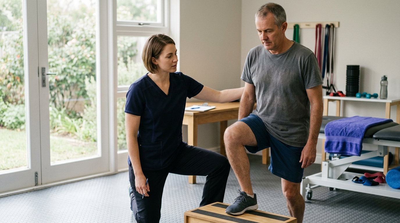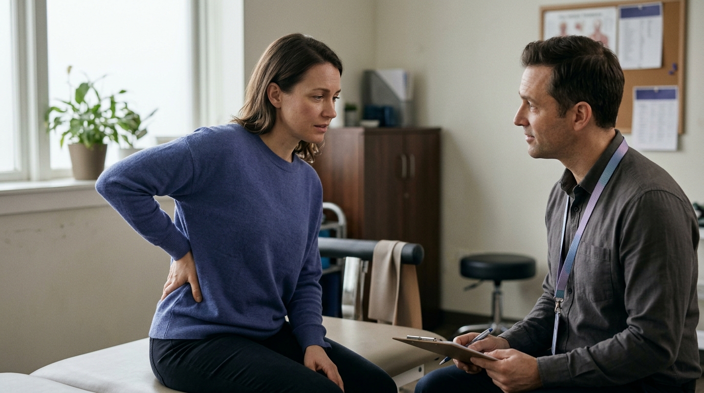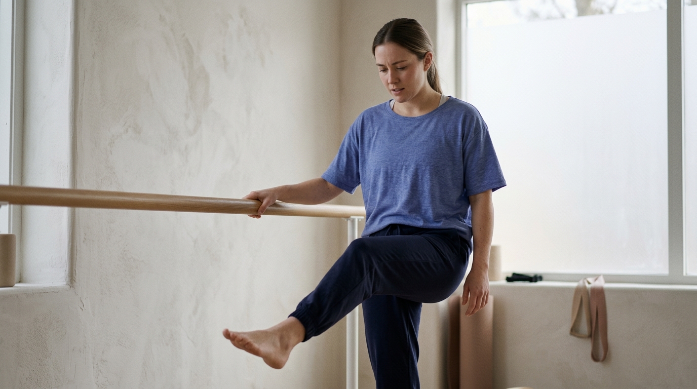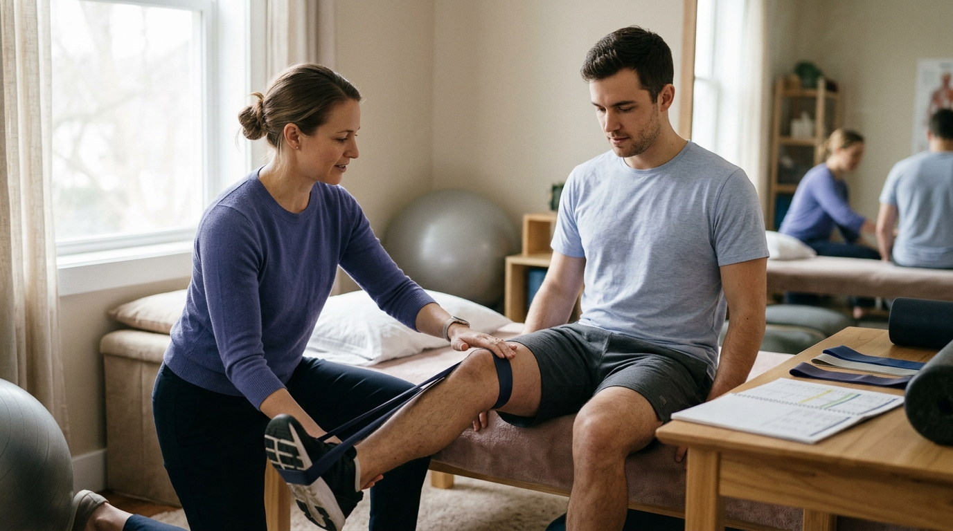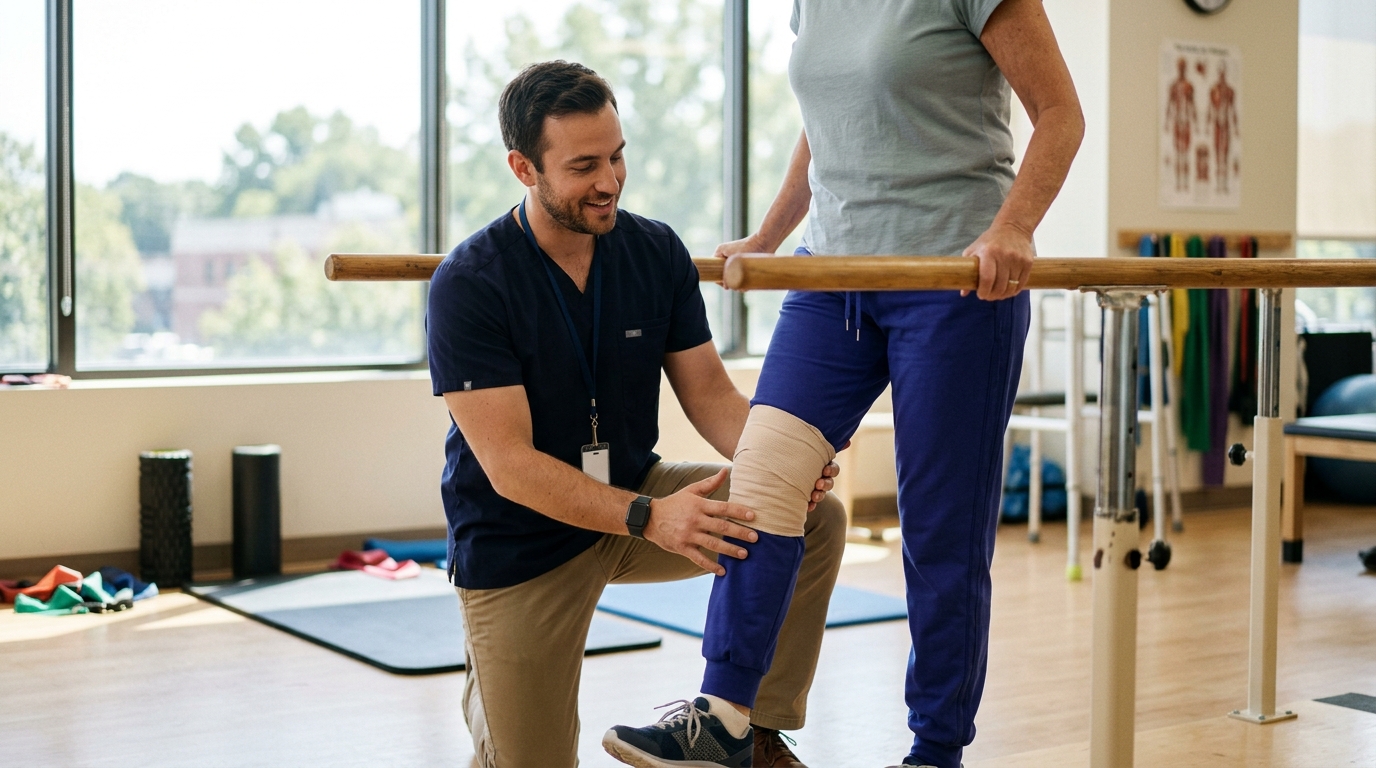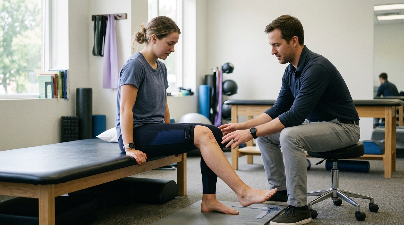

Three distinct assessment components work together to help clinicians interpret scan findings accurately by matching visible structural changes to your actual joint symptoms.





An advanced medical scan is often treated as the ultimate answer for joint or back pain. Many people assume that an MRI or X-ray will instantly reveal the exact source of their discomfort.
In clinical practice, an image is only a single piece of evidence. A scan can reveal detailed structural changes without explaining why a person feels pain. At the same time, severe pain and functional limitations can exist even when an image appears completely clear.
An orthopedic diagnosis is an informed interpretation rather than a label read from a screen. Clinicians combine a patient history, a thorough physical examination, and targeted imaging to understand what is happening. Relying on any single test in isolation can lead to misdirected treatments, unnecessary anxiety, and prolonged recovery times.
Understanding how these elements fit together allows you to engage with your healthcare team more effectively. It helps you ask better questions, set realistic recovery expectations, and make sense of complex medical reports.
A diagnosis is a working model built by combining three distinct clinical inputs. These inputs are your personal history, your physical examination, and your diagnostic images.
Your history describes what happened, when the problem started, and what positions make symptoms better or worse. It also captures the specific daily activities that are currently restricted. This story gives the clinician essential context about how your body responds to mechanical loads.
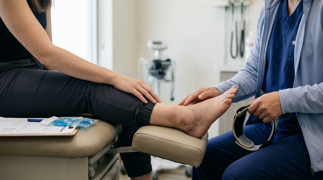
The physical examination allows the clinician to observe movement, test muscle strength, and check nerve reflexes. The clinician uses hands-on tests to see if specific motions reproduce your familiar pain. These findings narrow down the list of anatomical structures that might be involved.
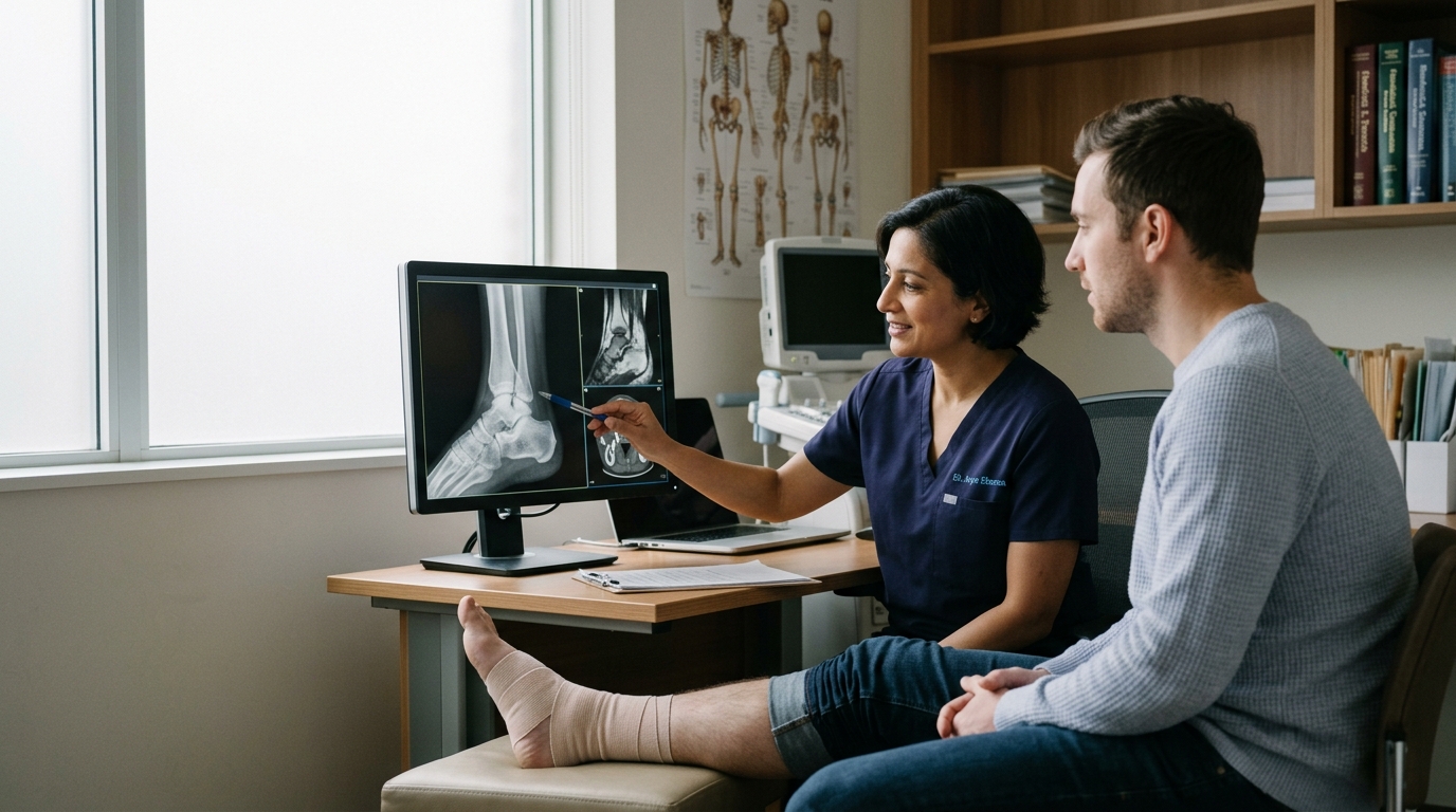
Imaging should be used to answer a focused question raised by the history and examination. A scan provides an anatomical picture, but it cannot measure pain, stiffness, or daily function. A clinician must determine whether the visible structural features match your actual physical complaints.
Clinicians rely on a concept known as pre-test probability before ordering any imaging study. This means they estimate the likelihood of a specific condition based purely on your history and physical tests.
If your history strongly points toward a specific tendon issue, the pre-test probability for that condition is high. An ultrasound or MRI is then used to confirm that specific suspicion. The scan serves to refine the diagnosis rather than search blindly for random abnormalities.
When imaging is ordered without a clear clinical suspicion, the results can be confusing. Scans often pick up minor structural changes that have nothing to do with your symptoms. Interpreting an image requires checking whether a visible finding matches your pain location and movement limitations.
Guidelines from the American College of Radiology emphasize that imaging choices must depend on specific clinical conditions. The decision to order a scan rests on the individual circumstances of your case. A scan is appropriate when its result will meaningfully change your care plan.
Extensive medical research demonstrates that structural changes are common in completely healthy, pain-free bodies. As we age, our tissues undergo natural alterations that appear on scans but do not always cause symptoms.
These visible features are often normal signs of aging rather than active injuries. When a scan reveals an anatomical change, it does not automatically prove that the feature is the source of your pain. Research across the spine, knee, and shoulder illustrates how common these findings are in the general population.
A major systematic review by Brinjikji and colleagues examined spinal MRI findings in people who had no back pain. The researchers discovered that degenerative features are exceptionally common across all age groups.
The review estimated that disk degeneration is present in 37% of pain-free 20-year-olds. By age 80, disk degeneration is visible in 96% of pain-free individuals. Disk bulges follow a similar pattern, rising from 30% in 20-year-olds to 84% in 80-year-olds.
Disk protrusions were identified in 29% of asymptomatic individuals at age 20 and 43% at age 80. Annular fissures were found in 19% of pain-free 20-year-olds and 29% of 80-year-olds. These numbers prove that a bulging or degenerated disk is often an incidental finding rather than an active medical problem.
Similar patterns appear when researchers scan pain-free knees. A systematic review and meta-analysis led by Culvenor evaluated MRI scans of asymptomatic, uninjured knees.
The researchers found a pooled prevalence of 24% for cartilage defects and 10% for meniscal tears across all pain-free adults. Among adults aged 40 and older, cartilage defects were found in 43% of uninjured knees. Meniscal tears were present in 19% of healthy, pain-free adults in that same older group.
A landmark study published in the New England Journal of Medicine by Englund and colleagues evaluated middle-aged and older adults. Among individuals with radiographic knee osteoarthritis, meniscal tears were present in 63% of people who reported frequent knee pain.
Crucially, meniscal tears were also present in 60% of people who had no knee pain, aching, or stiffness. The study concluded that meniscal damage is extremely common regardless of symptoms. This shows that finding a meniscal tear on an MRI does not automatically make it the primary source of pain.
Imaging studies of the shoulder show substantial variation in pain-free populations. A systematic review of rotator cuff imaging found that abnormal findings in asymptomatic shoulders ranged widely across published studies.
For full-thickness rotator cuff tears, prevalence estimates ranged from 0% to 14% in general population studies. In studies focused on pain-free athletes, estimates ranged between 0% to 11%. One small population study reported a 20% prevalence rate, though it evaluated only 20 shoulders.
Physical activity also influences what appears on a joint scan. A study of asymptomatic rock climbers found intra-articular shoulder abnormalities on MRI in 80% of participants. Furthermore, 57% of these pain-free climbers showed cartilage damage in the shoulder joint.
These findings reflect tissue adaptations to regular mechanical loading. They demonstrate why scan results must be interpreted in light of your specific physical activities. To learn more about how physical stress shapes joints, read about active aging and joint health.
Radiology reports are written in specialized technical language designed to describe every visible structural variation. When patients read these reports, descriptive anatomical terms can sound alarming and dangerous.
Words like degeneration, tear, fraying, or protrusion are often interpreted as catastrophic damage. In reality, these terms are simply anatomical descriptions of what the scanner detected. They do not describe your level of tissue health, your pain intensity, or your future physical potential.
from Treatment Indications
To understand an imaging report, it helps to distinguish three separate clinical concepts:
A radiology report only provides the first piece of this puzzle. It lists the anatomical findings visible in the joint or spine. Determining clinical significance requires your doctor to compare those findings with your physical examination.
Even when a finding is clinically significant, it does not mean surgery or invasive procedures are required. Many structural changes respond exceptionally well to progressive exercise, physical therapy, and load management. Understanding recovery science principles can help you view tissue changes with a calmer perspective.
Physical examination maneuvers are not pass-fail tests that provide instant, absolute answers. Instead, each test slightly increases or decreases the likelihood of a specific condition.
Clinicians combine several physical tests to build a diagnostic cluster. When multiple tests point in the same direction, the diagnostic certainty increases. A single positive or negative test rarely confirms or rules out an injury entirely.
Two statistical concepts help explain how physical examination tests function in a clinical setting:
Clinical assessments for suspected lumbar disc herniation provide a clear example of how tests differ. A standard straight-leg raise test has a reported sensitivity of 0.80 and a specificity of 0.40.
Because its sensitivity is high, a negative straight-leg raise makes an active disc herniation irritating the nerve root less likely. However, because its specificity is only 0.40, a positive test often occurs in people who have tight hamstrings or non-nerve back pain.
In contrast, the crossed straight-leg raise test evaluates the opposite, unaffected leg. This test has a reported sensitivity of 0.35 and a specificity of 0.90.
Because its sensitivity is low, a negative test does not tell you much. But if raising your unaffected leg reproduces nerve pain down your painful leg, the high specificity strongly supports nerve root irritation.
Clinicians never rely on just one maneuver. They combine these tests with reflex checks, muscle testing, and sensory maps to evaluate nerve health.
Diagnostic imaging is an exceptional tool when used at the right time for the right clinical question. However, ordering scans too early for uncomplicated musculoskeletal pain can actually lead to poorer patient outcomes.
When imaging is performed immediately for routine back or joint pain, it frequently reveals incidental age-related changes. These findings can create unnecessary worry and lead patients to pursue invasive treatments that do not resolve the problem.
Clinical reviews of mechanical low back pain show that routine early imaging is not recommended for most patients without red flags. American College of Radiology criteria suggest considering imaging after six weeks of conservative medical and physical therapies if symptoms fail to improve.
Research indicates that early routine MRI for uncomplicated back pain is associated with higher rates of spinal surgeries without improvements in long-term pain or physical function. A randomized trial found that early radiography was associated with worse overall health outcomes, likely because it highlighted minor, incidental abnormalities that alarmed patients.
This six-week timeline is a condition-specific guideline for uncomplicated low back pain. It is not a universal rule for every orthopedic condition or acute traumatic injury. Acute fractures, complete tendon ruptures, and high-energy joint trauma often warrant prompt diagnostic imaging.
Imaging is most valuable when the results will directly alter the clinical plan. Scans are essential under the following circumstances:
Clinical diagnosis is rarely a simple, linear process. Symptoms and imaging findings frequently diverge, creating edge cases that require careful clinical interpretation.
Recognizing these edge cases helps prevent hasty decisions based solely on scan results. It also ensures that urgent symptoms receive immediate medical attention.
There are three common clinical scenarios where symptoms and scans appear to disagree:
Certain symptom patterns indicate serious underlying conditions that require urgent medical evaluation rather than routine scheduling. In the spine, the most critical pattern is suspected cauda equina syndrome, which occurs when the nerve roots at the base of the spinal cord become severely compressed.
If you experience any of these warning signs, seek emergency medical care immediately. Do not wait for a scheduled clinic visit or a routine imaging appointment.
Looking at realistic clinical examples helps illustrate how symptoms, examination, and imaging connect in real life. These patterns demonstrate how clinicians work through conflicting evidence to find practical solutions.
An active 48-year-old develops dull lower back stiffness after working in the yard. An MRI report from an urgent care clinic notes a moderate L4-L5 disk bulge and mild disk degeneration.
The individual fears that their spine is unstable and stops all exercise. During a comprehensive physical examination, the clinician finds normal reflexes, full leg strength, and a negative straight-leg raise test. The pain is localized to the lumbar muscles and does not travel down the leg.
Clinical Synthesis: The disk bulge is an incidental, age-related finding that does not match the physical exam. The actual issue is a self-limiting muscular strain. The individual safely returns to daily walking and progressive core exercises, recovering fully without surgery or injections.
A 55-year-old fitness walker experiences generalized anterior knee ache after increasing daily mileage. An MRI reveals a small, degenerative tear of the medial meniscus.
The patient assumes arthroscopic surgery is necessary to trim the torn tissue. However, the physical examination reveals that the medial joint line is not tender. Instead, the pain is reproduced by loading the patellar tendon and the patellofemoral joint during deep squats.
Clinical Synthesis: The meniscal tear is an incidental structural change common in adults over 40. The true driver of symptoms is patellofemoral overload from a sudden spike in walking volume. The individual benefits from injury recovery and healing resources and targeted quadriceps strengthening, avoiding unnecessary knee surgery.
A 42-year-old recreational swimmer experiences pain when reaching overhead. An ultrasound shows minor fraying of the supraspinatus tendon.
The physical examination demonstrates good rotator cuff strength, but reveals significant stiffness in the thoracic spine and poor scapular upward rotation. When the clinician manually assists scapular movement, the swimmer can raise the arm without pain.
Clinical Synthesis: The tendon fraying reflects normal mechanical loading over time. The primary issue is a movement coordination deficit in the shoulder blade and upper back. Incorporating targeted rehabilitation and movement strategies resolves the overhead discomfort without invasive tendon treatments.
A 38-year-old runner experiences persistent, burning hip pain that limits running. An X-ray and MRI of the hip joint are completely normal, showing healthy cartilage and no labral tears.
The runner feels frustrated and confused because the images show nothing wrong despite noticeable pain. A detailed physical examination uncovers severe weakness in the deep hip rotators and tenderness over the trochanteric bursa and gluteal tendons.
Clinical Synthesis: The pain is driven by tendinopathy and muscle deconditioning, which may not show obvious structural defects on standard joint scans. A structured program focused on progressive resistance training restores hip capacity, allowing a gradual return to running.
Navigating an orthopedic consultation is much easier when you know what to ask. Preparing specific questions helps you understand the reasoning behind your diagnosis and prevents misunderstandings regarding your scan results.
Use these practical questions to guide your next medical appointment:
An orthopedic diagnosis is never a single label taken from an imaging report. It is a comprehensive clinical synthesis that brings together your personal history, hands-on physical examination tests, and targeted imaging.
Scans are powerful tools for visualizing anatomy, but they do not measure pain, tissue sensitivity, or physical capacity. Normal, age-related changes frequently appear on scans in people who feel zero pain. At the same time, real and limiting symptoms can occur without any visible structural damage on an image.
By focusing on how your symptoms, examination, and scans fit together, you can avoid unnecessary procedures and focus on treatments that restore real-world function.
Understanding the complete diagnostic picture allows you to navigate physical setbacks with clarity, patience, and confidence.

Use ReboundBody resources to understand common recovery stages, rehab terms, movement limits and strength rebuilding. Each guide is designed to make a complex comeback easier to understand.


Read practical guidance on injury recovery, rehabilitation, mobility and rebuilding strength as you work your way back to activity.
Explore Resources