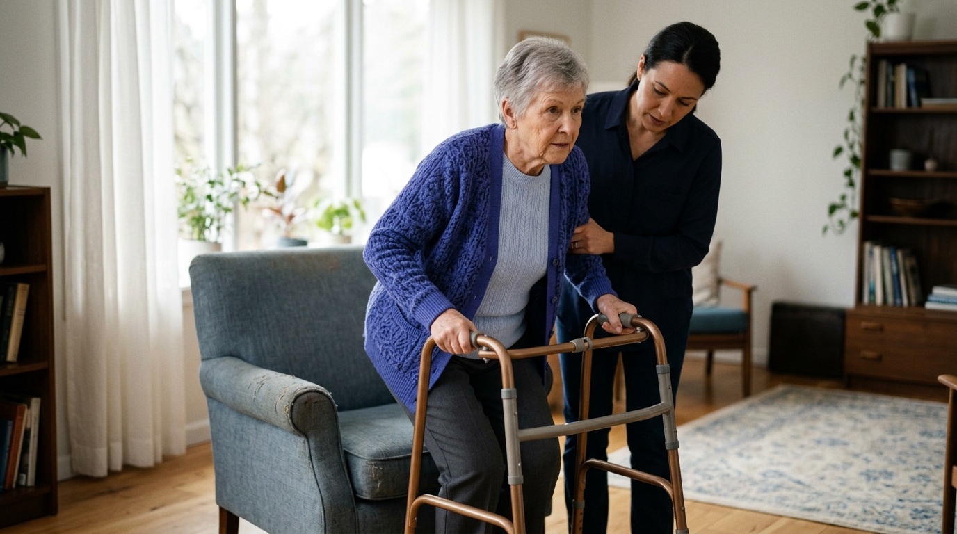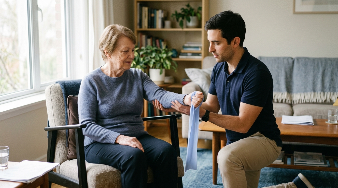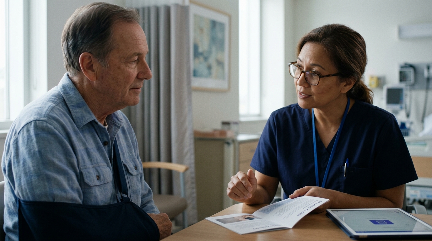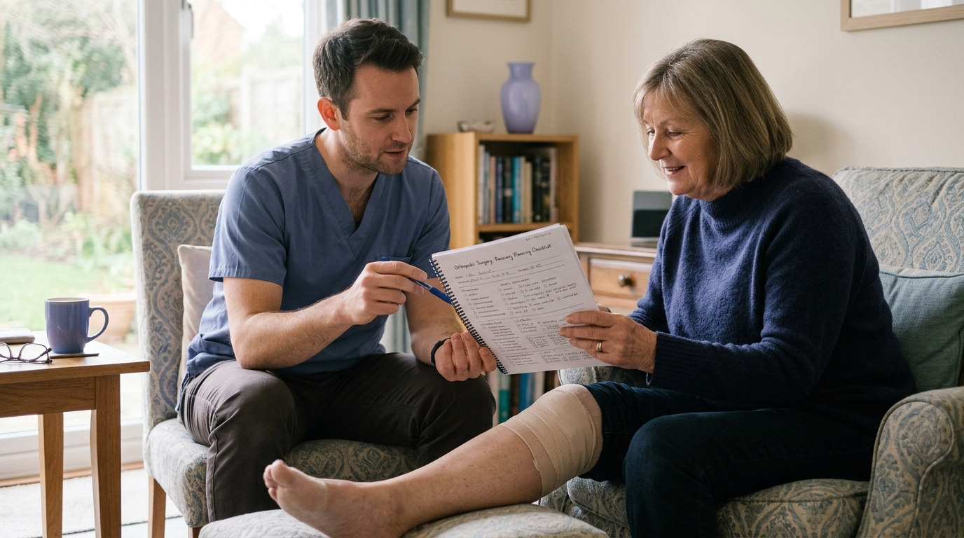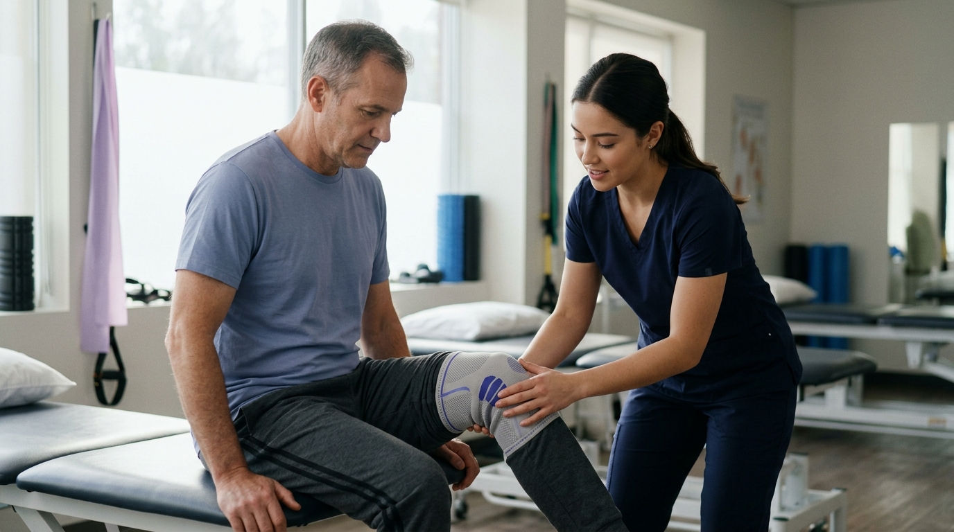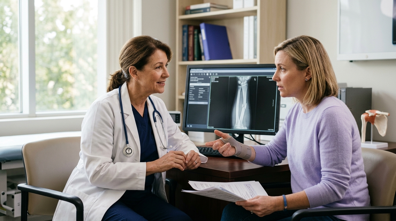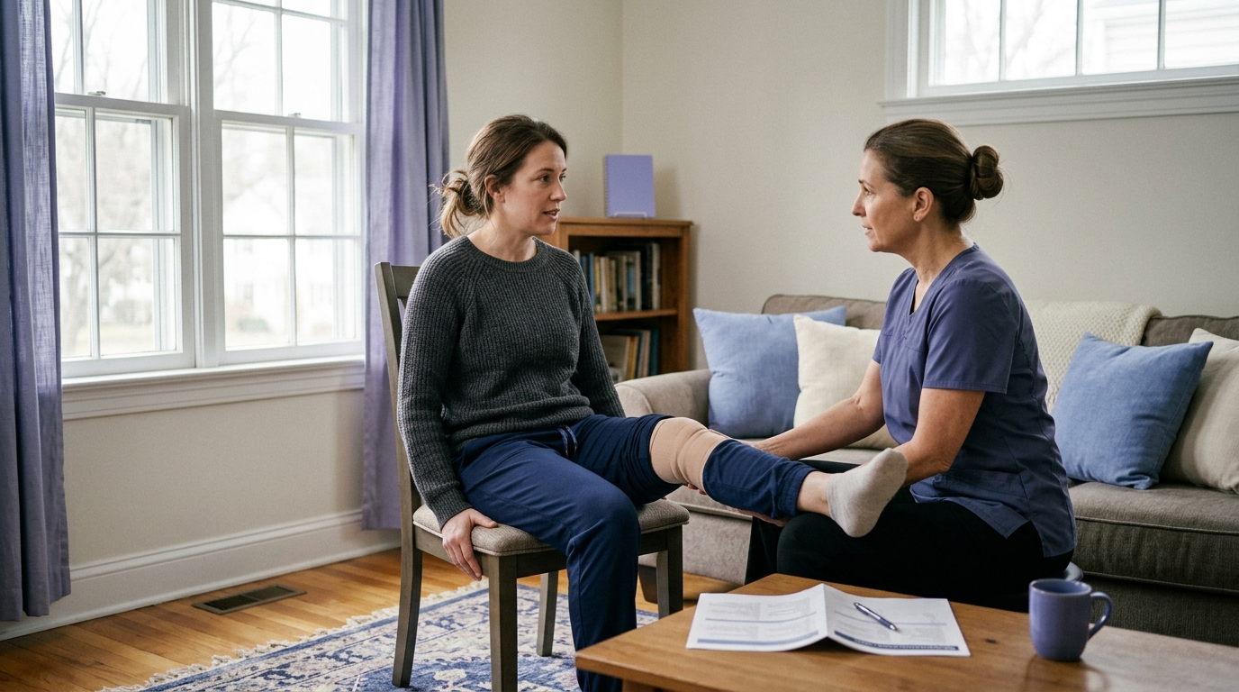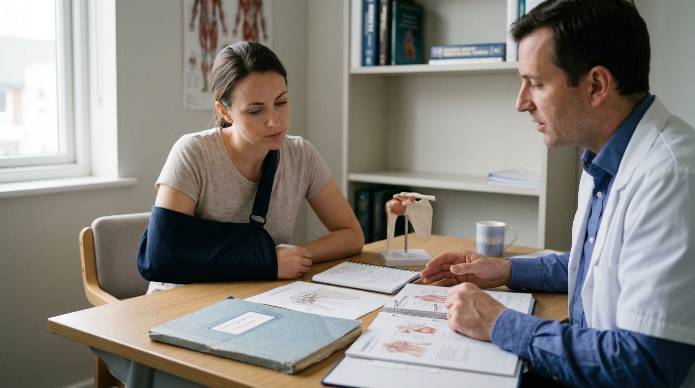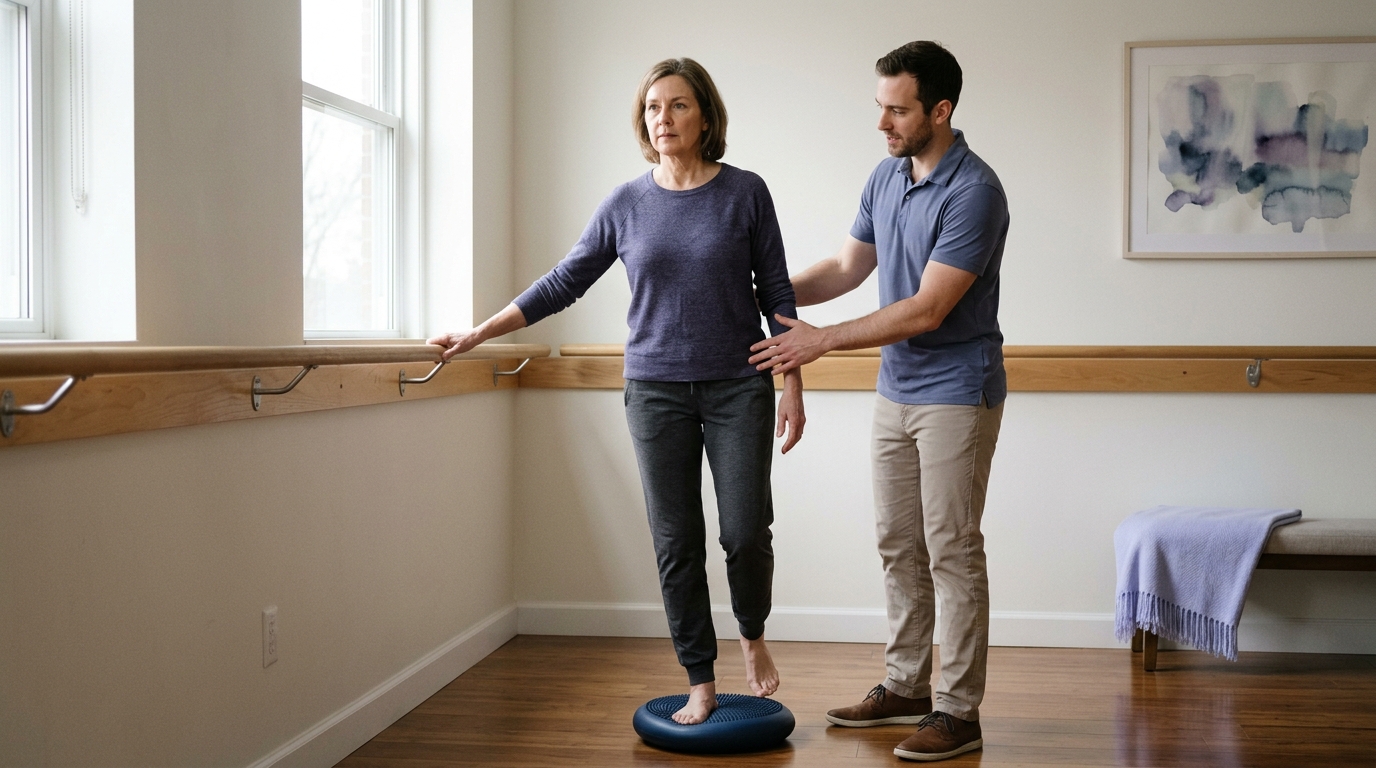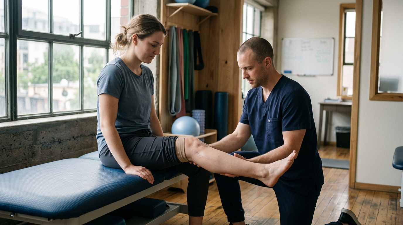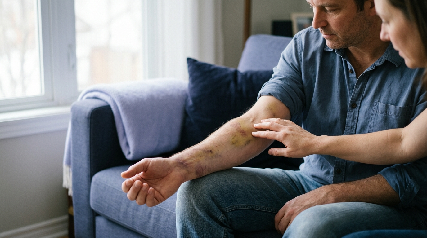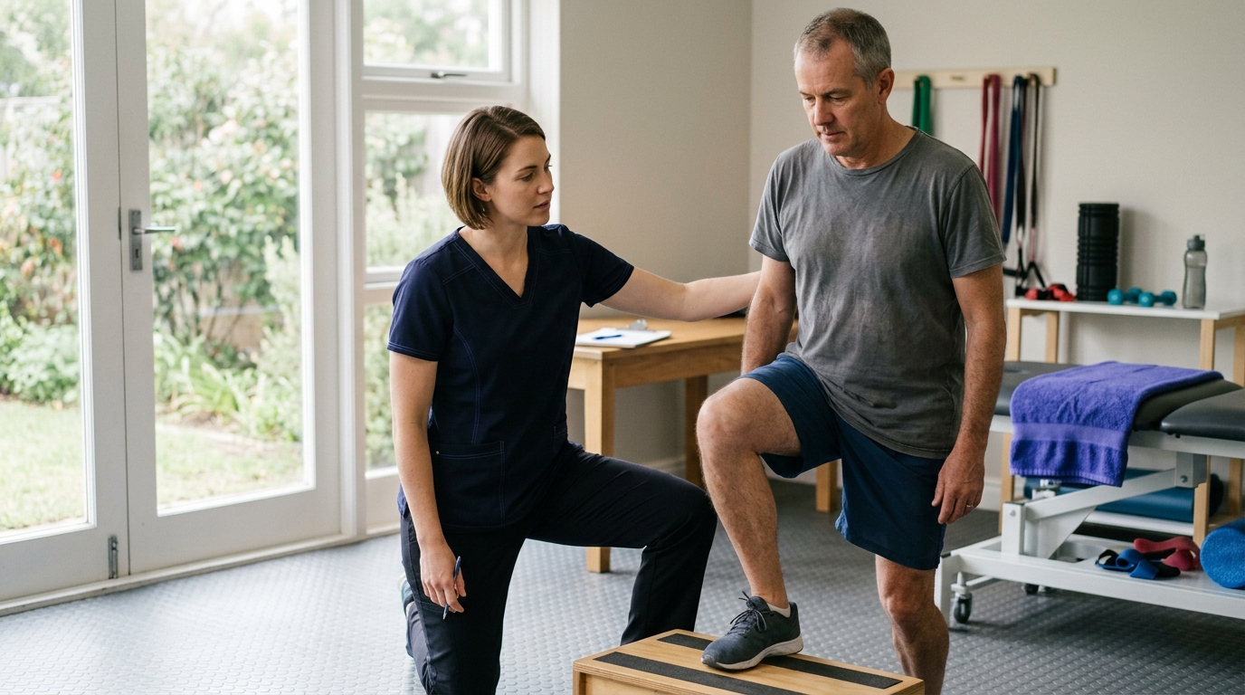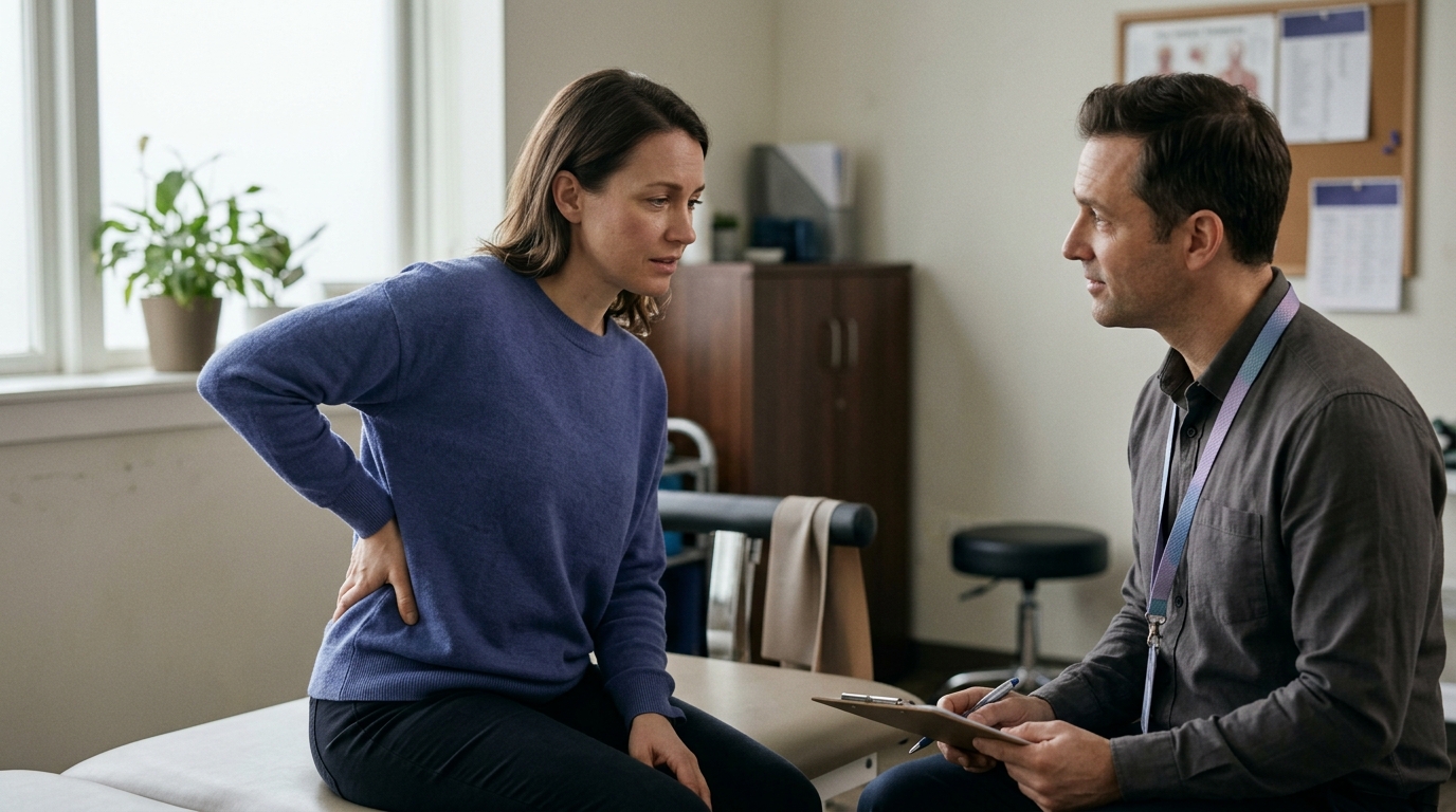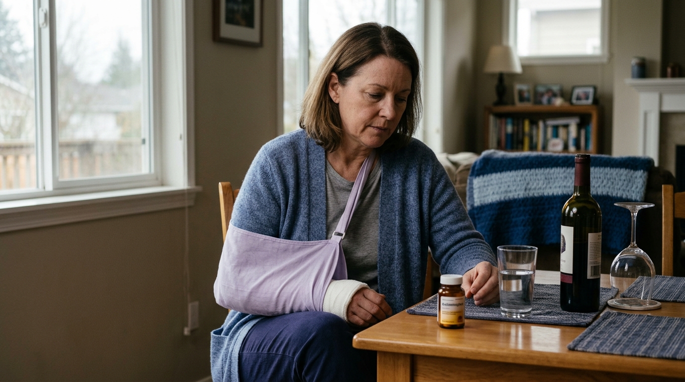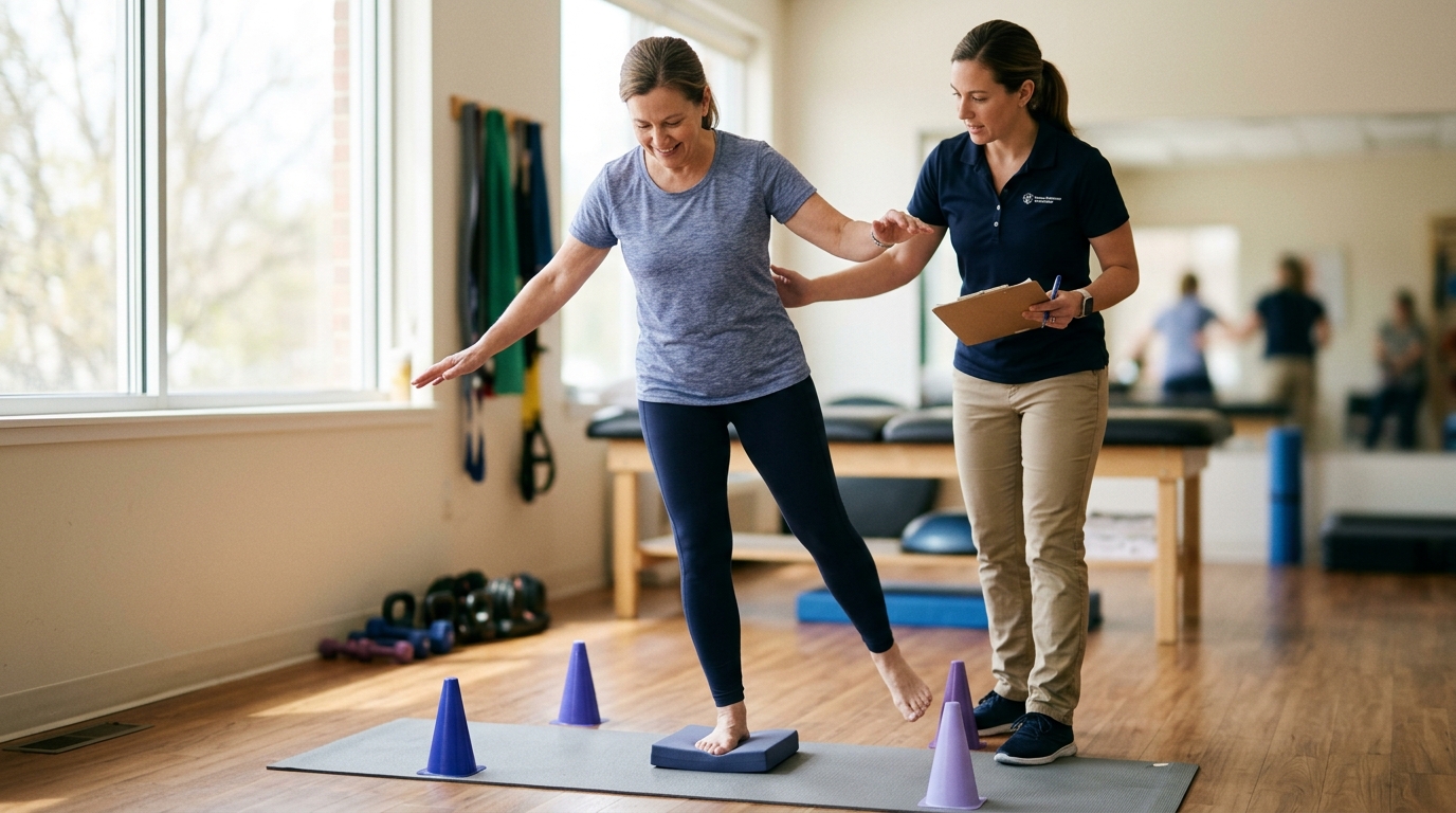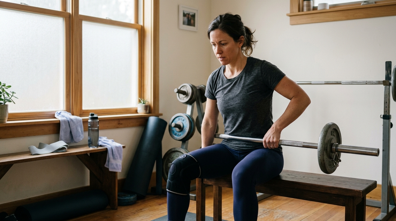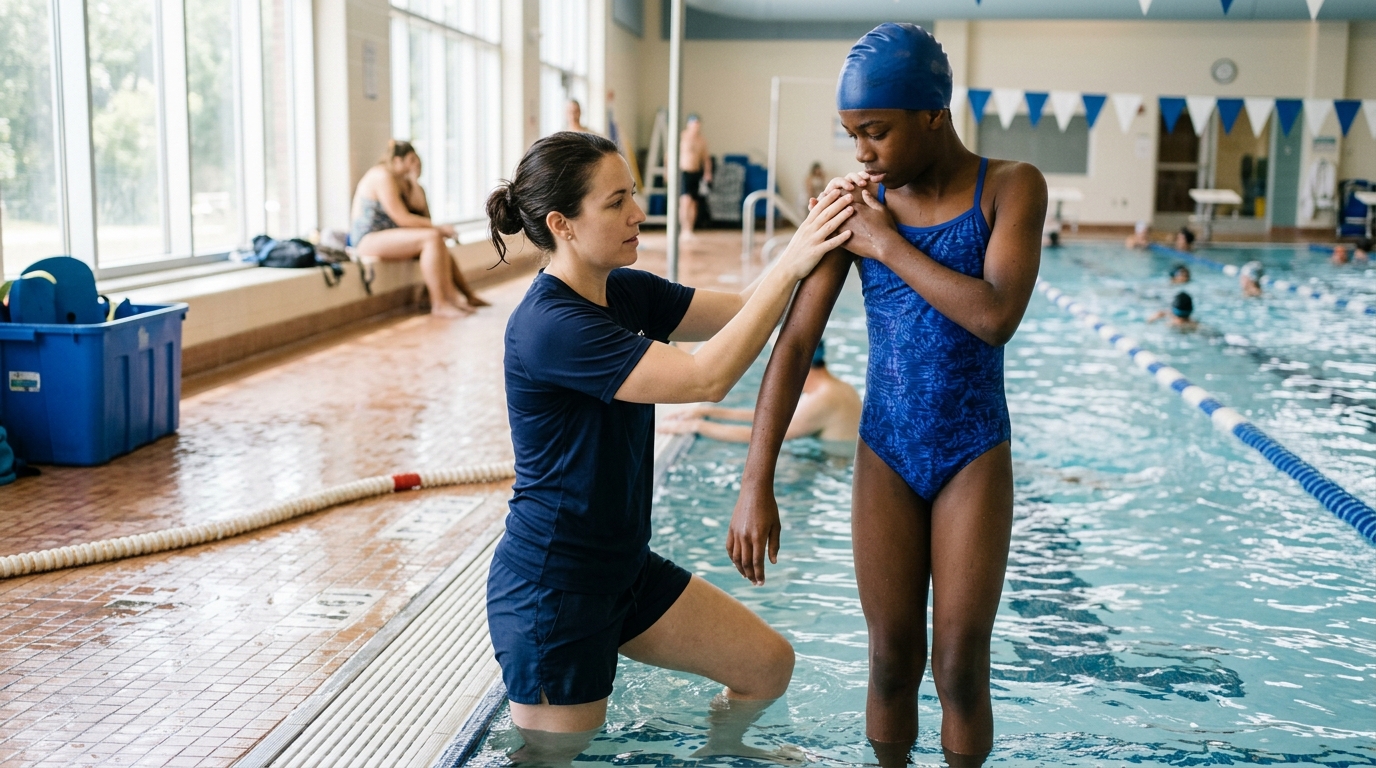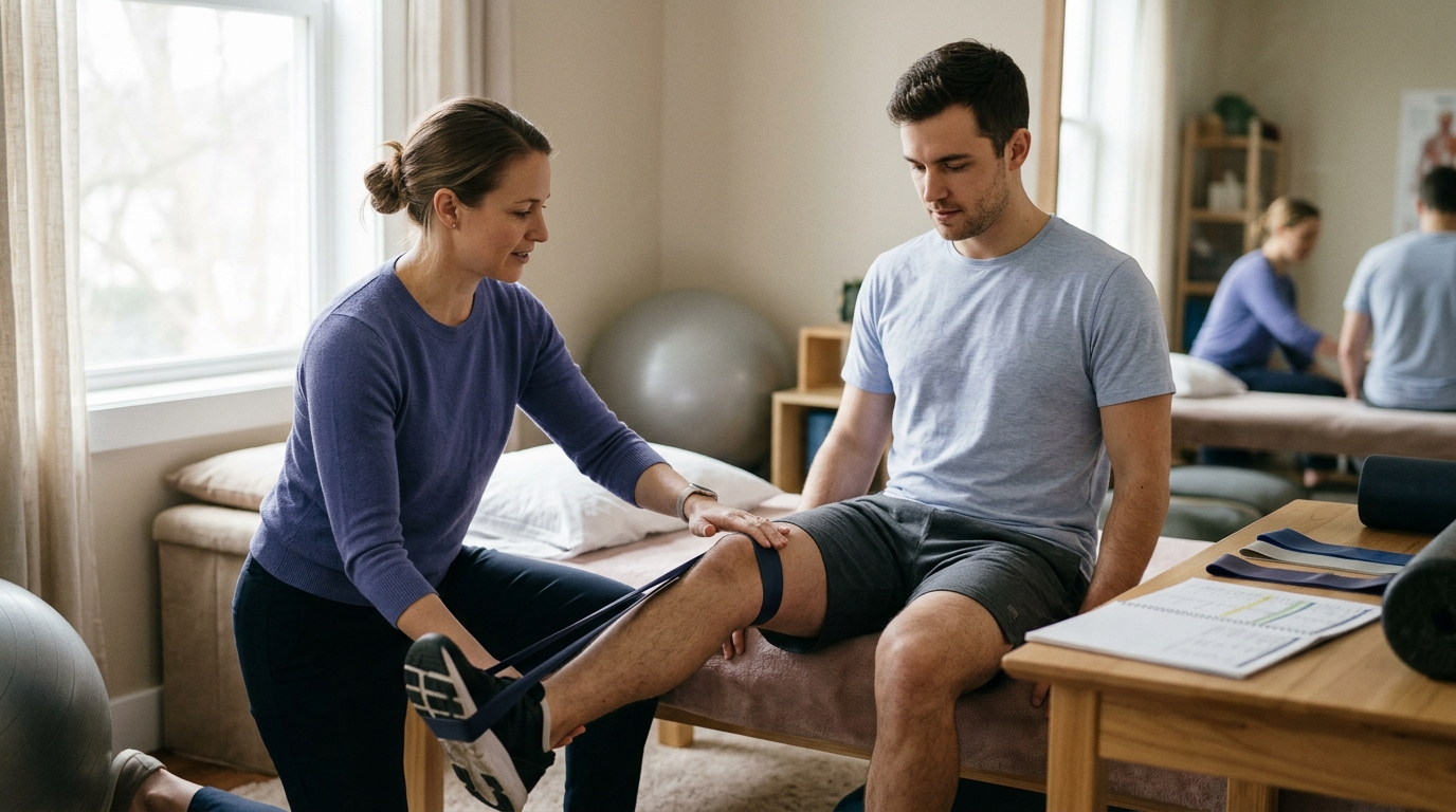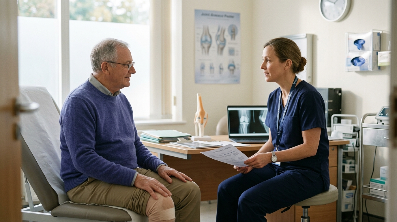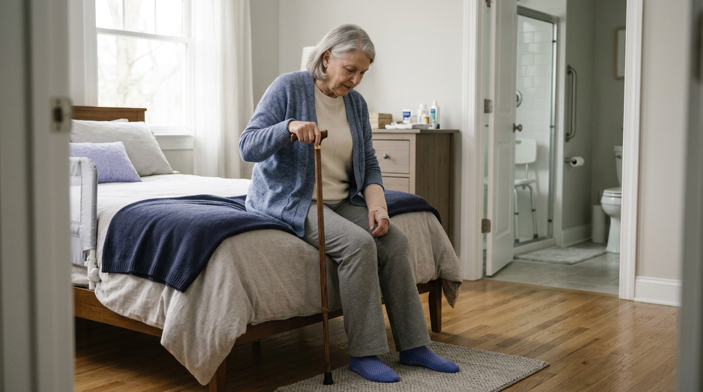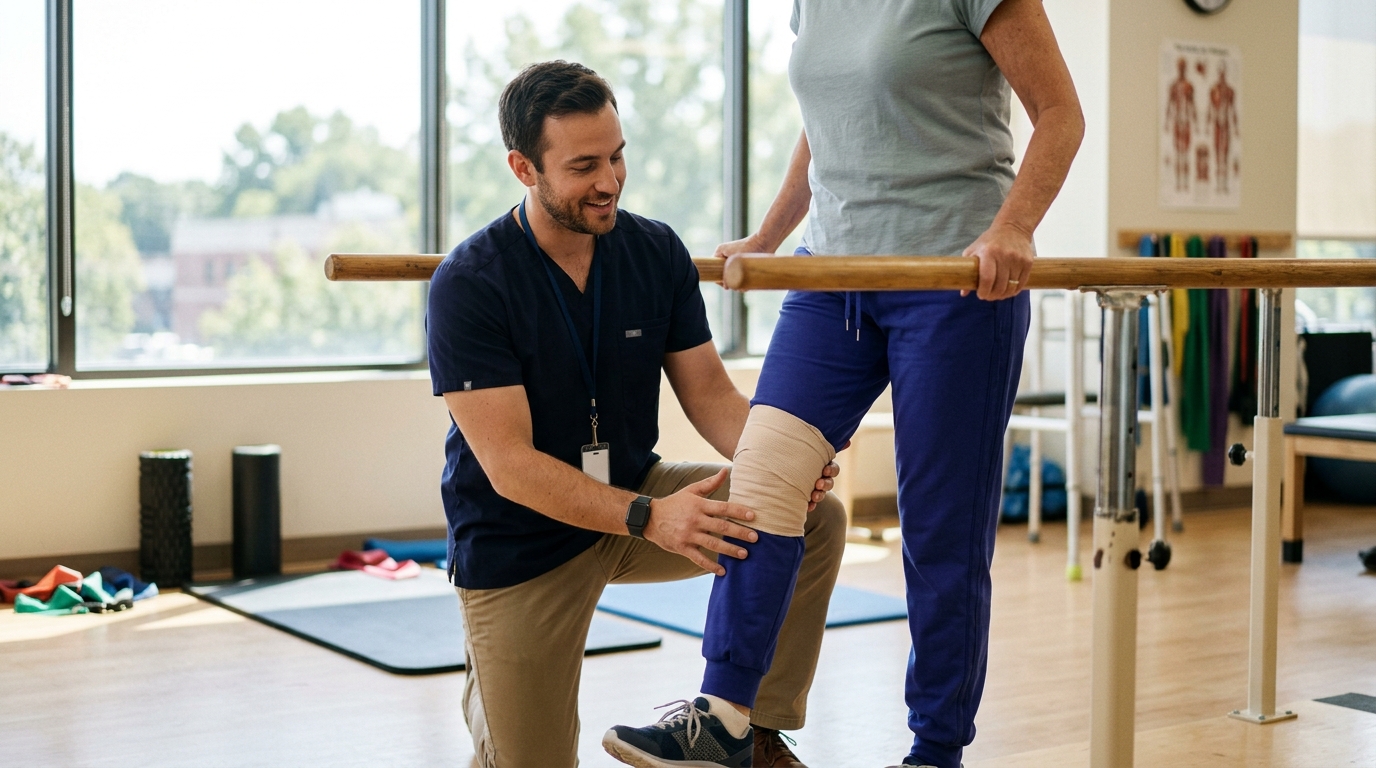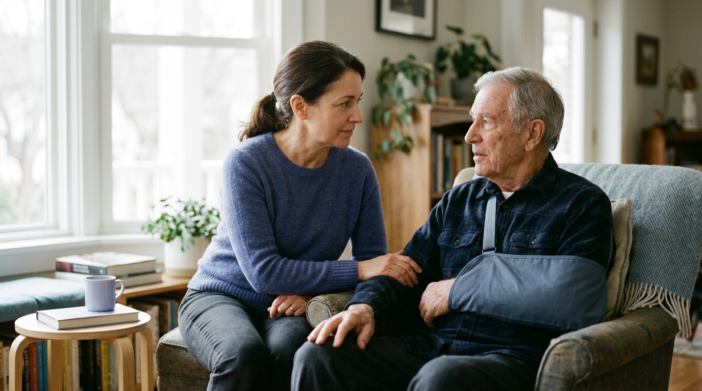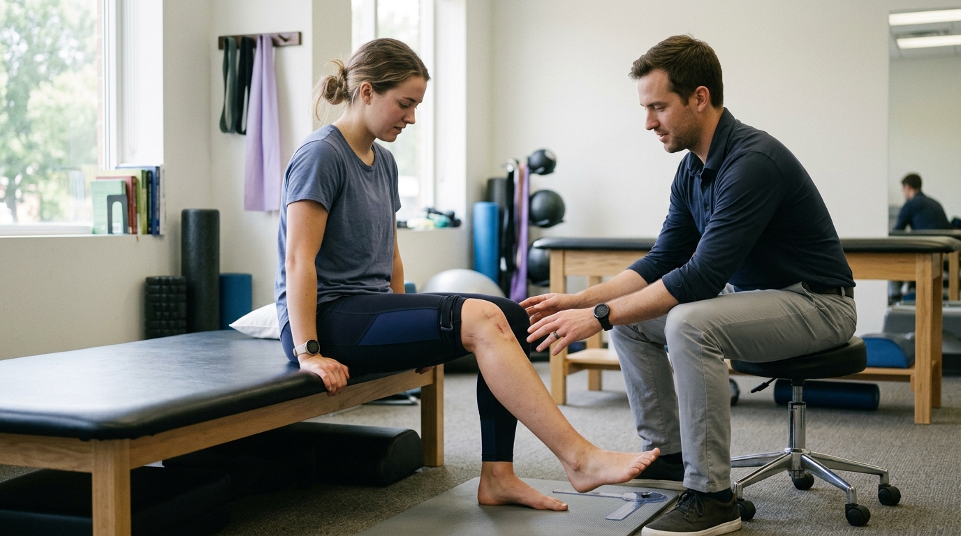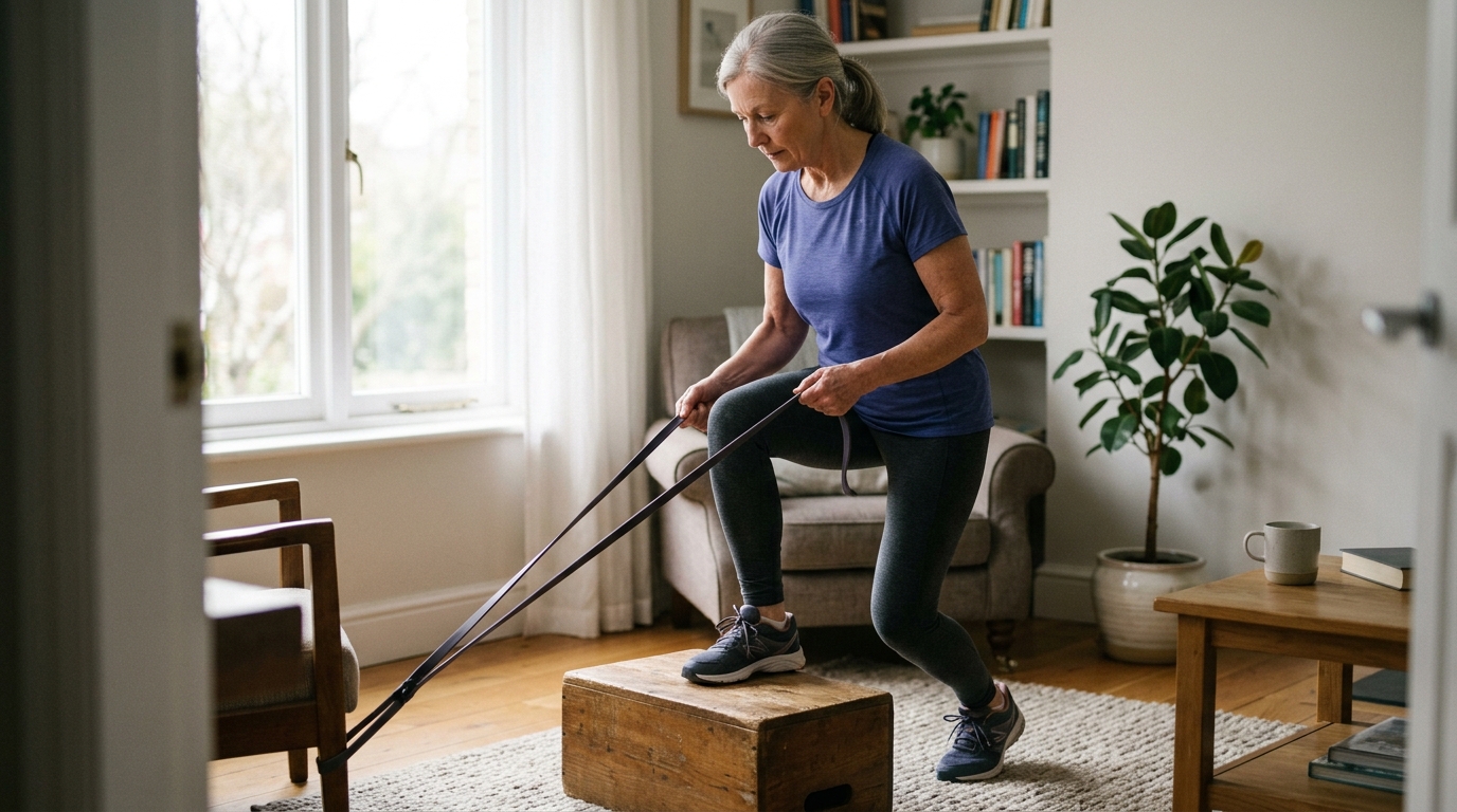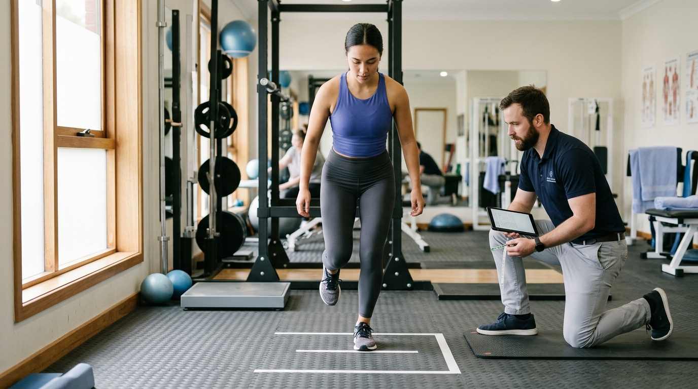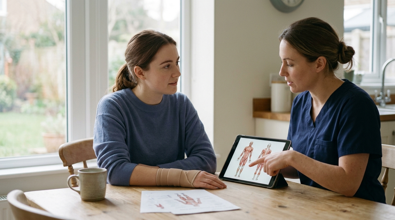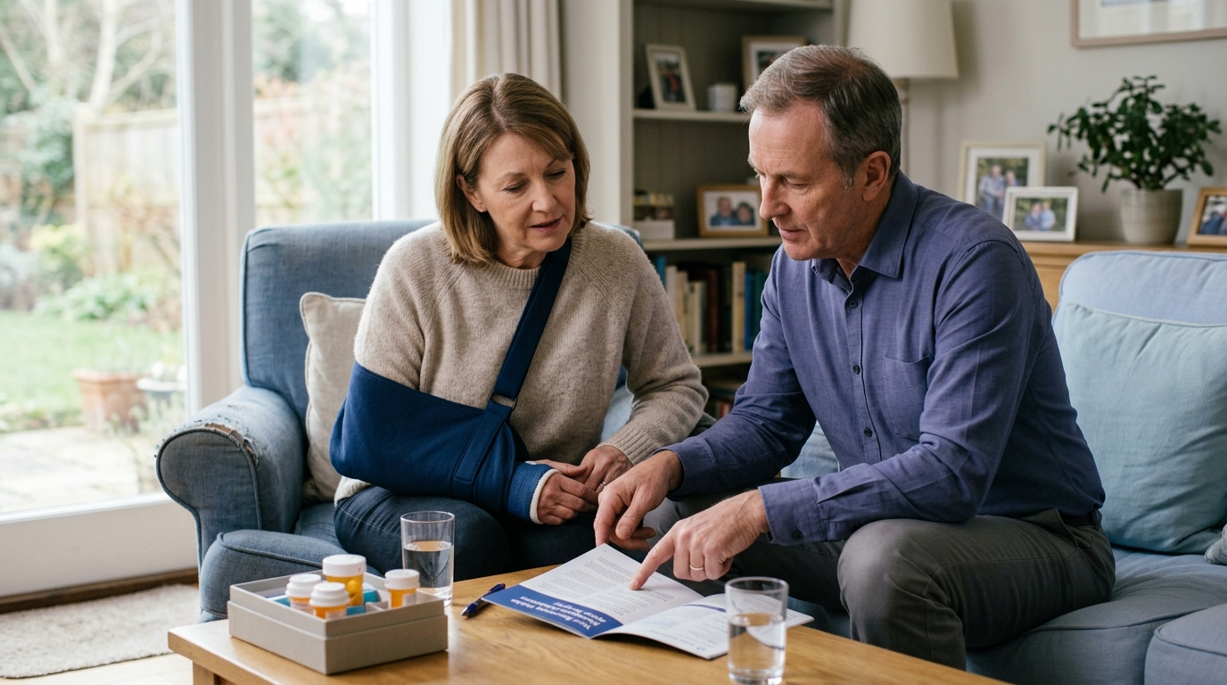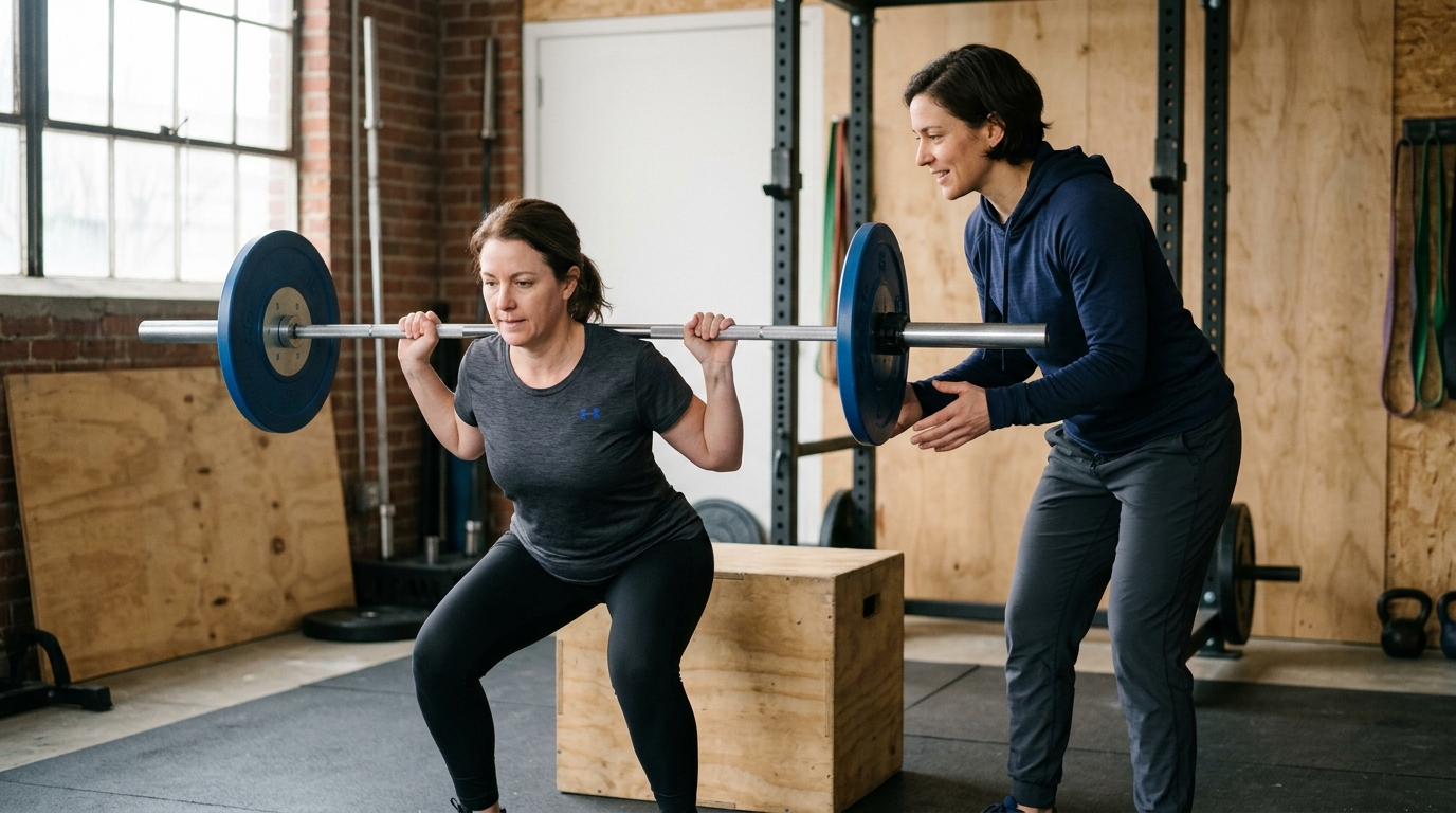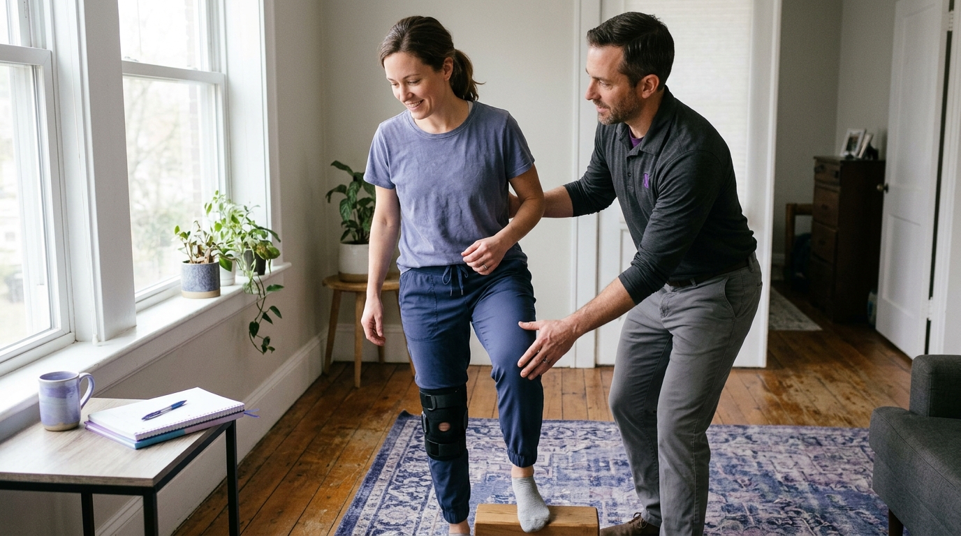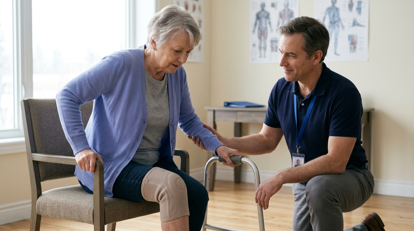

Biological tissue healing and functional recovery progress at different rates, showing why clear imaging and pain relief do not always guarantee full physical performance.





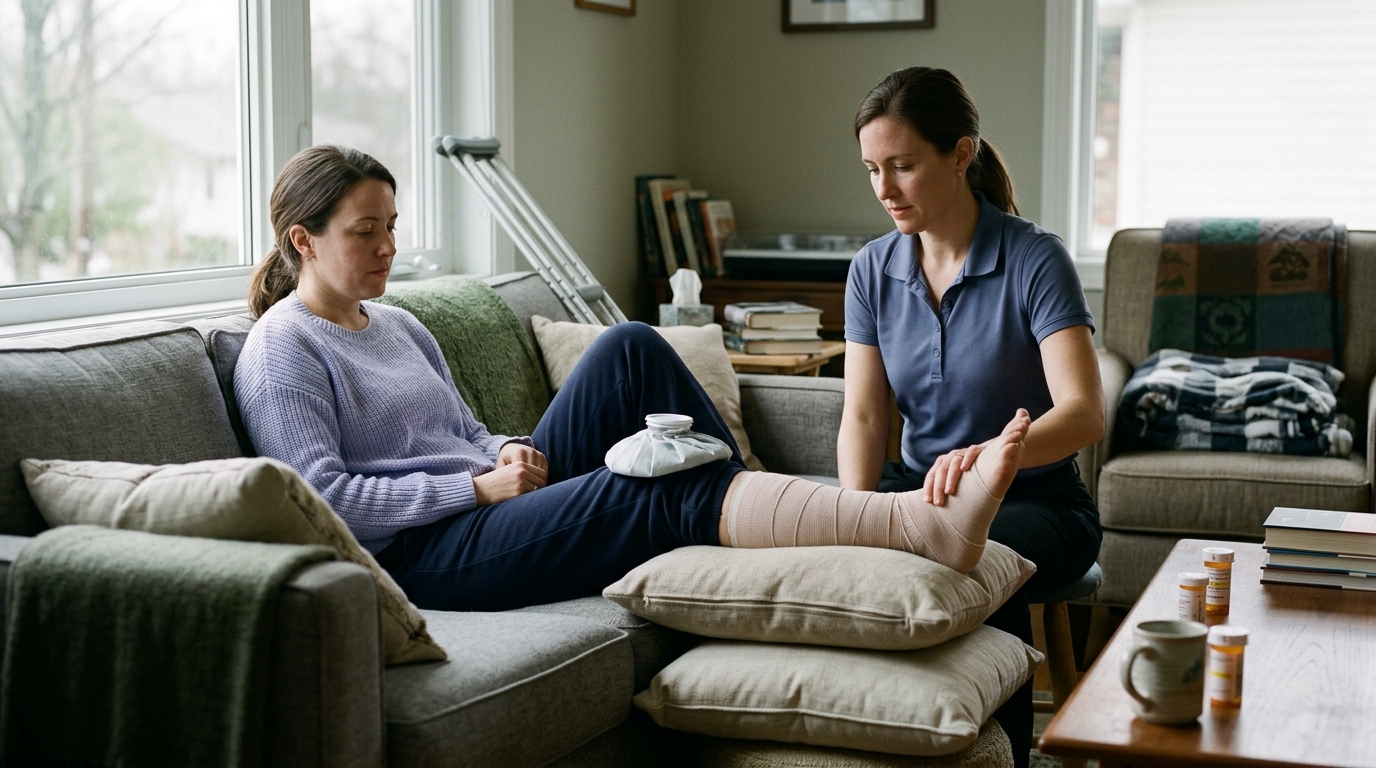
You finish six weeks of rehabilitation after a joint sprain or muscle strain. Your doctor reviews a follow-up scan, nods, and tells you that the tissue is healing as expected. Yet when you try to walk down a flight of stairs or carry a heavy bag of groceries, your limb feels awkward, stiff, and unsteady.
Another person might experience the exact opposite situation. They feel completely ready to play tennis or return to physical labor, but their physical therapist cautions that the repaired tissue inside their knee still needs months of quiet structural remodeling.
These common scenarios highlight a central truth of orthopedic recovery. Biological tissue healing and functional recovery are related processes, but they are not the same thing. They move on different schedules, respond to different inputs, and require different ways of measuring progress.
Understanding the distinction between these two tracks gives you a clearer framework for your rehabilitation. It helps you set realistic expectations, interpret symptoms accurately, and work more effectively with your medical team.
Biological tissue healing refers to the cellular and structural processes that repair damaged tissue in the body. When a bone breaks, a muscle tears, or a tendon is strained, the body activates a biological cascade. It clears cellular debris, lays down new structural proteins, grows fresh blood vessels, and slowly reorganizes the tissue matrix. Tissue healing is measured primarily through medical imaging, surgical inspection, and physical stability tests.
Functional recovery describes what you can actually do in your daily life. It encompasses your ability to move your joints, generate muscle force, maintain balance, and tolerate physical loads. It also includes whether you can perform specific tasks, such as climbing stairs, lifting a child, working a full shift, or playing a sport. Functional recovery is measured through movement assessments, strength tests, endurance metrics, and your real-world performance.
These two tracks often progress at noticeably different rates. A tissue can still be in the middle of long-term biological remodeling while you feel capable of performing basic tasks. In contrast, an injured structure might have regained enough baseline integrity to be labeled healed on a scan, while your neuromuscular coordination, local muscular endurance, and movement confidence remain significantly diminished.
No single calendar date, symptom check, or imaging report can capture your total recovery status. Complete recovery is multidimensional. Recognizing that biological repair and functional capability develop in parallel allows you to avoid two frequent mistakes: pushing too hard because a scan looks clear, or resting too long because a joint still feels unfamiliar. You can find more foundational concepts in our comprehensive injury recovery guides.
To understand why physical function can lag behind or run ahead of tissue status, it helps to examine how the body repairs structural damage. In human biology, the response to injury involves two main mechanisms: regeneration and replacement.
Regeneration replaces damaged tissue with the exact same specialized tissue type, restoring the original cellular architecture. Replacement fills the injured area with fibrous connective tissue, commonly known as scar tissue. Most adult musculoskeletal injuries heal through a combination of both mechanisms. Because scar tissue lacks the exact cellular arrangement of original tissue, a healed structure may not be an exact structural replica of what existed before the injury.
The biological healing process is traditionally described as a sequence of stages. While these stages are helpful for learning, they overlap continuously in real life rather than acting as separate steps.
The moment an injury occurs, the body initiates hemostasis to stop bleeding. This is immediately followed by an acute inflammatory response. During this stage, specialized cells such as neutrophils and macrophages move into the injured area.
These cells clear damaged proteins, dead cells, and bacteria. The acute inflammatory phase typically peaks within the first 72 hours after an injury or surgical procedure. It produces the classic signs of inflammation: localized swelling, redness, heat, and resting discomfort. While inflammation is often treated as something to eliminate entirely, this early biological phase is necessary. It releases growth factors and biochemical signals that tell the body to begin building new tissue.
As the initial inflammatory surge subsides, the proliferation stage begins. In many soft tissue injuries, this subacute phase starts around the third day and can continue for roughly three weeks.
During this stage, specialized cells called fibroblasts multiply rapidly. They produce an extracellular matrix composed mostly of disorganized type III collagen and proteoglycans. At the same time, new blood vessels sprout through a process called angiogenesis to bring oxygen and nutrients to the repairing site. The newly formed repair tissue, often called granulation tissue, is fragile and vulnerable to excessive mechanical stress.
The remodeling stage typically begins around the third week and can continue for several months, sometimes lasting up to a full year or longer. During remodeling, the cellular density in the repair site decreases.
The body gradually replaces the weak, disorganized type III collagen with stronger, more structured type I collagen. Mechanosensitive cells within tendons, ligaments, and bone detect physical forces. In response to these forces, the cells align new collagen fibers along the primary lines of mechanical stress.
Controlled, progressive mechanical loading acts as a direct physical signal for tissue remodeling. Without appropriate physical load, remodeling tissue remains disorganized and mechanically weak. However, applying excessive force too early can disrupt the delicate repair matrix. Finding the right balance between mechanical protection and progressive loading is a core objective of physical rehabilitation. For a deeper look into tissue biology, read our biological recovery science articles.
Just as tissue healing is more complex than a simple scar, physical function is more complex than a single strength test. The World Health Organization developed the International Classification of Functioning, Disability and Health, known as the ICF model. This framework breaks human functioning down into three distinct levels.
Understanding these three tiers prevents confusion about what being recovered actually means. Progress at one level does not automatically translate to success at the next level.
This is the most localized tier of the ICF model. It refers to the physical anatomy and physiological operation of body systems.
Examples of body structures include your knee cartilage, your Achilles tendon, or your lumbar vertebrae. Examples of body functions include the degrees of flexion in your elbow, the peak force your quadriceps can generate on a dynamometer, or your joint position sense. Clinical measurements in a physical therapy clinic often focus on this level. While restoring range of motion and baseline strength is necessary, these isolated measurements do not tell the whole story of your daily physical capacity.
The activity level describes your ability to execute a discrete physical task or action. It shifts the focus from isolated joint mechanics to whole-body movement patterns.
Examples of activities include:
A person can have normal isolated muscle strength on an examination table but still struggle with the complex balance and coordination required to walk down a steep hill. Conversely, an individual might learn to compensate for a stiff ankle and perform a basic squat, even though their joint structure remains limited.
Participation represents the highest and most meaningful level of function. It describes your involvement in real-world life situations, responsibilities, and social roles.
Participation includes:
This distinction explains why medical clearance should always be framed around a specific context. When a clinician asks if you have recovered, the appropriate response is to ask: recovered for what?
A person might regain the basic activity level needed for light desk work within a month of an injury. However, returning to full participation as an electrician or a competitive soccer player requires a much higher threshold of physical conditioning, fatigue resistance, and structural remodeling. For targeted movement ideas, check out our movement and mobility strategies.
Many people assume that physical recovery can be confirmed by two simple indicators: an image that looks clean and an absence of pain. While medical scans and symptom tracking provide valuable clinical clues, modern orthopedic science shows that neither measure gives a complete picture of recovery.
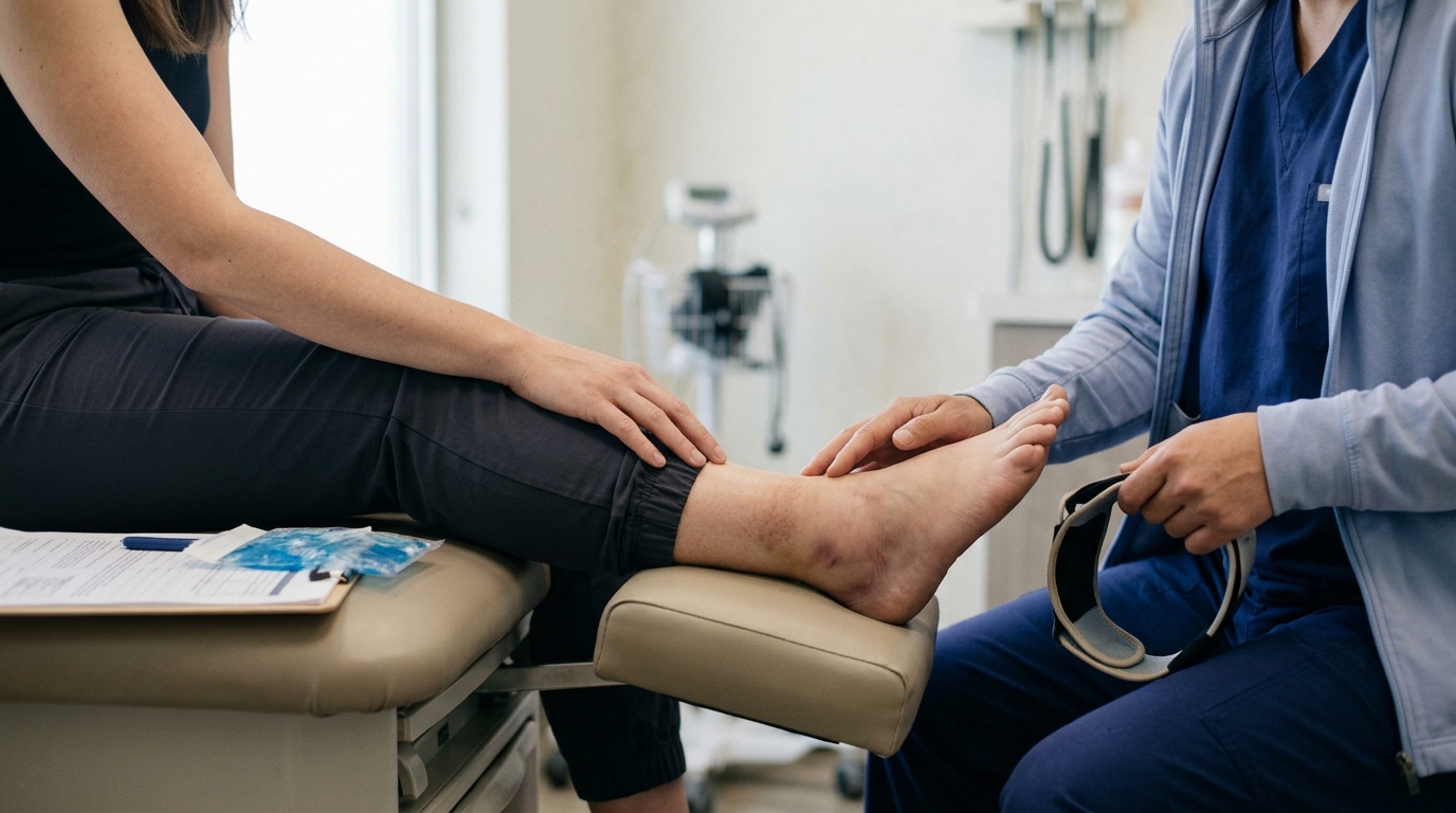
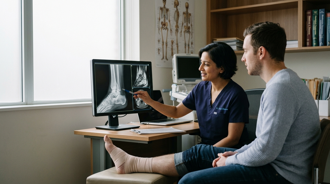
Magnetic resonance imaging (MRI), computed tomography (CT), and X-rays provide detailed views of internal anatomy. They allow clinicians to identify broken bones, torn ligaments, and joint degeneration. However, structural appearance on a scan does not correlate perfectly with functional capacity.
In sports medicine, research demonstrates that athletes recovering from acute muscle strains frequently show persistent abnormalities on MRI scans even after they are completely symptom-free. These structural findings can include localized fluid accumulation, connective tissue scarring, tendon thickening, or small residual gaps in the muscle architecture.
Systematic reviews confirm that complete normalization of an MRI signal is not required for a safe return to physical activity. Functional performance, clinical strength tests, and movement control consistently serve as more reliable guides for activity clearance than structural scans alone.
The reverse is also true. A joint or muscle can look structurally intact on an imaging study, but the surrounding neuromuscular system may remain inhibited, weak, or prone to rapid fatigue. Imaging reveals anatomy at a single frozen moment in time. It cannot show how that tissue handles continuous movement, heavy impact, or prolonged fatigue.
Pain is a vital biological alarm system designed to protect you from harm. However, contemporary pain neuroscience shows that pain is not a simple direct readout of physical tissue damage.
During the acute phase of an injury, pain levels generally match the severity of tissue trauma. As the weeks progress, the central and peripheral nervous systems adapt. Pain perception is influenced by several factors:
Because of this complex processing, you can experience noticeable discomfort in an area where structural healing is well underway. This type of symptom response often reflects tissue sensitivity and deconditioning rather than ongoing damage.
On the other hand, the complete absence of pain during rest does not prove that a tissue is ready for high-impact loads. Many degenerate or healing tendons cause no discomfort while sitting on a couch, but they may react with irritation when subjected to rapid jumping or sprinting. Evaluating your recovery requires looking at how your body handles physical tasks over time, rather than relying solely on resting pain.
Different tissues in the human body possess distinct cellular structures, blood supply levels, and metabolic rates. Because of these differences, a bone fracture, a muscle tear, and a tendon injury heal along fundamentally different biological timelines.
Bone has a rich blood supply and an exceptional capacity for true regeneration. When a bone fractures, the repair process proceeds through several distinct biological stages.
First, a fracture hematoma forms, providing a biological scaffold for incoming cells. Next, the body forms a soft cartilaginous callus, which provides initial mechanical stability across the fracture gap. Over the following weeks, this soft callus undergoes endochondral ossification, transforming into a hard bony callus visible on X-rays. Finally, osteoclasts and osteoblasts remodel the hard callus over months to years, restoring the bone's original shape and internal trabecular architecture.
In clinical research, defining when a bone has achieved union is surprisingly complex. A systematic review of fracture literature showed that researchers define union inconsistently:
A patient may have a stable bone that can bear weight without discomfort, even while the fracture line remains faintly visible on an X-ray. Clinical assessment of stability and tenderness must be weighed alongside imaging findings.
Skeletal muscle has a substantial blood supply and contains specialized stem cells known as satellite cells. When muscle fibers tear, these satellite cells activate, divide, and differentiate into new muscle fibers to repair the structural gap.
However, muscle healing is a delicate race between muscle fiber regeneration and fibrous scar formation. In larger or more severe tears, fibroblasts produce dense collagenous scar tissue to bridge the defect quickly. While this scar restores mechanical continuity, it does not contract like healthy muscle fibers.
Functional recovery after a muscle strain requires rebuilding the muscle's tensile strength, active contraction velocity, and passive flexibility. A person might regain normal walking mechanics quickly, but restoring high-velocity sprinting requires structured loading to strengthen the interface between the new muscle fibers and the scar tissue.
Tendons and ligaments connect muscles to bones and bones to other bones. Unlike muscle, these connective tissues have relatively low cellular density and a limited direct blood supply.
Tendon healing involves an extended remodeling phase. The initial repair tissue contains disorganized collagen with low mechanical strength. Because tendons must transmit enormous tensile forces, they depend heavily on mechanotransduction. As you apply progressive tension to the tendon during exercise, tendon cells align new collagen fibers parallel to the direction of pull.
Controlled mobilization promotes favorable collagen alignment and improves tensile strength. However, scientific reviews point out that the optimal timing and exact dosage of tendon loading remain debated within sports medicine. Tendons remodel slowly, which is why functional rehabilitation for tendon conditions often requires several months of consistent, progressive strength training. Explore structured loading ideas in our progressive strength and return to activity guidance.
Biological healing is not an automatic process that runs on an identical clock for every human being. A wide range of systemic, mechanical, and biological factors can speed up or slow down cellular repair.
The physical characteristics of the original injury play a major role in biological repair. A clean, non-displaced bone fracture heals much faster than a complex comminuted fracture with multiple bone fragments.
In traumatic fractures, open injuries, where the bone pierces through the skin, carry significantly higher risks of delayed healing and nonunion. A large systematic review of surgically managed fractures found that open fractures had 2.6 times the odds of nonunion compared to closed fractures. Severe soft tissue disruption damages local blood vessels, starving the repair zone of oxygen and necessary growth factors.
Systemic medical conditions directly affect cellular repair mechanisms throughout the body. Diabetes mellitus provides a clear example of this biological connection.
Elevated blood glucose levels and altered insulin signaling impair new blood vessel formation, alter collagen synthesis, and prolong the inflammatory phase. Meta-analyses examining fracture healing report that individuals with diabetes face approximately 1.6 times higher odds of experiencing delayed union or nonunion compared to those without diabetes. Managing systemic metabolic health supports the cellular environment required for tissue repair.
Nicotine and the chemical compounds found in tobacco smoke significantly impair biological tissue healing. Nicotine causes peripheral vasoconstriction, reducing microvascular blood flow to healing tissues.
Carbon monoxide from smoke binds to hemoglobin, reducing oxygen delivery to actively repairing cells. A meta-analysis analyzing 7,516 surgical and orthopedic procedures found that smokers had 2.2 times the risk of delayed healing or nonunion compared to nonsmokers. Quitting smoking or avoiding nicotine products during recovery improves tissue perfusion and reduces complications after orthopedic trauma.
Aging involves natural biological shifts, including decreased cellular turnover, reduced stem cell availability, and lower resting collagen synthesis rates. While healthy older adults retain robust healing capabilities, the biological timeline for tissue repair is often somewhat longer than that of an adolescent.
Nutritional status also provides the raw materials for cellular rebuilding. Severe protein deficiency, inadequate total caloric intake, and micronutrient deficiencies can compromise collagen production and immune function during the repair phase. Maintaining balanced nutrition supports normal tissue repair throughout rehabilitation. For post-operative recovery context, review our surgical rehabilitation insights.
While biological factors determine how quickly cells repair tissue, functional recovery is shaped by neuromuscular adaptation, physical conditioning, and psychological factors. An individual can have a fully united bone or a repaired ligament, yet remain functionally limited by secondary adaptations that occurred during the recovery period.
When a joint or limb is injured, the central nervous system alters its motor output to protect the area. This phenomenon, known as arthrogenic muscle inhibition, prevents full activation of muscles surrounding an injured or inflamed joint.
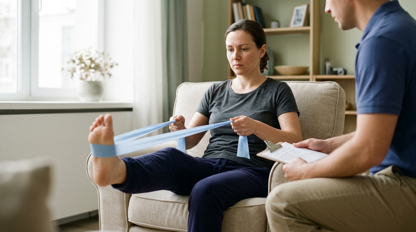
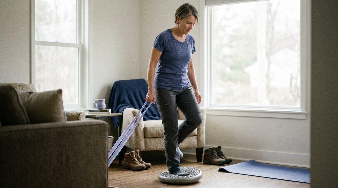
Even after tissue repair has progressed, this neurological inhibition can persist. Furthermore, periods of limb immobilization or reduced activity cause rapid muscle atrophy and loss of local muscular endurance. Restoring functional capacity requires structured strength training to retrain the nervous system, restore motor unit recruitment, and rebuild muscle mass.
Physical recovery does not occur in a purely mechanical vacuum. Extensive clinical research shows that psychological factors strongly influence functional outcomes and the speed of returning to normal activities.
A comprehensive systematic review investigating biopsychosocial predictors of recovery found strong evidence that personal recovery expectations directly predict return-to-work outcomes. Patients who maintain realistic, positive recovery expectations achieve better long-term functional results than those with high levels of fear avoidance or catastrophizing.
These findings do not suggest that physical symptoms are imaginary. Instead, they demonstrate that fear of reinjury, movement anxiety, and low recovery confidence can lead individuals to avoid helpful physical activities. When people avoid moving, their physical deconditioning worsens, which prolongs disability. Structured rehabilitation helps people rebuild movement confidence through safe, gradual physical exposure.
The gap between biological healing and functional readiness depends heavily on the physical demands of your target activities.
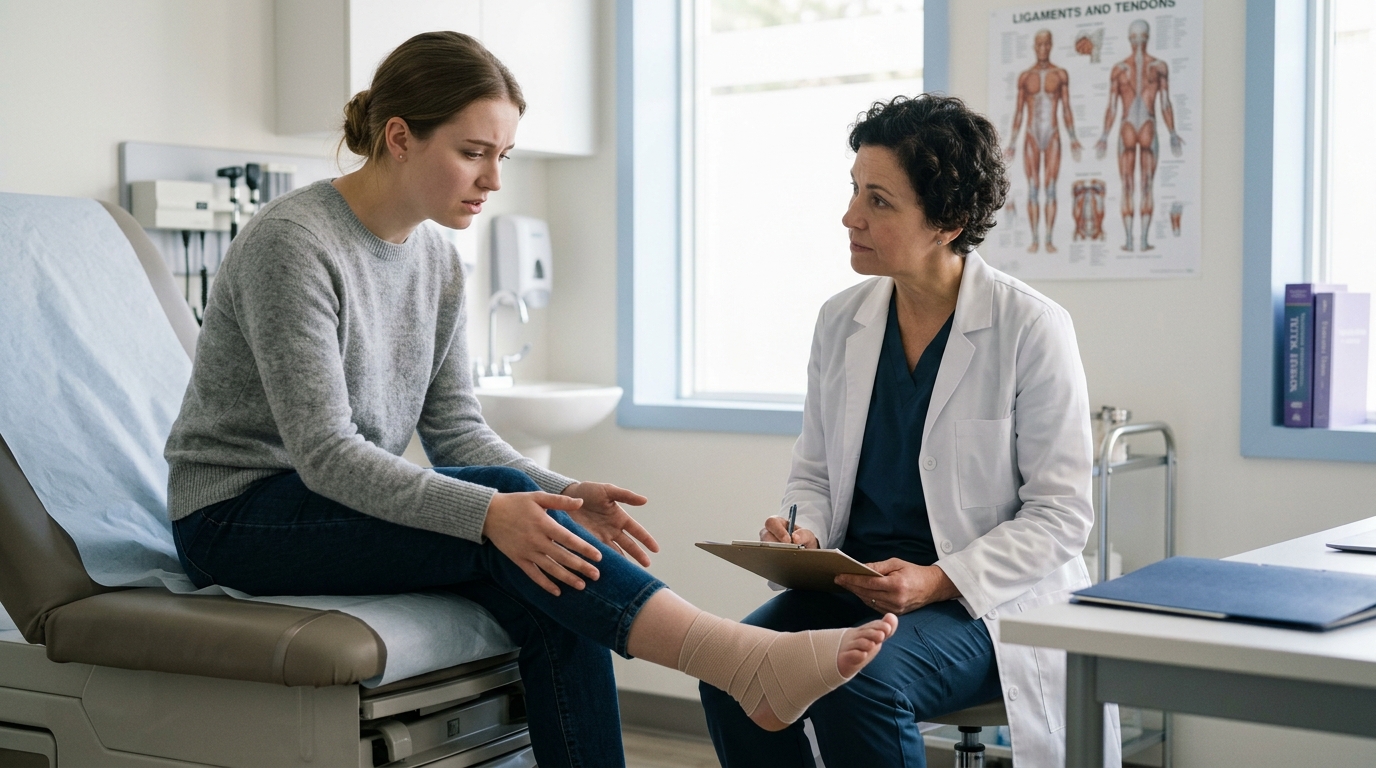
An office worker recovering from an ankle injury might achieve the functional capacity needed for their job within three weeks, while the tissue is still in the early proliferative stage. An industrial construction worker with the same injury requires high levels of balance, uneven ground tolerance, and heavy carrying capacity before they can safely work a full shift. Functional recovery must always be evaluated relative to the specific physical demands of your daily environment.
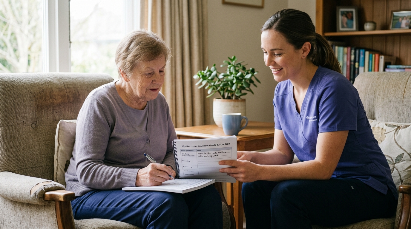
Rather than asking the simple question, "Is my injury healed?", you can evaluate your recovery across several interrelated dimensions. This comprehensive framework helps you distinguish steady progress from complete recovery, allowing you to make sensible choices about physical activity.
What is the current biological state of the injured tissue? Review what your surgeon or physician has documented regarding the injury severity, surgical repair stability, or fracture union. Be clear about any specific medical precautions, such as weight-bearing limits or range-of-motion restrictions that protect healing tissue.
How do your symptoms behave during different situations? Track what you feel at rest, during physical tasks, and 24 hours after an activity.
Mild, temporary discomfort during or after an exercise session is often a normal part of tissue adaptation. However, sharp pain, spreading symptoms, or joint swelling that persists into the next morning indicates that the mechanical load exceeded your current physical tolerance.
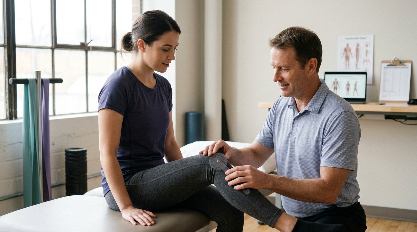
What can your musculoskeletal system do under controlled testing conditions? Evaluate your isolated joint range of motion, muscle strength, balance, and tissue endurance. Comparing your injured side to your uninjured side provides a helpful objective benchmark for identifying residual deficits.
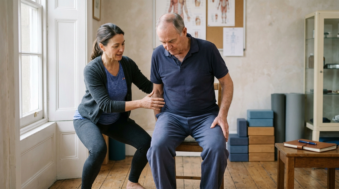
Can you perform the specific movement patterns required for your daily life? Look at how you walk, negotiate steps, squat, carry loads, or reach overhead. Are you moving smoothly, or are you limping, favoring one side, or using awkward compensations to get through the task?
To what extent have you resumed your meaningful life roles? Track your ability to complete a workday, manage household responsibilities, participate in social activities, or engage in recreation. Note whether you can sustain these activities for normal durations without excessive fatigue or symptom flares.
Is your physical tolerance expanding, plateauing, or declining over multi-week blocks? Recovery rarely follows a straight line every single day. Look at the general trend over two- to four-week periods to ensure you are steadily handling more activity with fewer setbacks.
What broader lifestyle and environmental factors are influencing your physical system? Assess your sleep quality, daily stress levels, nutritional intake, and workplace physical demands. Addressing barriers in your daily environment creates the biological and psychological conditions needed for continued physical progress.
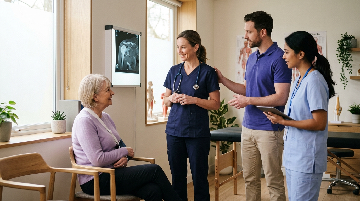
Collaborating with your healthcare team helps ensure that your activity choices align with both your biological healing stage and your functional goals. Here are practical questions to guide your clinical discussions:
Biological tissue healing and functional recovery are two distinct aspects of orthopedic rehabilitation. Tissue healing restores cellular integrity, repairs structural defects, and remodels the extracellular matrix over weeks and months. Functional recovery restores your strength, movement control, physical endurance, and ability to participate fully in daily life.
These two tracks do not always progress at the same speed. A scan showing structural repair does not guarantee that your body is ready for heavy physical demands. Similarly, mild residual symptoms or ongoing tissue remodeling do not mean you must remain inactive.
Tracking your recovery across multiple dimensions gives you a realistic view of your progress. By respecting biological healing constraints while steadily challenging your physical capacity, you can rebuild strength, restore functional performance, and return to your valued activities with confidence.
Revisit this framework whenever you transition between different phases of rehabilitation, such as moving from protective rest to active exercise, or from clinic-based therapy back to work or sport. It is also helpful to review these concepts if your progress seems to plateau, or if you feel uncertain about how to interpret changes on a medical imaging report.
Recovery is a gradual process of physical adaptation that unfolds over time through consistent, thoughtful effort.

Use ReboundBody resources to understand common recovery stages, rehab terms, movement limits and strength rebuilding. Each guide is designed to make a complex comeback easier to understand.


Read practical guidance on injury recovery, rehabilitation, mobility and rebuilding strength as you work your way back to activity.
Explore Resources