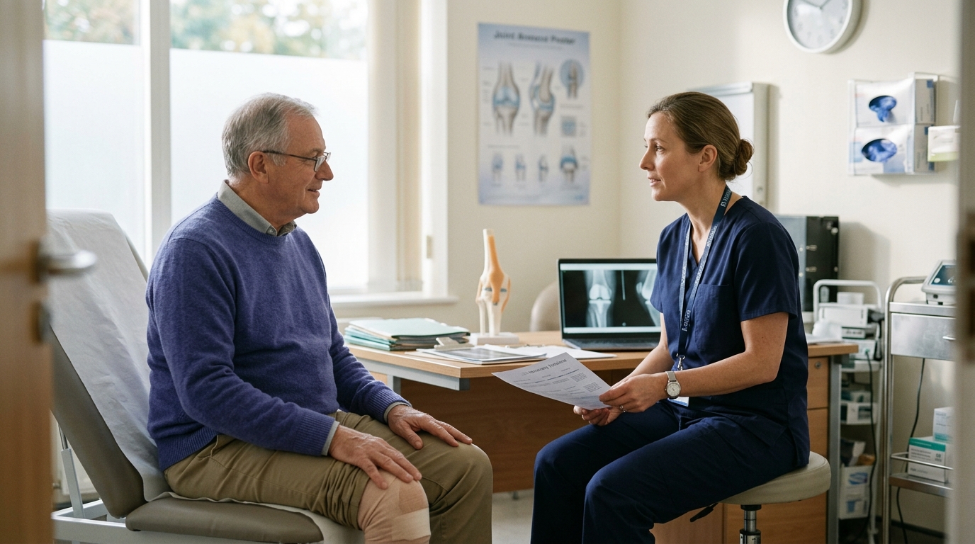

Clear expectations for orthopedic recovery emerge from understanding how tissue biology, procedure types, and clinical restrictions shape long-term joint rehabilitation timelines.





Many people assume that a smaller surgical cut means a faster recovery. They expect a procedure done through two tiny punctures to heal much sooner than an operation requiring a long incision.
This assumption often leads to surprise and frustration during rehabilitation. The true speed and difficulty of recovery are not dictated by the size of the skin incision. They are shaped by the biological task happening deep inside the joint.
Recovery depends on what tissue was cut, repaired, reconstructed, or replaced. It also depends on what mechanical loads that healing tissue must tolerate as you return to normal movement. Two operations that look identical on the surface can require completely different rehabilitation timelines.
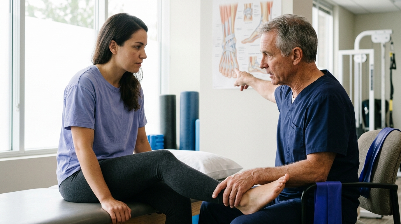
Understanding these structural differences helps you set realistic expectations. It also allows you to work more effectively with your physical therapist and surgical team throughout your recovery journey.
The word arthroscopy describes how a surgeon gets inside a joint. It refers to a surgical technique where a small camera and slim instruments enter through narrow incisions. It does not describe a single, uniform operation.
According to National Health Service (NHS) guidance, recovery following arthroscopic surgery can range from one week to several months. The timeline depends entirely on what the surgeon did once the camera was inside.
A simple diagnostic examination or a minor cleanup of loose joint tissue leaves the underlying structures intact. In contrast, reattaching a torn tendon or securing a replacement ligament through those same small incisions creates a delicate biological repair. That repair requires months of strict protection to heal securely.
When evaluating any surgical recovery and comeback path, you must look past the entry method. You need to focus on the biological repair.
Every orthopedic operation involves one of four primary mechanical tasks:
Each of these four tasks creates a unique environment for healing. Each task places different demands on your body during rehabilitation.
Orthopedic procedures generally fall into four broad categories. Each category follows a distinct rehabilitation logic based on how the treated tissue repairs itself.
In an uncomplicated arthroscopic debridement or diagnostic procedure, the surgeon removes loose fragments, trims torn cartilage edges, or inspects joint surfaces. No torn tissue is stitched together. No new tissue is grafted into place.
Because there is no delicate structural repair to protect, rehabilitation can often progress quickly. For uncomplicated knee arthroscopy, NHS physiotherapy guidance notes that full weight-bearing is often permitted right away unless your surgeon instructs otherwise. Many people return to light daily activities within a few weeks once initial swelling and post-surgical soreness settle down.
The main challenge in this category is managing joint irritation. Even without a structural repair, the joint capsule was distended with fluid during surgery. Soft tissues still need time to calm down before handling heavy exercise or high-impact loading.
Tendon repairs involve reattaching a fibrous tendon back to its bony insertion point. Common examples include rotator cuff repairs in the shoulder and Achilles tendon repairs in the ankle.
Tendon-to-bone healing is a slow biological process. The tendon must form strong microscopic connections into the bone anchor before it can withstand forceful muscle contractions. Because of this slow biological timeline, early rehabilitation focuses heavily on protecting the repair.
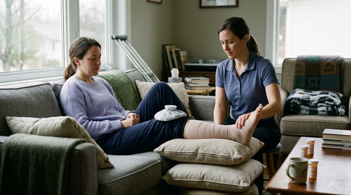
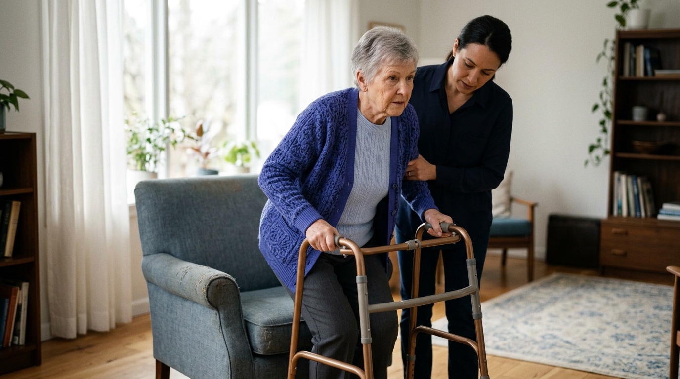
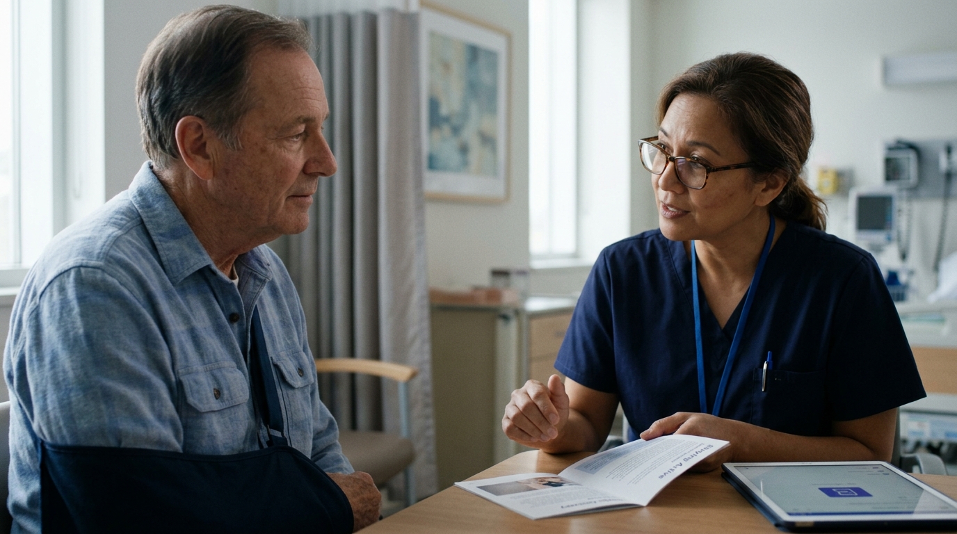
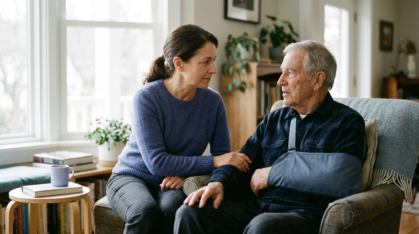
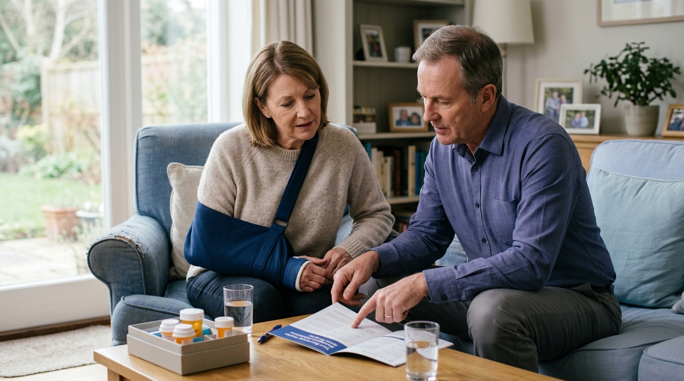
You may need to wear a sling or a rigid walking boot for several weeks. During this early phase, active movement of the repaired muscle is strictly restricted to prevent pulling the repair apart. Strengthening exercises are introduced gradually, often waiting months until the anchor site has gained structural stability.
Ligaments connect bone to bone and provide passive stability to joints. When a major ligament like the anterior cruciate ligament (ACL) tears completely, it usually cannot simply be stitched back together. Instead, the surgeon reconstructs it using a tendon graft taken from your own body or a donor.
Ligament reconstruction requires a complex biological process known as graft remodeling. Over many months, the body transforms the transplanted tendon graft into a functioning ligament. During this extended period, the graft actually weakens before it gradually becomes stronger.
Guidelines published in the British Journal of Sports Medicine emphasize that ACL rehabilitation must be staged and criterion-based. It must balance early joint mobility with long-term graft protection. Rushing back into pivoting or high-impact activities before the graft matures dramatically increases the risk of a secondary tear.
Joint replacement involves removing worn or damaged bone ends and replacing them with metal, ceramic, or high-density plastic implants. Common examples include total hip, total knee, and total shoulder replacements.
Unlike tendon or ligament repairs, joint replacement does not rely on soft tissue re-anchoring to establish immediate mechanical stability. The prosthetic components are firmly fixed to the bone with specialized cement or press-fit biological coatings during surgery. Because the joint surfaces are mechanically stable on day one, movement can start right away.
The National Institute for Health and Care Excellence (NICE) recommends offering rehabilitation within 24 hours of surgery for elective hip, knee, or shoulder replacements. Early walking helps restore blood circulation, rebuilds basic mobility, and reduces post-operative complications. However, early walking does not mean recovery is finished. Full recovery of soft tissue endurance, balance, and deep muscular strength often takes six to twelve months.
Understanding published clinical evidence helps separate realistic recovery milestones from casual assumptions. Clinical studies demonstrate why different procedures follow such vastly different timelines.
Tendon repairs highlight the clear difference between feeling better and achieving full structural healing. A systematic review published in BMC Musculoskeletal Disorders examined re-tear rates following rotator cuff surgery. The review found that patient age and initial tear size significantly influenced structural healing.
Patients over the age of 60 had a re-tear rate of 24.3 percent, compared to 14.4 percent in patients under 60. Larger tears also showed significantly higher re-tear rates than small tears. A comprehensive clinical review noted that re-tear rates across published surgical literature have ranged from 13 percent to over 90 percent in complex cases.
Interestingly, many patients with structural re-tears still report good pain relief and improved everyday shoulder function. This proves an essential recovery principle: symptom improvement does not always equal complete structural healing. Protecting the repair during early phases remains vital to give the tendon its best chance to knit securely to bone.
At the same time, rehabilitation protocols continue to evolve based on new research. Clinical guidelines from the American Academy of Orthopaedic Surgeons (AAOS) note that selected patients with specific types of arthroscopic rotator cuff repairs may safely begin weaning from a sling early to perform light daily tasks. This does not mean every patient can discard their sling immediately. It shows that protection guidelines are tailored to specific tear characteristics, tissue quality, and surgical fixation strength.
Ligament reconstructions show why calendar timelines alone cannot guarantee a safe return to strenuous activity. For years, athletes assumed that reaching the six-month mark after ACL surgery meant they were ready for full competition. Modern sports medicine research has challenged that assumption.
A prospective study highlighted by the Journal of Orthopaedic & Sports Physical Therapy examined young athletes returning to knee-strenuous sports after ACL reconstruction. The researchers found that athletes who returned to competition before nine months had approximately three to seven times the rate of a new ACL injury compared to those who delayed their return until at least nine months.
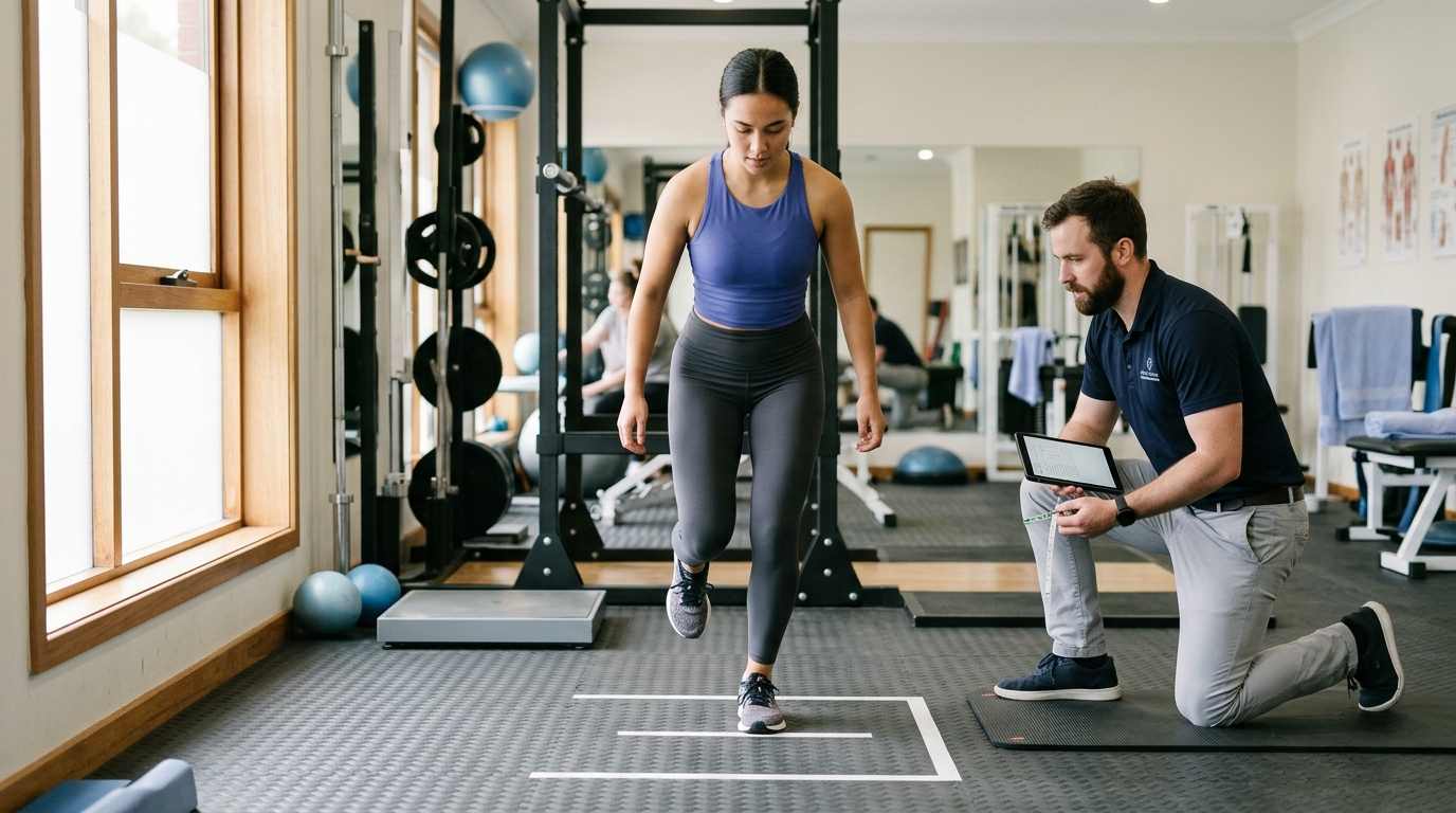
An international consensus statement published in the British Journal of Sports Medicine recommends continuing structured postoperative rehabilitation for nine to twelve months. The consensus panel emphasizes that passing objective functional tests is essential before resuming unrestricted sports. These tests evaluate muscle strength symmetry, jump landing quality, agility control, and psychological readiness.
Passing these physical criteria takes time and structured effort. A calendar date provides a rough safety baseline, but proven movement competence ensures long-term joint health. You can read more about structured progression in our guide to strength and return to sport strategies.
Joint replacements present a unique recovery pattern. They often feature rapid early progress in daily activities followed by a long, slow plateau of tissue remodeling.
Guidance from the AAOS indicates that most total knee replacement patients can resume basic activities of daily living within three to six weeks. Physical therapists teach specific exercises immediately after surgery to restore leg extension and encourage early walking. Serious joint infections occur in fewer than two percent of total knee replacement patients, making the procedure generally safe and predictable.
However, regaining deep knee flexion, resolving localized warmth, and rebuilding full walking endurance takes much longer. NHS information on recovering from total hip replacement states that full recovery typically takes between six and twelve months. The artificial joint is mechanically solid immediately, but surrounding muscles, cut nerve endings, and stretched joint capsules require many months to adapt to the new joint mechanics.
Surgeons give specific postoperative restrictions to protect vulnerable tissues while they heal. Understanding why a restriction exists helps you follow it consistently.
Restrictions should never feel like arbitrary rules. They are precise mechanical boundaries designed to prevent excessive tension on healing anchors, grafts, or incisions.
Weight-bearing instructions tell you how much body weight you can safely place through your operated leg. These guidelines range from non-weight-bearing to full weight-bearing as tolerated.
Progression between these levels depends on bone healing, swelling control, and quadriceps muscle control.
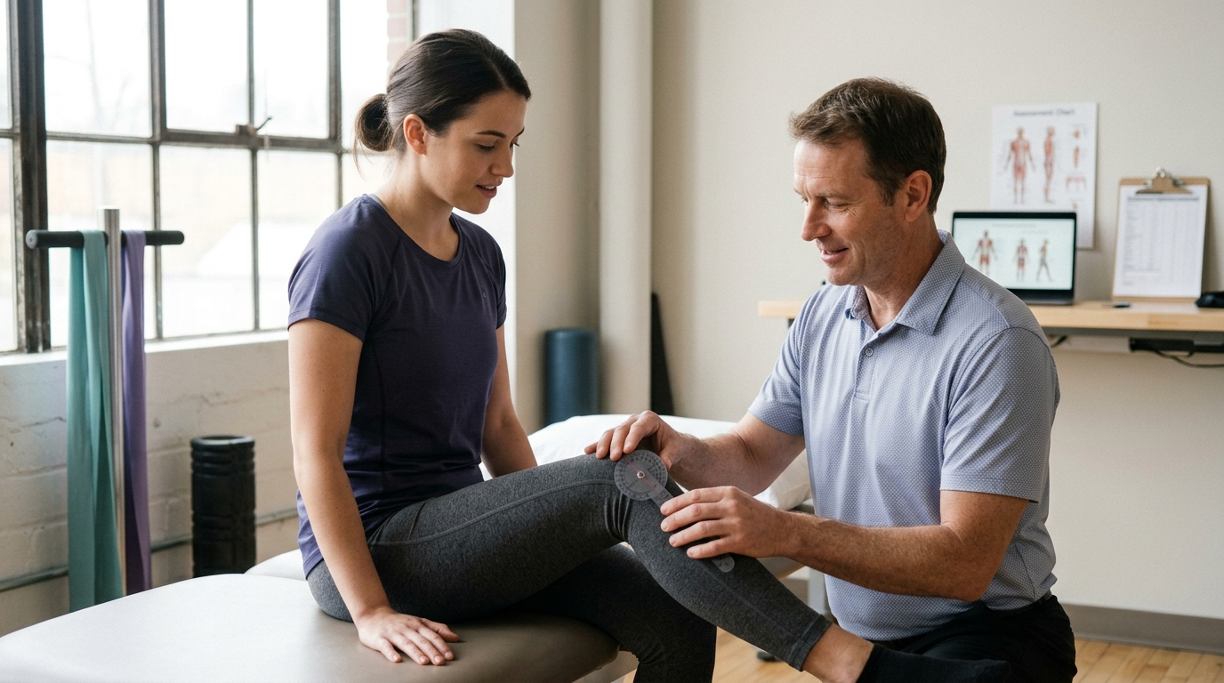
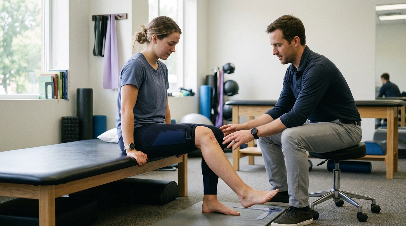
Range-of-motion restrictions limit how far you can bend or straighten an operated joint. These limits prevent stretching repaired tissues past their mechanical tolerance.
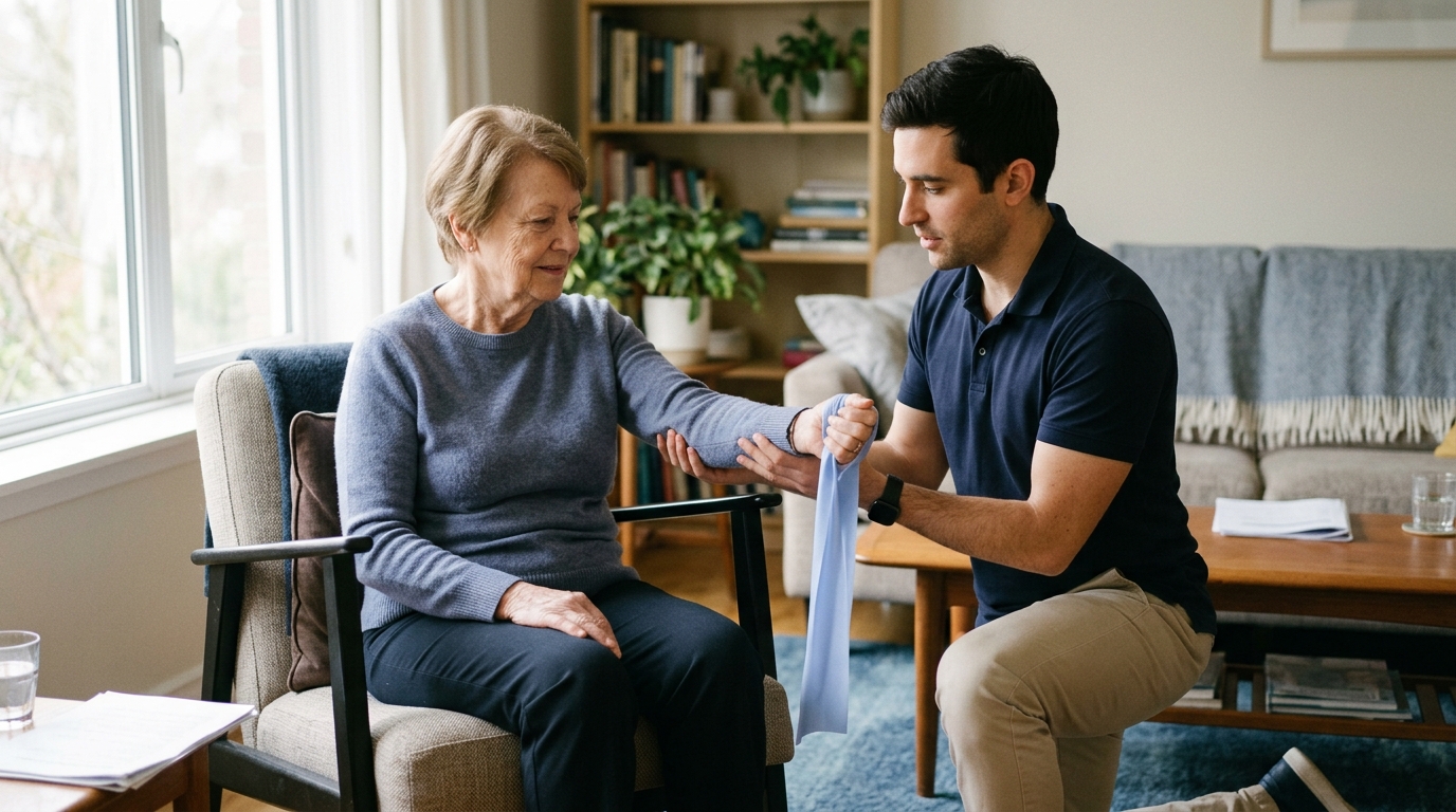
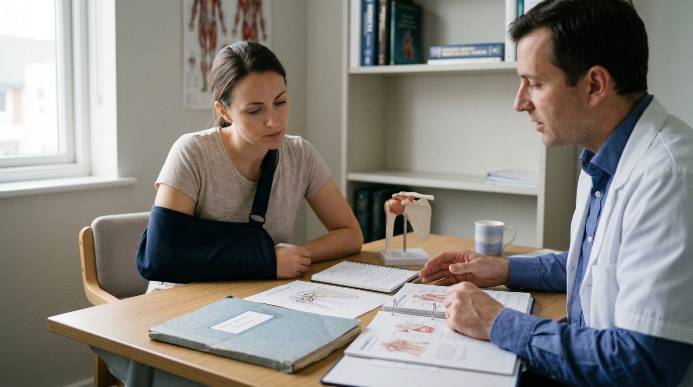
After a rotator cuff repair, you might be restricted to passive motion for the first month. In passive motion, a physical therapist or pulley moves your arm while your shoulder muscles stay
completely relaxed. This preserves joint mobility without allowing the muscle to pull against its new suture anchor.
Following a meniscus repair in the knee, deep bending past 90 degrees is often restricted for several weeks. Deep knee flexion squeezes the back of the meniscus, which could tear through the fresh repair stitches. Learning how to move within safe joint angles is an essential part of mobility and movement rehabilitation.
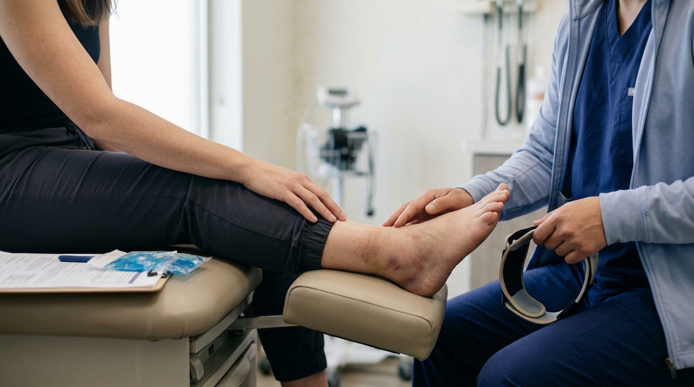
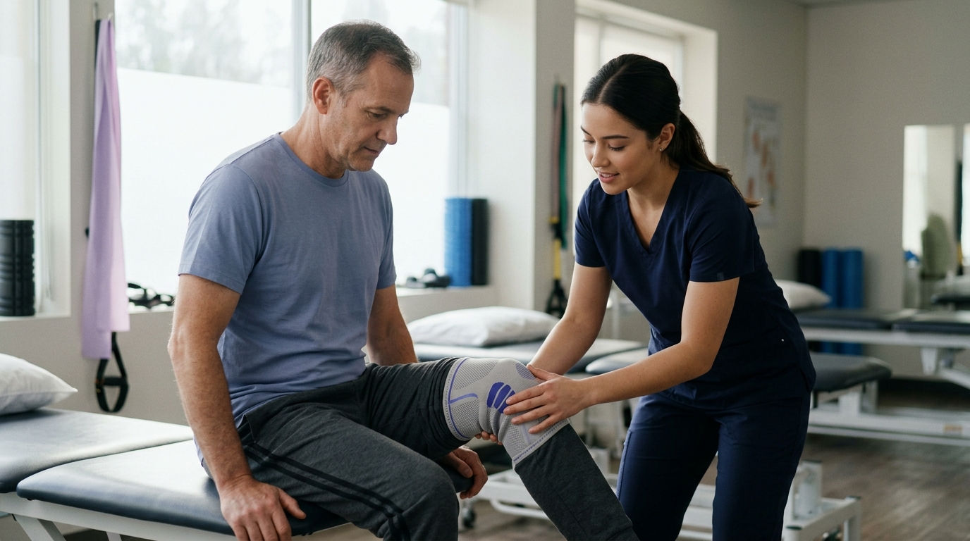
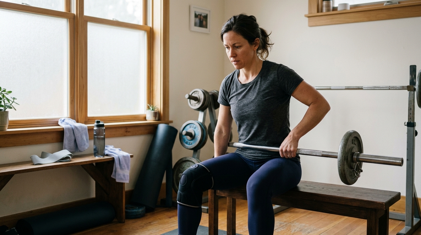
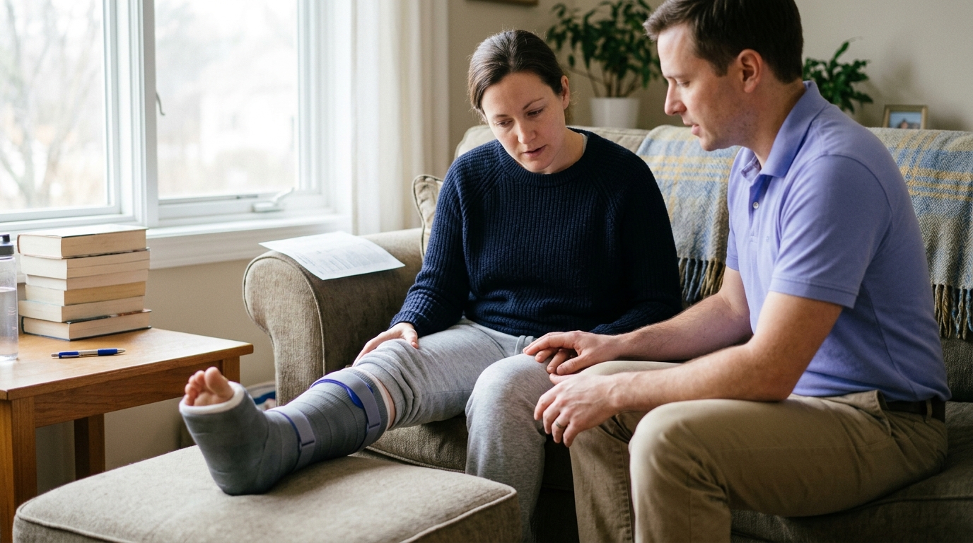
Slings, braces, and splints act as external bodyguards for surgical repairs. They hold the joint in a position of minimal tension during the initial healing window.
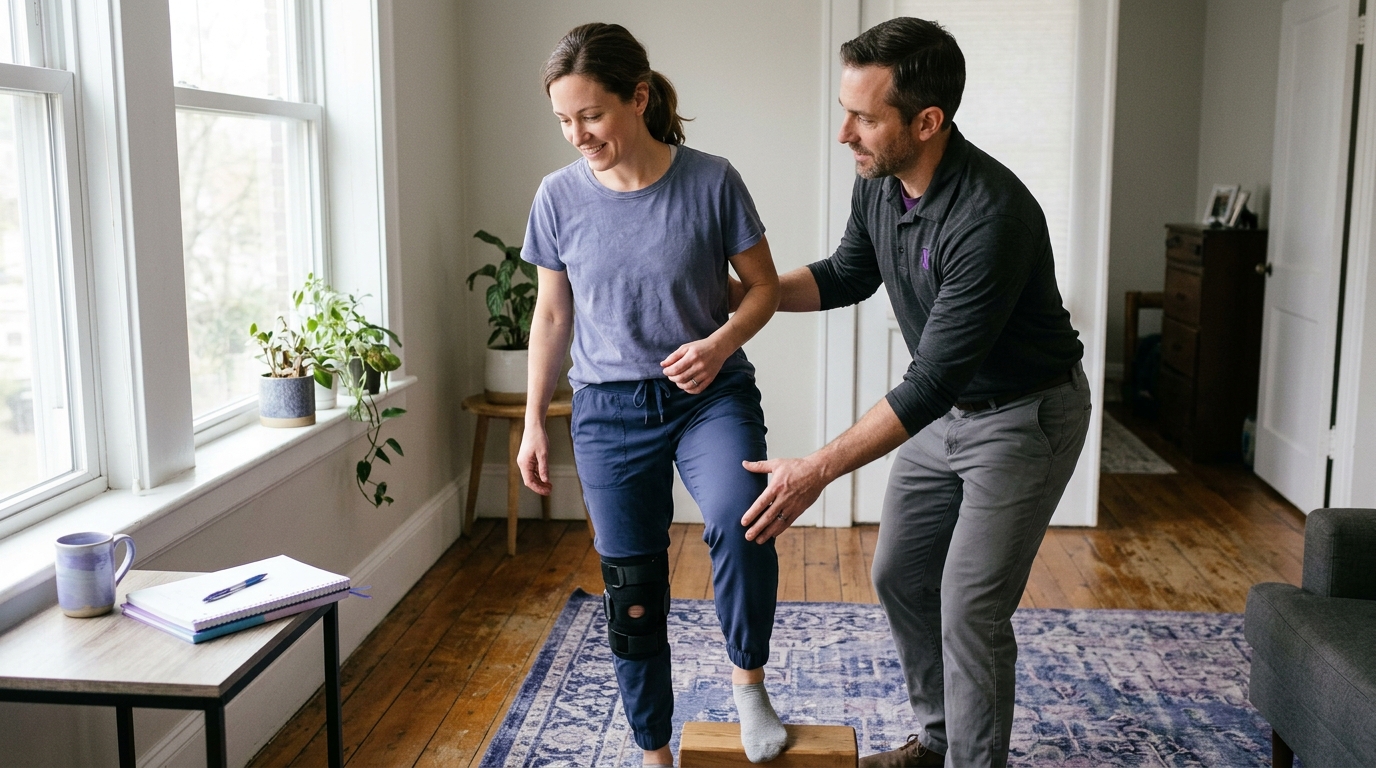
An abduction pillow sling holds the arm slightly away from the side to reduce pull on the supraspinatus tendon. A hinged knee brace locked in full extension keeps the leg straight while walking. This protects a healing patellar tendon or knee ligament from giving way under unexpected loads.
Immobilizers also protect the joint from accidental movements during sleep or sudden stumbles. Weaning off a brace requires careful coordination with your physical therapist once your active muscle control returns.
Recovery timelines are estimates, not rigid guarantees. Multiple individual and surgical factors can accelerate or lengthen your path back to full activity.
The biological health of your tissue directly affects how fast it repairs itself. Crisp, healthy tendon tissue from an acute injury in a younger adult holds surgical sutures more firmly than chronic, degenerated tissue.
Large, retracted tendon tears require higher tension to bring back to the bone. This extra tension creates higher mechanical stress on the suture anchors, requiring a slower, more protective rehabilitation program. Smaller, clean tears often permit earlier progression because the surrounding tissue remains robust.
Many orthopedic surgeries involve more than one procedure at the same time. A surgeon might perform an ACL reconstruction while simultaneously repairing a torn meniscus or smoothing damaged articular cartilage.
When multiple procedures are combined, your rehabilitation must follow the rules of the most restrictive repair. While an isolated ACL reconstruction might allow early progressive weight-bearing, adding a meniscus repair often requires crutches and limited knee flexion for several weeks. Always ask your surgical team which component of your operation dictates your current restrictions.
Your physical condition before surgery strongly influences your rehabilitation speed. Greater pre-operative strength, cardiovascular fitness, and joint range of motion provide a higher starting baseline for recovery.
Overall metabolic health plays a major role in tissue regeneration. Well-managed blood sugar levels, adequate nutrition, non-smoking status, and good sleep habits support optimal collagen formation and bone remodeling. Chronic inflammation or poor circulatory health can slow down the biological repair phase, requiring extended protection periods.
Understanding these broader health factors is central to modern surgery and rehab preparation.
Relying solely on a calendar to decide when you are ready for the next recovery phase is outdated and potentially risky. Modern orthopedic rehabilitation relies on criterion-based progression.
Criterion-based rehabilitation means you must achieve specific physical milestones before moving to harder tasks. A calendar gives a general estimate of biological healing, but objective physical testing confirms functional readiness.
The first phase of recovery focuses on settling the acute surgical trauma. Moving to higher-level strength work requires passing essential baseline checks:
Attempting to build heavy strength on an inflamed, swollen joint often leads to persistent pain and setbacks.
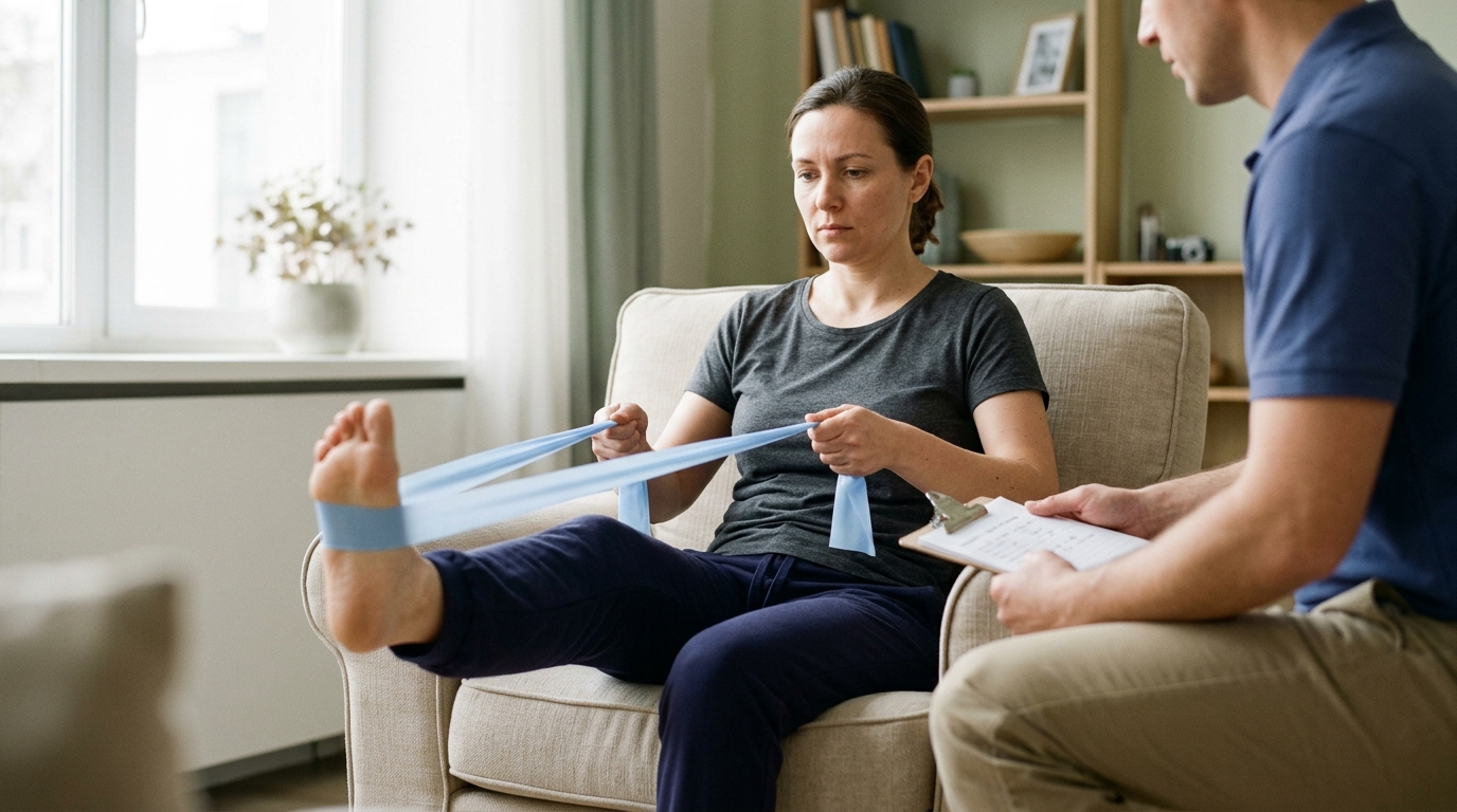
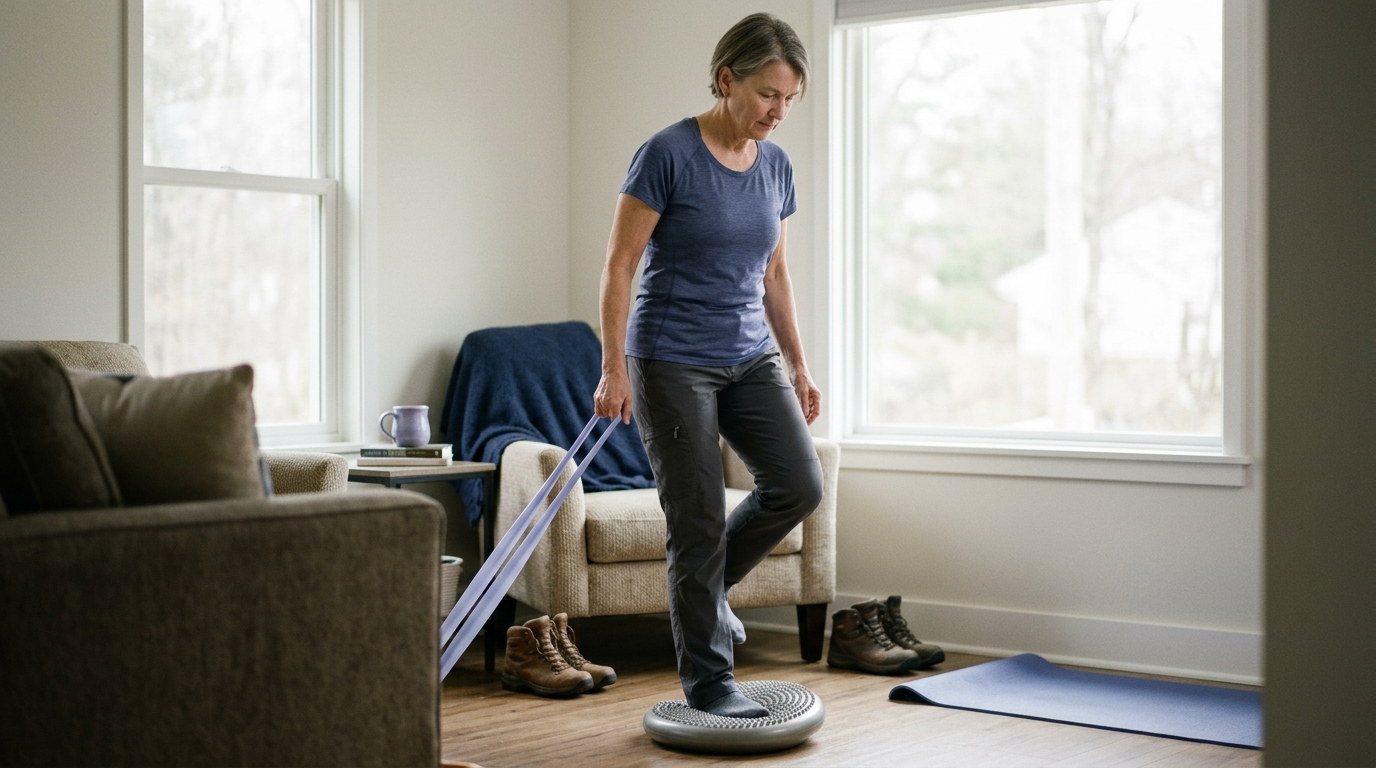
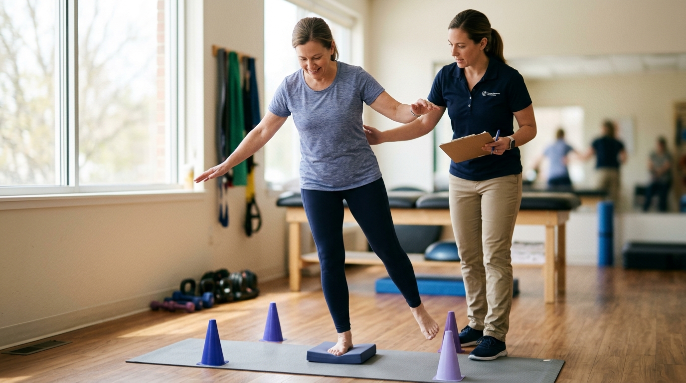
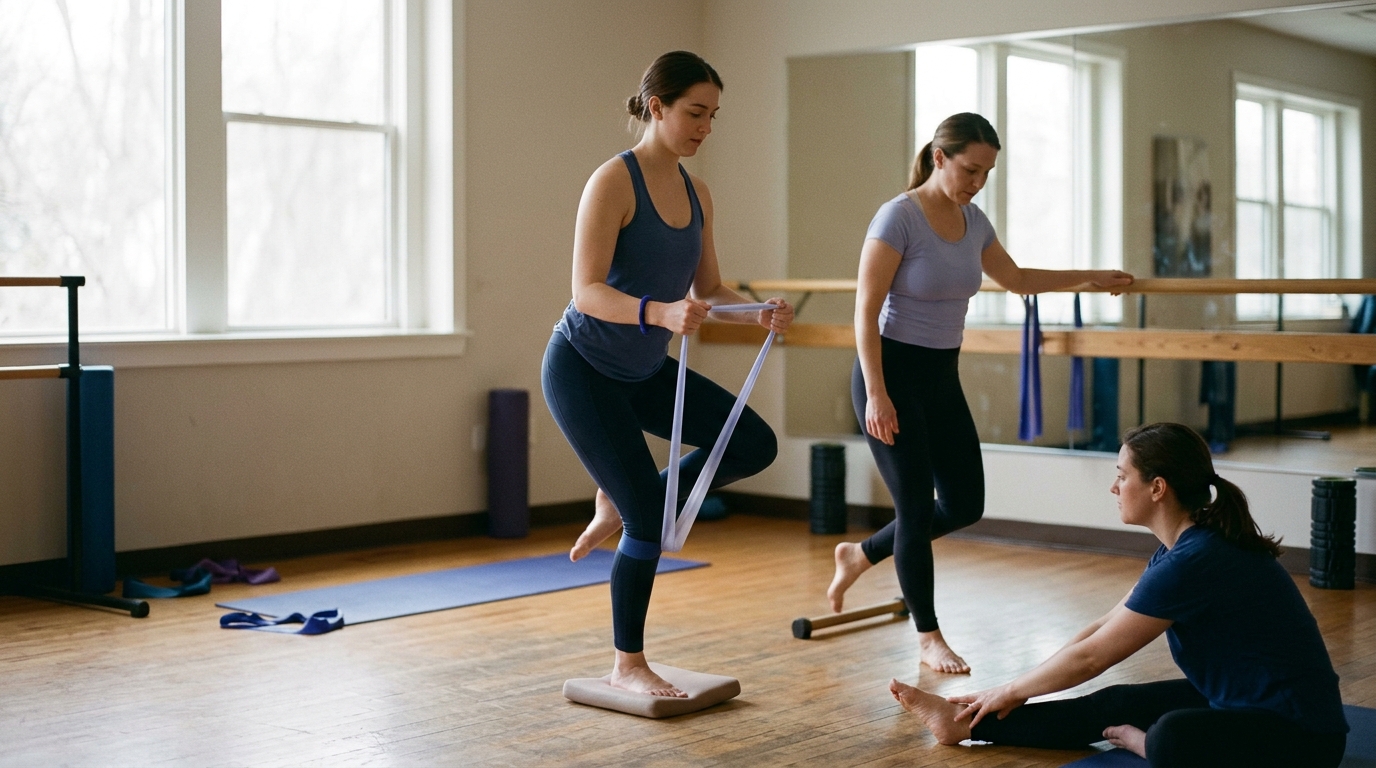
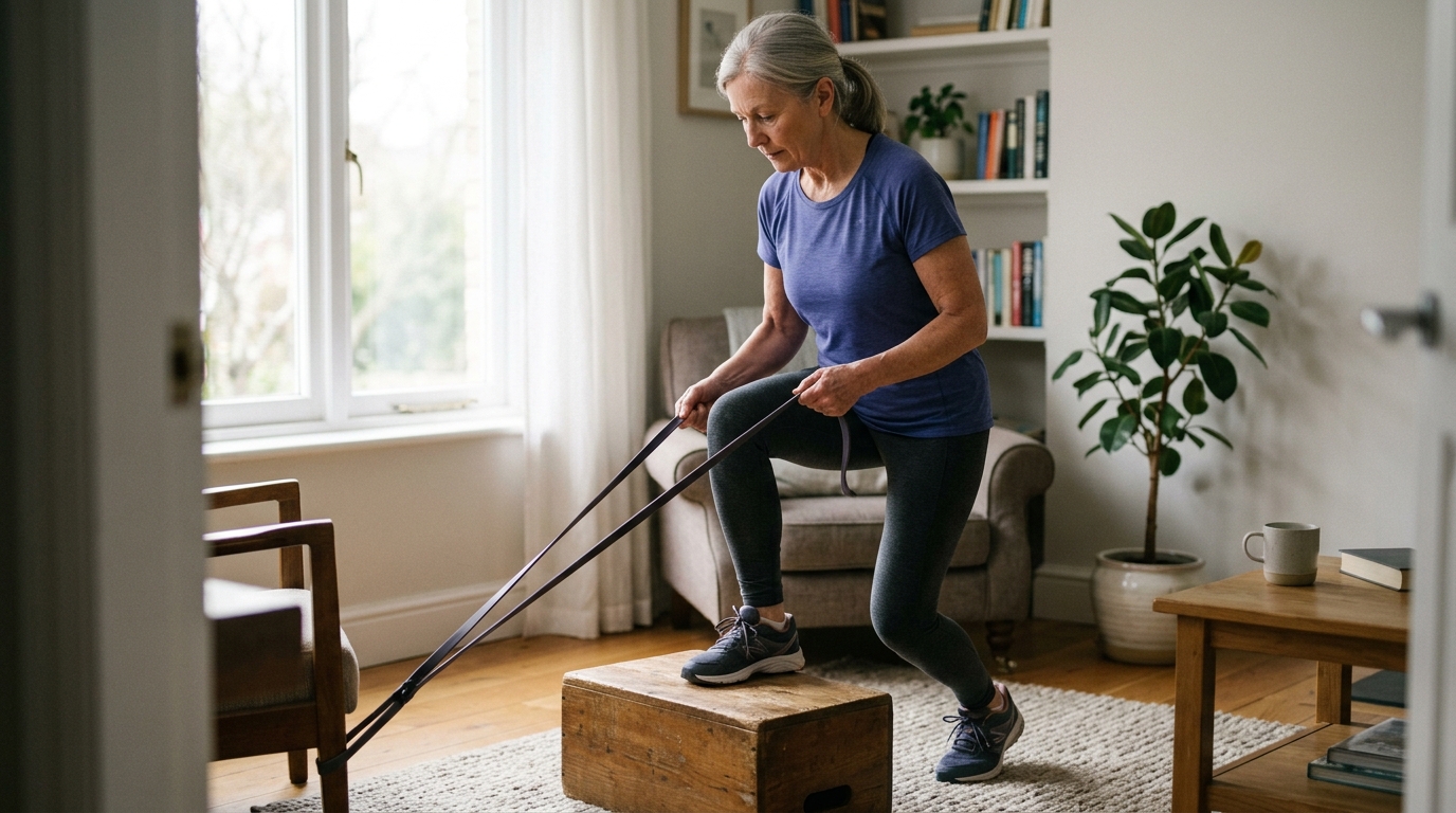
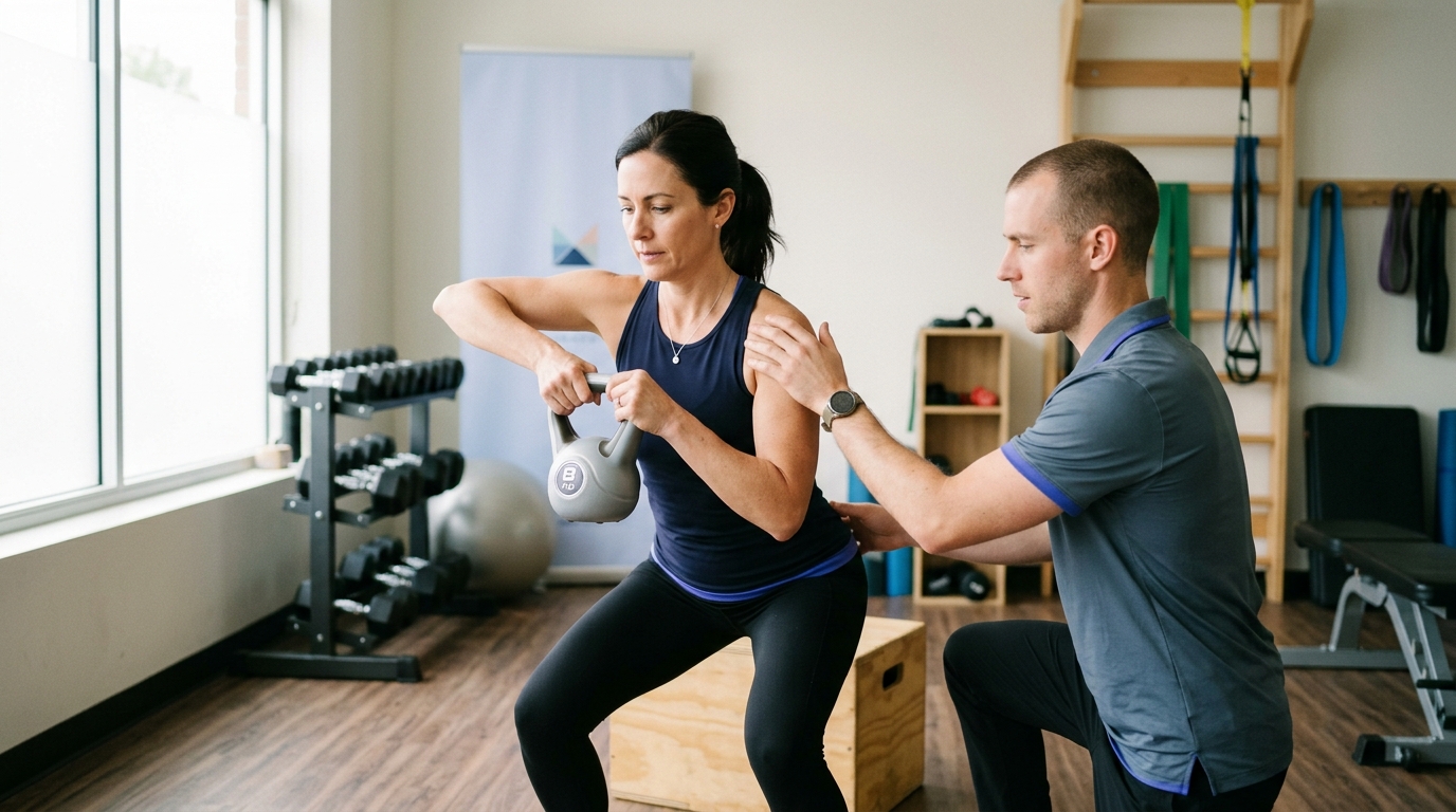
Once pain and swelling settle, the focus shifts to restoring muscular strength, joint control, and movement coordination:
Reaching these milestones ensures your joint can handle higher mechanical forces safely.
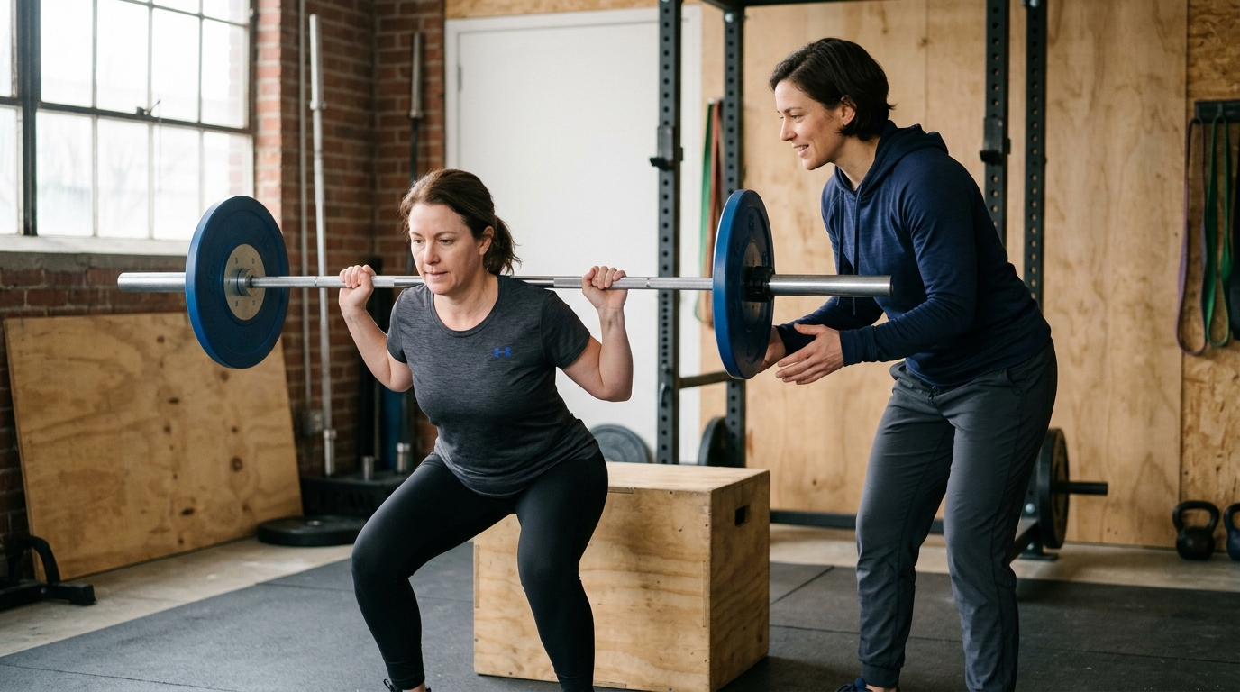
Returning to running, heavy manual labor, recreational sports, or athletic competition requires passing comprehensive discharge criteria:
Progression is not an all-or-nothing event. It is a gradual, tested series of steps designed to build durable physical capability.
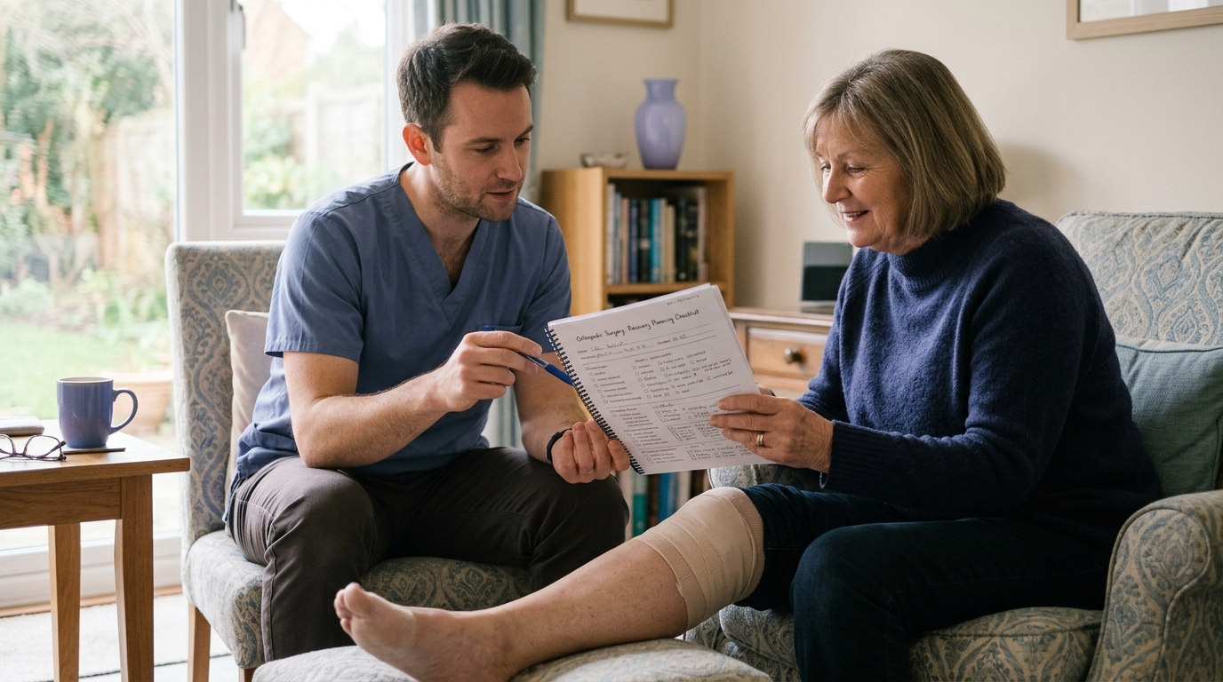
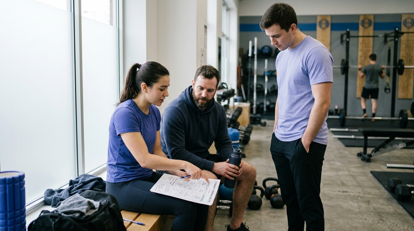
Navigating orthopedic recovery is easier when you ask specific, informed questions. Bring these practical questions to your appointments with your surgeon and physical therapist:
Orthopedic recovery is a structured biological progression that rewards patience, informed expectations, and consistent rehabilitation.

Use ReboundBody resources to understand common recovery stages, rehab terms, movement limits and strength rebuilding. Each guide is designed to make a complex comeback easier to understand.


Read practical guidance on injury recovery, rehabilitation, mobility and rebuilding strength as you work your way back to activity.
Explore Resources