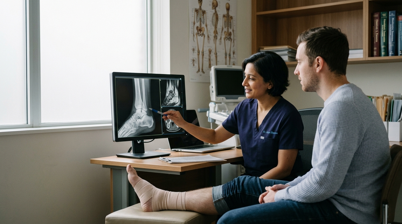

Orthopedic imaging tests like X-rays, MRIs, and CT scans help clinicians identify structural damage, evaluate complex joint injuries, and guide treatment plans.





Medical imaging is a diagnostic tool used to view internal body structures after an accident, sports mishap, or sudden joint pain. It is not a complete measurement of your recovery, pain levels, or physical capability.
Imaging tests do not replace a thorough physical examination or a detailed clinical history. Instead, imaging helps healthcare providers answer specific questions about what happened inside your body.
After an acute musculoskeletal injury, an X-ray, magnetic resonance imaging (MRI) scan, computed tomography (CT) scan, or ultrasound can confirm or rule out structural damage. Plain radiographs are usually the primary first step to check for broken bones or misaligned joints.
Advanced tests like an MRI or CT scan are reserved for specific clinical questions about soft tissues, ligaments, or complex joint surfaces. However, imaging results do not always match how you feel or function. Abnormal findings appear regularly on scans of people who have no pain at all.
Understanding how clinicians select these tests helps you interpret your medical reports with realistic expectations. This comprehensive guide explains what each test reveals, how clinical examination works alongside scans, and why finding a structural flaw does not automatically determine your long-term outcome.
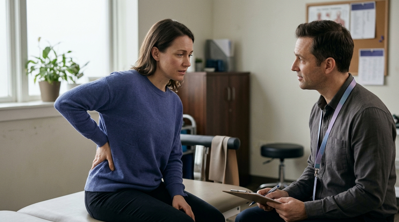
A physical assessment begins with your injury history and a hands-on physical exam. Your clinician asks about what happened, the direction of any impact, and the immediate onset of pain or swelling.
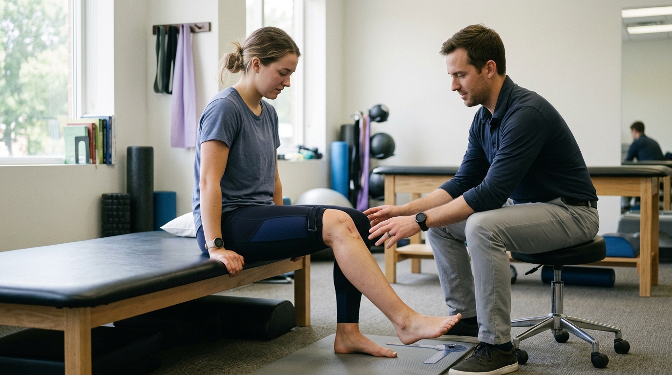
They also evaluate joint stability, tenderness along bony landmarks, range of motion, and nerve or blood vessel function. This hands-on process helps narrow down which anatomical structures might be damaged.
Medical imaging is ordered when the findings from the physical examination create questions that can only be answered by seeing internal structures. For example, if a patient cannot bear weight after a fall and has pinpoint tenderness over a bone, a doctor may order an X-ray to look for a fracture.
If a knee joint repeatedly gives way after a high-speed pivoting injury, an MRI may be considered to assess the stability of internal ligaments. The primary goal is to determine if a structural injury exists that would alter the treatment plan.
Imaging is not ordered simply because an area hurts. Pain severity does not always correspond to the degree of structural damage. A mild muscle strain or joint sprain can cause intense, disabling pain without showing major structural failure on a scan.
Conversely, serious structural changes can sometimes produce mild discomfort. Clinicians select tests to determine if surgery, immobilization, protected weight-bearing, or specific physical therapy is required.
In some cases, imaging is deferred during the initial visit. When an examination suggests a simple soft tissue sprain without red-flag symptoms, conservative management is often the first step.
Resources for injury recovery and healing emphasize that allowing natural tissue repair to begin while tracking functional improvement is a standard clinical strategy. Ordering scans too early can lead to unnecessary procedures or cause worry about normal age-related findings.
A radiology report describes physical structures captured by a machine at a single point in time. It cannot show the biological sensitivity of local nerve endings or the psychological stress that amplifies pain perception.
Relying entirely on an image without considering physical strength and mobility can lead to misdirected care. A structural abnormality seen on a screen is only relevant if it matches the physical symptoms discovered during your examination.
Professional guidelines from organizations such as the American College of Radiology stress that imaging should always be combined with clinical context. Clinicians treat the patient, not the picture.
When scans are interpreted alongside a physical examination, they provide valuable insight. When used alone, they can create confusion about what is actually causing the pain.
Plain radiography, commonly known as an X-ray, is the most accessible and widely used imaging tool in orthopedic care. It uses low doses of ionizing radiation to produce two-dimensional images of dense internal structures.
Because dense tissues like bone absorb more radiation, they appear white or light gray on the radiograph. Softer tissues allow more radiation to pass through and appear dark gray or black.
An X-ray is the standard initial imaging test when an acute bone injury or joint dislocation is suspected. It provides clear detail regarding bone continuity, major fractures, bone alignment, and joint spaces.
If a joint has slipped out of its socket, an X-ray reveals the position of the bones and helps the clinician plan a reduction. It can also detect severe joint space narrowing, bone spurs, and loose bone fragments.
While X-rays excel at showing dense bone, they provide limited information about soft tissues. Muscles, tendons, ligaments, and cartilage share similar densities, meaning they appear as indistinct gray shadows on a standard film.
An X-ray cannot directly show a torn anterior cruciate ligament, a ruptured Achilles tendon, or a damaged meniscus. It cannot detect early stress fractures, which often take weeks to show visible bone remodeling on plain film.
A normal X-ray provides reassurance that a displaced bone fracture is not present, but it does not mean the joint is uninjured. Significant soft tissue trauma can occur alongside a completely normal bone radiograph.
If your pain, swelling, or instability persists despite a clear X-ray, your healthcare team may re-evaluate the joint or recommend different testing. Understanding this difference prevents the false assumption that a normal X-ray means everything is fine.
Magnetic resonance imaging uses strong magnetic fields and radio waves to generate detailed cross-sectional pictures of your body. Unlike X-rays and CT scans, MRI does not use ionizing radiation.
It maps the water and fat content of tissues, making it exceptionally effective for evaluating soft structures that do not show clearly on plain radiographs. MRI can display thin tissue layers from multiple angles.
An MRI is often used when a clinician suspects damage to internal soft tissue structures that cannot be diagnosed by physical examination alone. In the knee, it provides views of the menisci, cruciate ligaments, collateral ligaments, and articular cartilage.
In the shoulder, it is frequently used to assess the rotator cuff tendons, the labrum, and the biceps tendon anchor. It can also identify occult fractures, which are subtle bone bruises or micro-fractures hidden beneath the outer cortex of the bone.
Despite its diagnostic detail, an MRI is rarely the primary test for acute joint trauma. Major clinical guidelines, including those from the American College of Radiology, recommend starting with plain radiographs for acute knee, ankle, or shoulder injuries.
An X-ray quickly rules out urgent bone displacement and is much more cost-effective. Furthermore, immediate MRI scans can capture widespread swelling and fluid that make subtle anatomical tears harder to evaluate.
Advanced imaging is most helpful when the scan result directly influences your treatment plan. If discovering a ligament tear would change the choice between surgery and structured physical therapy, an MRI is highly valuable.
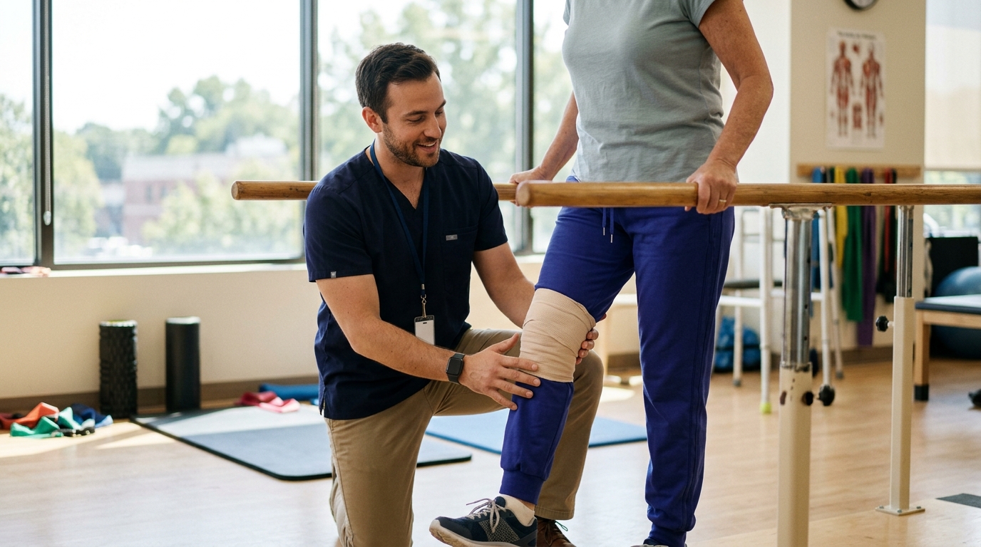
However, if the recommended treatment remains conservative rehabilitation regardless of what the scan shows, an immediate MRI may not be necessary. You can read more about structured rehabilitation concepts in our guide to rehabilitation, mobility, and movement.
While X-rays and MRI scans are the most common orthopedic imaging tools, computed tomography and diagnostic ultrasound serve important roles in specific situations. Each modality offers unique advantages depending on the type of injury and the anatomical area involved.
A CT scan combines multiple X-ray measurements taken from different angles to create cross-sectional, three-dimensional images of bones and joints. While standard X-rays compress a three-dimensional bone into a flat image, CT scans allow clinicians to view the structure in thin slices.
This makes CT the ideal imaging choice for complex, high-energy fractures that involve joint surfaces, such as intra-articular fractures of the tibial plateau, pelvis, or ankle.
Surgeons frequently rely on CT scans for preoperative planning. The three-dimensional reconstructions allow them to measure the exact displacement of bone fragments, identify loose pieces within a joint, and select surgical hardware.
CT is also valuable for evaluating subtle fracture healing when metal plates or screws create interference on an MRI. However, CT scans use higher amounts of radiation than standard X-rays and offer limited detail for soft tissue injuries.
Musculoskeletal ultrasound uses high-frequency sound waves to create real-time images of muscles, tendons, ligaments, and fluid collections. It is safe, non-invasive, and does not use radiation.
One of the unique strengths of ultrasound is its dynamic capability. A clinician can move your joint, contract a muscle, or stress a ligament while actively viewing the tissue on a screen.
Ultrasound is commonly used to evaluate superficial tendons, such as the rotator cuff in the shoulder, the long head of the biceps, the Achilles tendon, and the patellar tendon. It can detect tendon tears, dynamic snapping of tissues, and fluid build-up inside bursae.
It is also widely used to guide needle placement for therapeutic joint injections or fluid aspiration.
However, ultrasound has notable limitations. Sound waves cannot penetrate dense cortical bone, meaning ultrasound cannot see inside a joint to evaluate the cruciate ligaments or deeper cartilage surfaces.
The accuracy of an ultrasound exam is also highly dependent on the skill and experience of the person operating the machine. Because of this, it is considered a focused diagnostic tool rather than a universal replacement for MRI.
To avoid unnecessary radiation and reduce healthcare costs, medical researchers have developed validated clinical decision rules. These rules use specific physical examination findings to help clinicians decide whether an X-ray is necessary after an acute injury.
They provide an evidence-based framework that balances the need to catch serious fractures with the desire to avoid low-value testing.
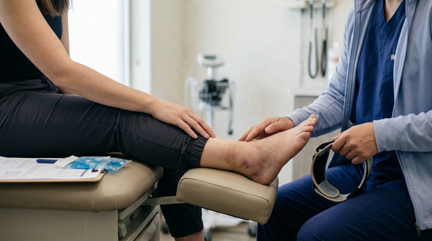
The Ottawa Ankle Rules are among the most thoroughly researched decision aids in emergency medicine and orthopedics. They guide whether an X-ray is needed after an acute ankle or midfoot injury.
Under these guidelines, an ankle X-ray series is only indicated if there is pain in the malleolar zone along with one of the following findings:
For midfoot pain, radiographs are recommended only if there is bone tenderness at the base of the fifth metatarsal, tenderness at the navicular bone, or an inability to take four steps.
A systematic review and meta-analysis published in the British Journal of Sports Medicine demonstrated that the Ottawa Ankle Rules have a pooled sensitivity of 99.4% and a specificity of 35.3%.
This high sensitivity means the rules are exceptional at ruling out fractures. If a patient does not meet the criteria, the likelihood of a significant bone fracture is less than 1%.
However, the lower specificity means that meeting the criteria does not guarantee a fracture is present. A positive rule simply means an X-ray is warranted to find out.
Similar evidence-based guidelines exist for knee trauma. The Ottawa Knee Rule suggests that a knee radiograph series is only required for patients with acute knee pain who meet one or more of these criteria:
According to clinical reviews on acute knee trauma, the Ottawa Knee Rule shows a sensitivity of 97% and a specificity of 27%. Another tool, the Pittsburgh Knee Rule, uses a history of blunt trauma or fall combined with age criteria or inability to walk four steps.
It has demonstrated a sensitivity of 99% and a specificity of 60%. These rules help emergency clinicians determine who needs an immediate X-ray without exposing every patient with a mild twist to unnecessary imaging.
One of the most surprising concepts in orthopedic medicine is that structural findings on a scan do not always correlate with pain or physical limitation. Patients often assume that a clear scan means zero pain, while a scan showing tears or degeneration guarantees severe disability.
Clinical research shows this is not the case. Structural abnormalities are common in active, healthy adults who experience no symptoms at all.
As joints age and adapt to life, internal structures naturally develop wear, fraying, and minor tears. These changes are a normal part of the aging process, much like gray hair or skin wrinkles.
When a person experiences a sudden injury, an MRI may detect one of these pre-existing changes. It is easy to mistakenly assume that the finding on the screen is the direct cause of the current pain.
A comprehensive systematic review of 63 studies examined MRI findings in asymptomatic, uninjured knees. The researchers found that the overall pooled prevalence of meniscal tears in symptom-free knees was 10%.
When broken down by age groups, the differences were striking:
The same systematic review analyzed articular cartilage defects in asymptomatic knees. The overall pooled prevalence of cartilage damage across pain-free individuals was 24%.
For adults aged 40 and older, the prevalence of asymptomatic cartilage defects was 43%, compared to 11% for adults under 40.
These numbers prove that a tear or cartilage flaw on an MRI report does not automatically explain why a joint hurts. Understanding these age-related baselines is vital for adults following active aging and prevention strategies.
When an incidental structural change is treated as an acute crisis, patients may undergo unnecessary interventions. A person might receive invasive surgery to trim a meniscal tear that was present and pain-free for years before a simple muscle strain caused their acute discomfort.
Research consistently indicates that addressing muscle strength, joint mobility, and neuromuscular control often resolves pain even when the structural "flaw" remains visible on a scan.
A scan report is a catalog of physical anatomy, not a measurement of function. Pain is processed by the central nervous system and influenced by localized inflammation, tissue load tolerance, and movement mechanics.
Therefore, clinicians evaluate whether the exact location of your tenderness and mechanical symptoms align with the scan findings before recommending targeted treatments.
The choice of imaging is never one-size-fits-all. Multiple clinical factors guide your healthcare provider when deciding which test to order, when to order it, or whether to wait.
These factors help ensure that imaging is performed safely and provides actionable data.
The exact physics of an injury provide essential clues. A high-energy impact, such as a motor vehicle collision or a direct fall from a ladder, creates high suspicion for complex bone fractures and structural dislocations.
In contrast, a low-energy twist while walking across an uneven lawn points toward a soft tissue sprain or strain. High-energy trauma almost always warrants immediate plain radiography or CT, while low-energy trauma may be managed with clinical observation first.
Patient age significantly influences diagnostic choices. Older adults are at higher risk for low-trauma fractures due to reduced bone mineral density or osteoporosis.
A minor slip that causes a mild bruise in a 25-year-old could cause a hip or wrist fracture in a 70-year-old. For older adults, clinicians have a lower threshold for ordering initial X-rays and may use MRI or CT earlier to look for subtle insufficiency fractures.
Certain physical signs indicate an urgent need for advanced diagnostic evaluation. These red flags include:
When red flags are present, clinicians bypass standard waiting periods and order urgent imaging to evaluate the joint integrity.
When an initial examination shows no red flags, conservative management is typically recommended for the first few weeks. If an injury does not follow the expected recovery timeline, diagnostic imaging becomes a logical next step.
For example, provincial clinical guidelines for acute ankle injuries recommend that if pain or weight-bearing ability has not improved within five to seven days, the patient should seek follow-up medical assessment.
At that stage, an X-ray or ultrasound may be ordered to check for missed micro-fractures, syndesmotic sprains, or tendon subluxations. Tracking your progress through rehabilitation science principles helps you know when a plateau warrants a second look.
Looking at realistic clinical scenarios helps illustrate how doctors choose imaging pathways. These illustrative examples demonstrate how physical exams, injury mechanisms, and imaging tools interact in standard clinical practice.
An active adult plants their foot and twists their knee while playing tennis. They hear a slight pop, experience immediate swelling within two hours, and struggle to bear weight.
At the initial clinic visit, the provider performs an examination and orders a standard two-view knee X-ray.
The X-ray shows normal bone structure and rules out an acute fracture. Because swelling and guarding limit the doctor from performing a full ligament stability test, the knee is placed in a supportive brace, and the patient uses crutches.
Two weeks later, once the initial swelling subsides, a repeat examination reveals ongoing joint laxity. An MRI is then ordered, which confirms an anterior cruciate ligament tear.
The initial X-ray ensured safety, while the delayed MRI provided the precise soft tissue roadmap needed to guide surgical and physical therapy choices. You can read more about this journey in our overview of surgery and rehabilitation.
A runner steps off a curb awkwardly, rolling their ankle inward. They experience moderate lateral pain, but they are able to walk back home and take four steps in the urgent care clinic.
During the exam, the clinician checks the posterior edges of both malleoli and the base of the fifth metatarsal. There is zero bone tenderness; the pain is located entirely over the anterior talofibular ligament.
Applying the Ottawa Ankle Rules, the clinician determines that an X-ray is not necessary. The patient is diagnosed with a grade I ankle sprain and advised to begin early weight-bearing and gentle range-of-motion exercises.
By avoiding an unnecessary X-ray, the patient avoids unnecessary radiation and focuses immediately on active rehabilitation. As long as steady improvement occurs over the next five to seven days, no imaging is ever required.
A 52-year-old develops sharp shoulder pain after lifting a heavy suitcase into an overhead compartment. They have difficulty raising their arm above shoulder height due to pain.
The clinician begins with plain shoulder radiographs to evaluate the glenohumeral joint, the acromioclavicular joint, and bone morphology. The X-rays show mild subacromial spurring but no fracture or dislocation.
Because the patient has persistent weakness during rotator cuff muscle testing, the provider considers next steps. Depending on local clinical access, an ultrasound or an MRI is ordered to check for a rotator cuff tendon tear.
The scan confirms a small partial-thickness supraspinatus tear. Armed with this knowledge, the physical therapist develops a targeted rotator cuff strengthening program that protects the healing tendon while restoring pain-free movement.
A 60-year-old trips over a rug and experiences bruised ribs and mild knee discomfort. An MRI of the knee is ordered by an overcautious provider.
The MRI report describes a complex tear of the posterior horn of the medial meniscus. However, upon clinical examination, the patient has no joint line tenderness, no swelling, and full pain-free knee flexion. Their knee discomfort was simply a superficial contusion over the patellar tendon.
Because the meniscal finding did not match the physical examination, the clinician wisely advises against knee arthroscopy. The patient is reassured that meniscal tears are present in roughly one out of five pain-free adults in their age group.
Within two weeks, the superficial bruise heals completely, and the patient returns to normal walking with zero knee symptoms. The structural finding on the scan was an incidental, age-related passenger rather than the driver of pain.
Following a heavy fall during a skiing trip, a patient arrives at the clinic with acute hip pain. When the clinician attempts to test hip mobility, intense muscle spasms prevent any movement.
The initial physical examination is inconclusive because the patient cannot tolerate joint stress tests.
Rather than guessing, the clinician immediately orders plain radiographs to rule out an occult femoral neck fracture. When the X-rays are negative, the patient is given short-term pain relief and advised to rest with protected weight-bearing for 48 hours.
At the follow-up appointment two days later, the acute muscle spasms have relaxed. The clinician can now perform a complete physical exam, confirming a deep muscle contusion that requires gentle progressive loading rather than surgery.
When reviewing imaging with your healthcare provider, asking structured questions helps you understand your diagnosis. It allows you to participate actively in your recovery plan and prevents misunderstandings regarding scan results.
Open communication ensures that diagnostic imaging serves its intended purpose: acting as a helpful tool to guide your physical rehabilitation. You can find more comprehensive recovery resources across our injury recovery guides.
An X-ray only evaluates dense bone tissue and general joint alignment. It cannot show damage to soft tissues such as ligaments, tendons, joint capsules, or muscles.
Severe sprains, muscle tears, and local inflammation can produce intense pain and swelling while leaving the underlying bone completely undamaged. If symptoms fail to improve with conservative care, your doctor may consider soft tissue imaging.
Yes. An MRI captures anatomical structures as they appear on the day of the scan.
Old ligament tears that healed with scar tissue, chronic tendon fraying, and long-standing cartilage thinning will all appear on an image. This is why your doctor must correlate your MRI findings with your current symptoms to avoid treating an old, asymptomatic injury.
Immediately after an acute trauma, widespread fluid accumulation and muscle guarding can make scans difficult to interpret. Furthermore, many acute soft tissue sprains begin resolving quickly with basic conservative care.
Waiting a short period allows acute swelling to subside, gives minor strains time to improve, and lets the clinician perform a more accurate physical exam to see if advanced imaging is truly necessary.
No. An MRI is substantially better at showing soft tissue structures, including tendons, ligaments, nerves, and cartilage.
A CT scan is primarily used to evaluate complex bone fractures, joint surface congruity, and three-dimensional skeletal alignment. Each test has specific strengths, and they are chosen based on whether bone or soft tissue is the main clinical concern.
Medical imaging is a powerful diagnostic tool, but it is only one part of an orthopedic assessment. An X-ray remains the frontline standard for evaluating fractures and joint alignment, while MRI, CT, and ultrasound provide targeted answers for complex bone or soft tissue questions.
A scan does not measure pain, and structural abnormalities appear regularly in healthy, active adults who have no symptoms.
The most accurate diagnosis relies on combining your injury story, a hands-on physical exam, and imaging results when appropriate.
Focusing on how your body moves and functions, rather than fixating solely on a scan report, creates the clearest path forward for your long-term recovery.

Use ReboundBody resources to understand common recovery stages, rehab terms, movement limits and strength rebuilding. Each guide is designed to make a complex comeback easier to understand.


Read practical guidance on injury recovery, rehabilitation, mobility and rebuilding strength as you work your way back to activity.
Explore Resources