
Most people instinctively reach for ice and complete rest, but modern recovery relies on early assessment, appropriate protection, and recognizing when injuries need professional evaluation.





The first 72 hours after an acute injury represent a critical window for assessment, symptom tracking, and early protection. This period is not a universal biological deadline where healing starts and finishes. It is also not a guaranteed timeframe in which every soft tissue problem resolves. Instead, the first three days serve as an editorial and practical benchmark. During this time, you must identify urgent medical risks, prevent further tissue damage, and determine whether professional care is necessary.
A general guide cannot replace an individualized clinical evaluation or emergency medical services. Symptoms between simple bruises, ligament sprains, muscle strains, bone fractures, and joint dislocations often overlap. Early care requires a clear sequence of priorities: check for immediate danger, protect the injured body part, observe how symptoms change, and seek medical attention when red flags appear.
Understanding early injury care helps active adults manage setbacks with realistic expectations. In the sections below, you will learn how to assess acute physical trauma safely. You will also learn what current medical evidence says about early interventions, what factors influence recovery, and how clinicians determine the next steps.
When an accident happens, the immediate impulse is often to examine the specific body part that hurts. However, clinical first aid guidelines prioritize the whole person and the environment before focusing on a single joint or muscle. First responders follow a principle called triage before treatment. This approach ensures that life-threatening hazards and severe physical trauma are addressed first.
Guidance from the Resuscitation Council UK and the European Resuscitation Council emphasizes a structured sequence. Before touching an injured area, check the surroundings for hazards such as traffic, falling objects, or unstable ground. Next, assess whether the injured person is conscious, responsive, and breathing normally. If the person is unresponsive or struggling to breathe, call emergency medical services immediately.
Catastrophic bleeding is another critical priority in the initial minutes. If an open wound is spurting blood or bleeding heavily, apply continuous, firm pressure with a clean pad or dressing. First aid guidelines from the American Heart Association and the American Red Cross highlight direct pressure as the primary method for controlling serious bleeding. Do not remove embedded objects from a wound, as they may be plugging deeper damaged blood vessels.
Once immediate safety, responsiveness, breathing, and major bleeding are accounted for, evaluate the risk of head, neck, or spinal trauma. If the injury involved a high-impact fall, a vehicle collision, or direct force to the head, keep the person still. Avoid moving their neck or spine unless they are in immediate physical danger from their environment. Only after these basic safety checks are complete should you focus on localized limb or soft tissue injuries.
Not every injury can be managed at home or evaluated at a routine clinic visit days later. Certain warning signs indicate severe tissue trauma, structural instability, nerve disruption, or compromised blood flow. Recognizing these red flags early prevents serious long-term complications.
Head impacts require careful observation. According to the Centers for Disease Control and Prevention (CDC), a suspected concussion or mild traumatic brain injury must be evaluated by a healthcare professional. Emergency medical care is required immediately if danger signs appear after a blow to the head.
Head injury danger signs include:
Severe musculoskeletal injuries also have specific red flags that require urgent medical evaluation. If a bone is visibly protruding through the skin, this is an open fracture that requires emergency surgery to prevent deep bone infections. Obvious joint deformity, where a limb looks bent at an unnatural angle, indicates a possible fracture or complete joint dislocation. Mayo Clinic and Red Cross guidance state that you should never attempt to straighten, push back, or realign a deformed bone or joint.
Circulation and nerve function below the injury site must also be monitored. If a hand or foot becomes cold, pale, blue, or grey, blood flow may be restricted by bone fragments or severe swelling. Numbness, loss of sensation, or a persistent pins-and-needles feeling below the injury point indicates potential nerve compression. These neurovascular changes require immediate emergency room assessment rather than a wait-and-see approach.
When emergency danger signs are absent, you can perform a gentle visual and functional assessment. The proper approach is to look, ask, compare, and monitor. You should never aggressively manipulate, pull, or force an injured body part through its range of motion to test its integrity.
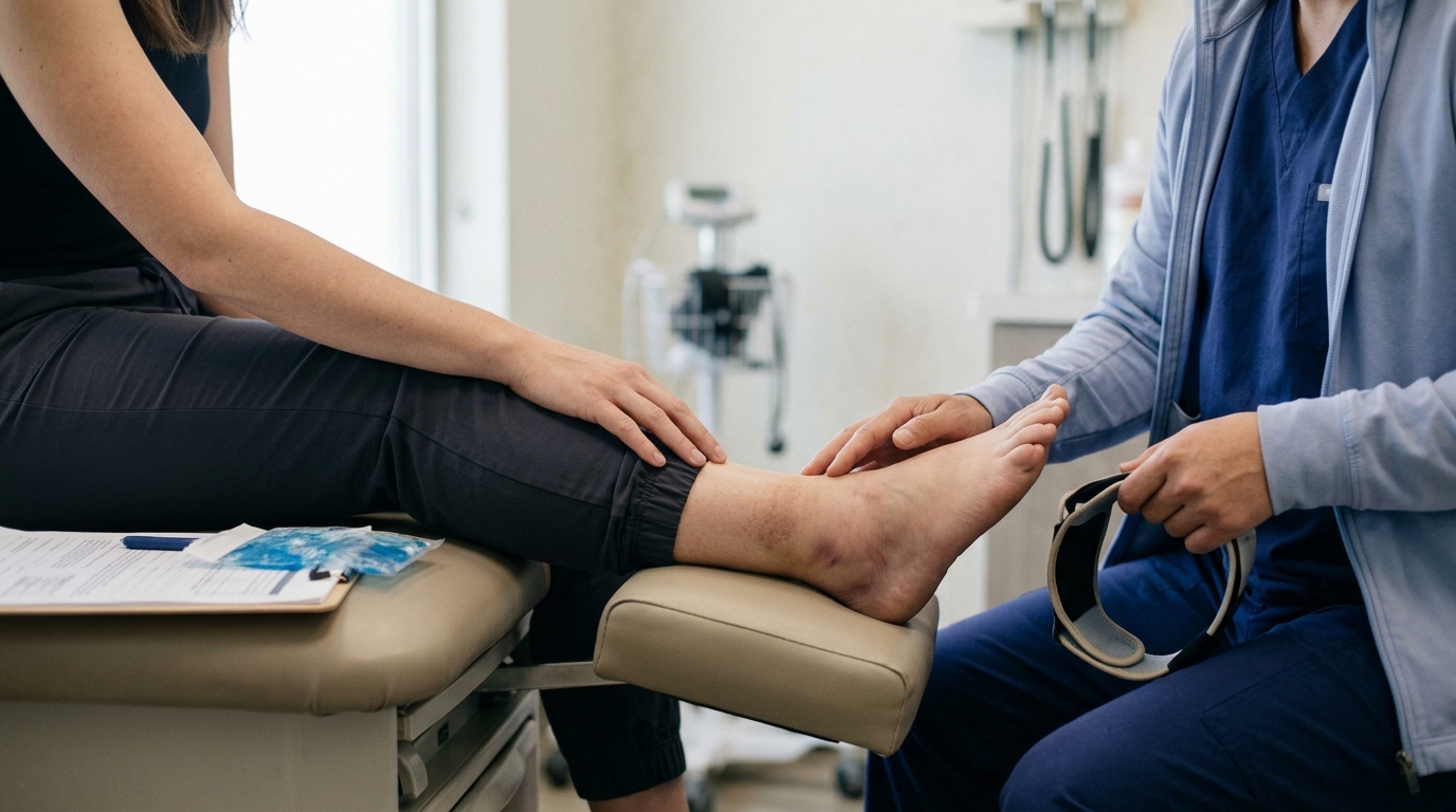
Start by comparing the injured limb to the uninjured side. Look for swelling, surface redness, skin discoloration, or subtle changes in shape. Minor swelling often develops gradually over several hours, whereas rapid, massive swelling within minutes may indicate a more severe ligament tear or bone fracture. Check the skin integrity to see if there are open scrapes, puncture wounds, or skin breaks near the swollen area.
Ask the person to describe the exact nature and location of their pain. Did they hear or feel a distinct pop, crack, or snapping sensation at the moment of impact? A loud pop accompanied by immediate pain often occurs with high-grade ligament ruptures or tendon tears. A sensation of bone grinding against bone, known as crepitus, suggests a structural break.
A major pitfall in early self-assessment is the myth that an ability to move a limb means it cannot be broken. Bone fractures can still allow partial movement, especially if the break is hairline, impacted, or located in a non-weight-bearing bone. Forcing a person to move a painful limb or trying to walk off a suspected fracture can displace bone fragments and damage nearby nerves. If basic movement causes sharp, severe pain, protect the limb and seek a professional medical diagnosis.
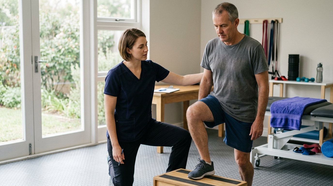
Observing functional capacity gives valuable clues without causing further tissue damage. For example, observe whether the person can gently wiggle their toes or fingers without severe discomfort. Check whether they can bear any weight on an injured leg, or if placing the foot on the floor causes immediate, intolerable pain. If bearing weight is impossible, immobilize the joint and arrange for prompt clinical assessment.
Early management of soft tissue injuries, such as sprains and strains, has evolved over decades of sports medicine research. For many years, the standard approach was encapsulated by the RICE acronym, which stands for Rest, Ice, Compression, and Elevation. While basic protection and elevation remain useful, newer clinical frameworks highlight a more nuanced balance between protection and early movement.
Modern protocols, such as the PEACE & LOVE framework published in sports medicine literature, categorize recovery into immediate care and subsequent management. The initial phase emphasizes Protection, Elevation, Avoiding anti-inflammatory modalities where appropriate, Compression, and Education. Later stages emphasize Load, Optimism, Vascularisation, and Exercise. You can learn more about structured tissue loading in our guide to rehabilitation and mobility resources.
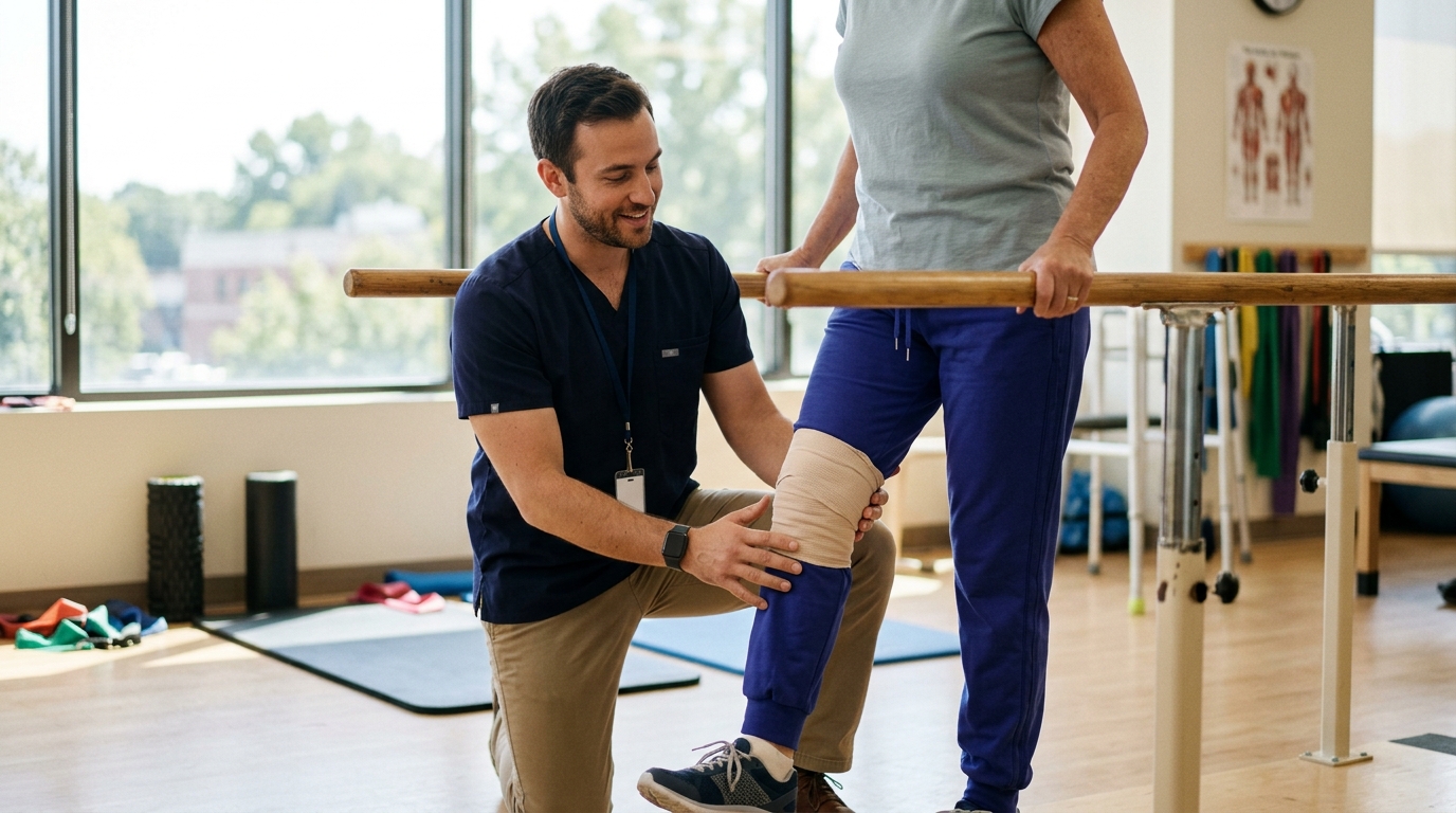
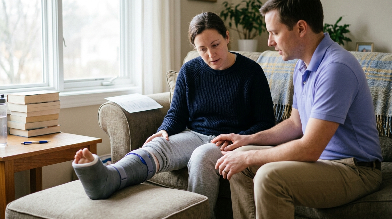
Protection means minimizing movement and unloading the injured tissue for the first few days. This prevents further fiber disruption in torn muscles or stretched ligaments. However, prolonged, complete immobilization is rarely ideal for uncomplicated minor sprains.
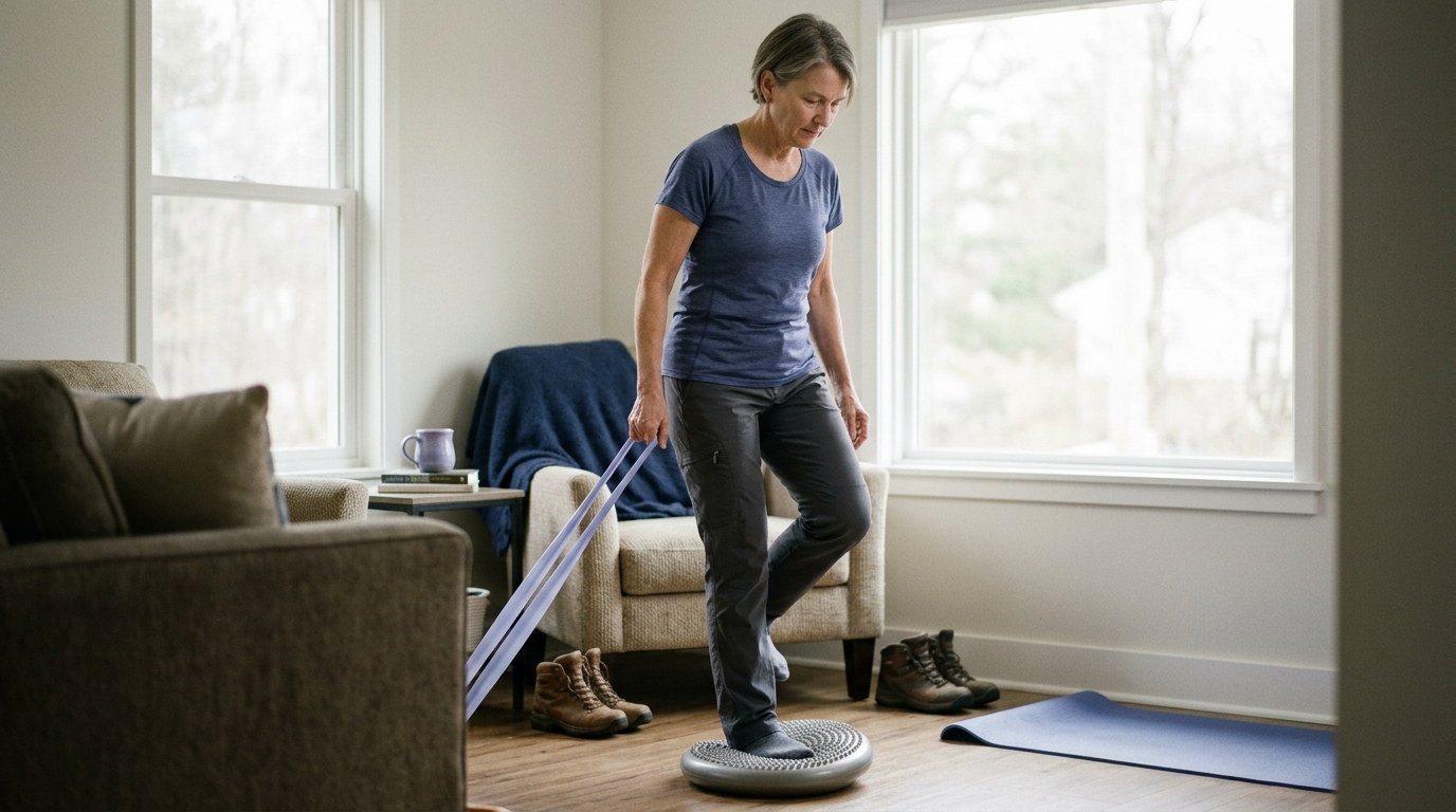
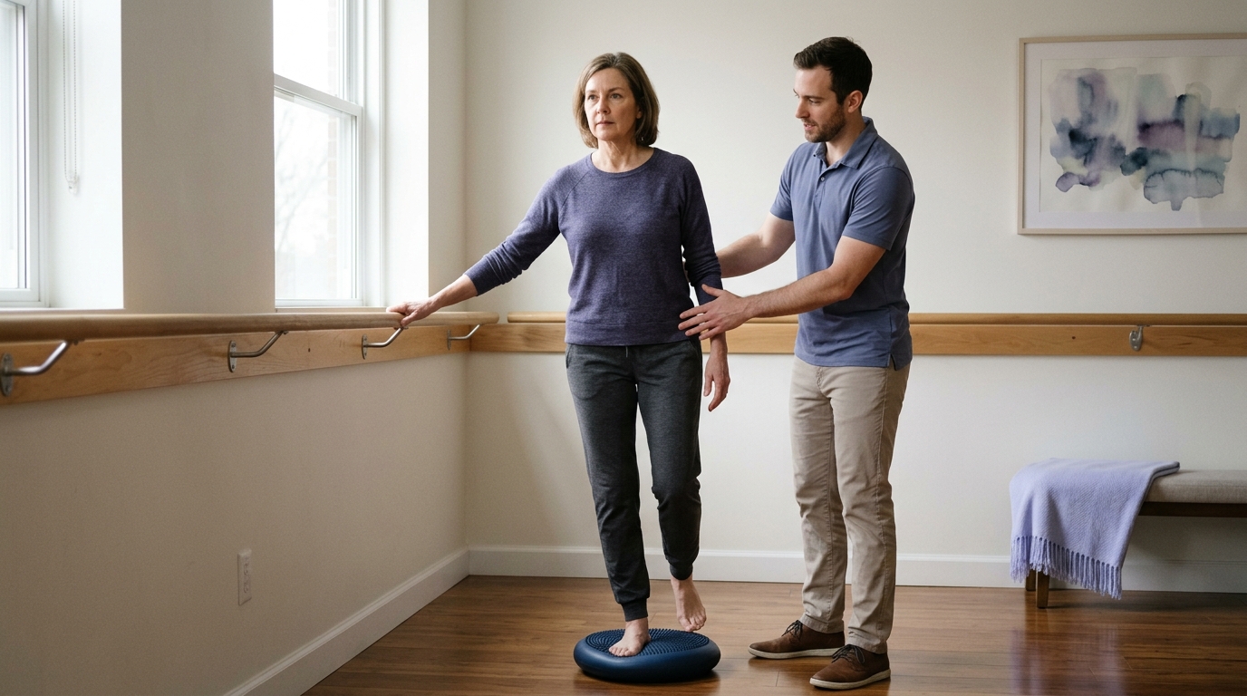
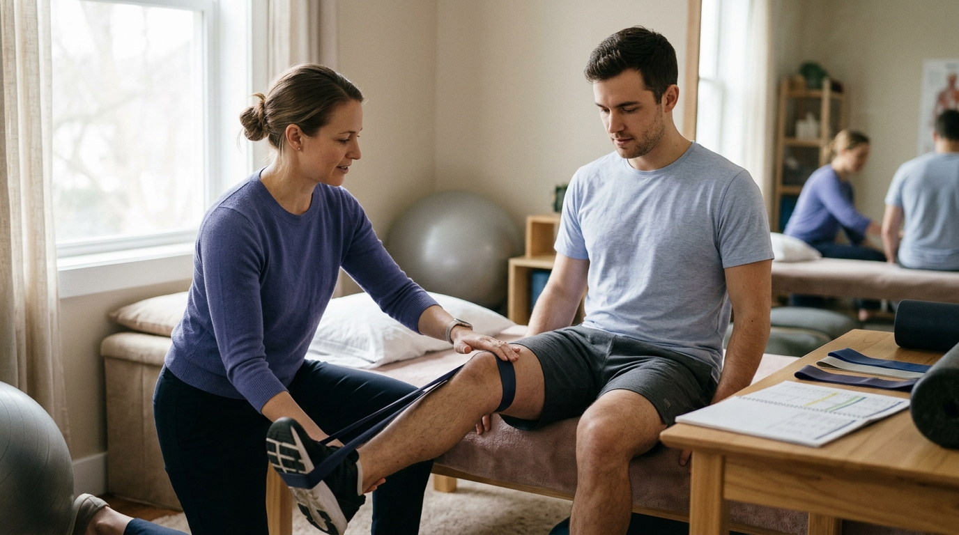
Current evidence shows that prolonged bed rest or total joint immobilization can lead to muscle atrophy, joint stiffness, and delayed tissue remodeling. Once severe injuries are ruled out, early protected movement within a pain-free range supports cellular repair. Gentle muscle contractions help clear local swelling through the lymphatic system. Protection should prevent tissue aggravation without leading to unnecessary stiffness.
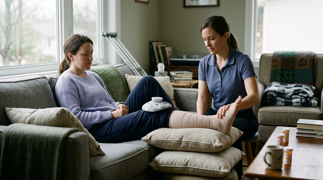
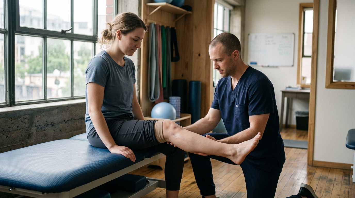
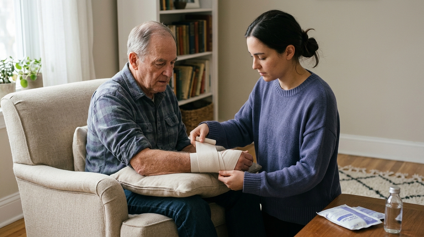
Compression and elevation are widely used to manage acute swelling. Applying a snug, elastic bandage helps limit excessive fluid accumulation in the interstitial tissues. The wrap must be applied evenly and must never be tight enough to cut off circulation, cause throbbing, or induce numbness.
Elevation involves positioning the injured limb above the level of the heart whenever practical. This uses gravity to assist venous and lymphatic return, reducing localized pooling of fluid. While elevation alone does not speed up structural tissue healing, it can relieve throbbing pressure and improve comfort during the first 48 to 72 hours.
The routine application of ice and non-steroidal anti-inflammatory drugs (NSAIDs) during early recovery is currently debated in sports medicine. Traditionally, ice was applied aggressively to shut down the inflammatory response. Modern physiological research shows that early acute inflammation is a necessary biological step for tissue repair.
Inflammatory cells, such as macrophages and neutrophils, release growth factors and clear cellular debris from the injury site. Suppressing this natural cascade completely through heavy ice application or high-dose NSAIDs might delay optimal tissue remodeling in some soft tissue injuries. However, ice remains an effective, non-pharmacological tool for acute pain relief.
If ice is used for comfort, apply it wrapped in a thin towel for 10 to 15 minutes at a time rather than placing bare ice directly on the skin. Understanding the biological mechanisms of tissue repair is discussed further within recovery science.
First aid protocols are not interchangeable across different injury categories. Applying soft tissue sprain guidelines to an open wound or a burn can cause harm. Each injury type requires specific, evidence-based initial actions:
Every injury occurs within a unique individual context. Two people with an identical ankle roll can experience entirely different recovery paths based on anatomical, biological, and health factors. Recognizing these variables prevents false comparisons and helps set realistic expectations for early healing.
The specific anatomical tissue involved changes the baseline timeline. Muscles have an abundant blood supply, which allows minor strains to begin cellular remodeling relatively quickly. Tendons and ligaments have a more limited vascular network, meaning moderate to severe sprains often take significantly longer to regain baseline tensile strength.
Injuries are generally classified into three clinical grades:
A grade 1 muscle strain may allow light, protected activity within several days. In contrast, a grade 3 ligament rupture may require weeks of targeted protection, bracing, or orthopedic surgical consultation. Practical strategies for managing movement after setbacks are covered in our overview of mobility and movement articles.
Chronological age and baseline physical fitness influence how tissues respond to trauma. As people age, tendons and ligaments naturally lose some of their water content and collagen elasticity. This structural change makes connective tissues more susceptible to strain under sudden, unexpected loads.
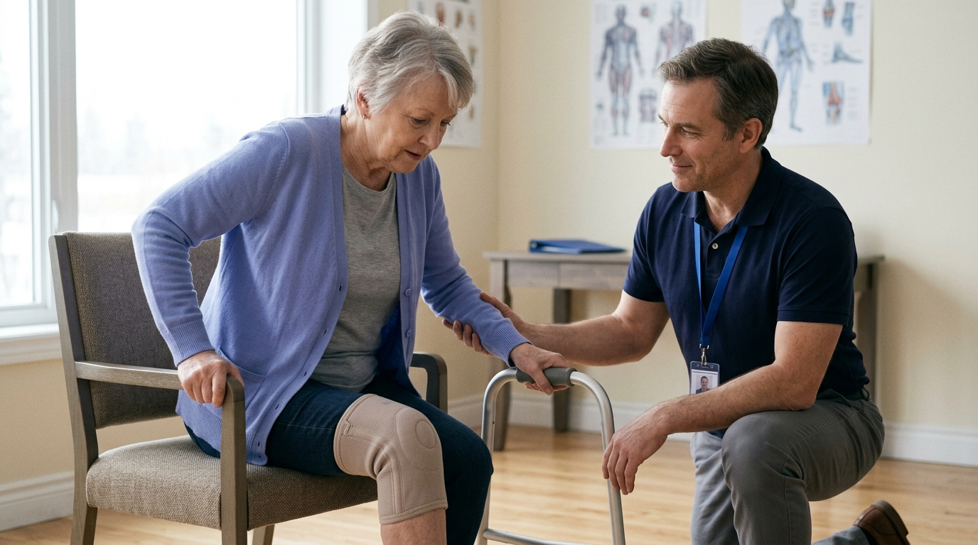
Older adults also experience slower cellular turnover and microvascular changes, which can prolong the acute swelling phase. A physical setback in a 65-year-old may require more deliberate pacing and structured protection during the first 72 hours than the same trauma in a younger adult. However, consistent pre-injury physical conditioning provides a strong foundation for recovery across all age groups.
Systemic health conditions alter the body's inflammatory response, circulation, and tissue repair capacity. Diabetes, for example, can impair peripheral blood flow and slow down both wound healing and soft tissue recovery. Peripheral neuropathy can reduce pain sensitivity, making it difficult for an individual to judge whether an injury is worsening.
Cardiovascular disease, chronic kidney disease, and autoimmune conditions also affect fluid balance and inflammatory resolution. People taking prescription blood thinners or anticoagulant medications face a higher risk of extensive bruising and internal joint bleeding after minor trauma. Anyone with chronic medical conditions should maintain a lower threshold for seeking early clinical advice after an injury.
When an injury involves an open cut, scrape, or puncture, wound characteristics dictate immediate medical priorities. The CDC categorizes wounds based on contamination levels to determine whether medical intervention, such as a tetanus vaccine booster, is needed.
Higher-risk wounds include:
A simple clean scrape from a kitchen surface carries a low risk of systemic infection. A puncture wound from a rusty outdoor nail, however, requires prompt medical evaluation regardless of how minor the surface mark appears. Clinicians review your immunization history to decide if a tetanus toxoid vaccine or tetanus immune globulin is warranted.
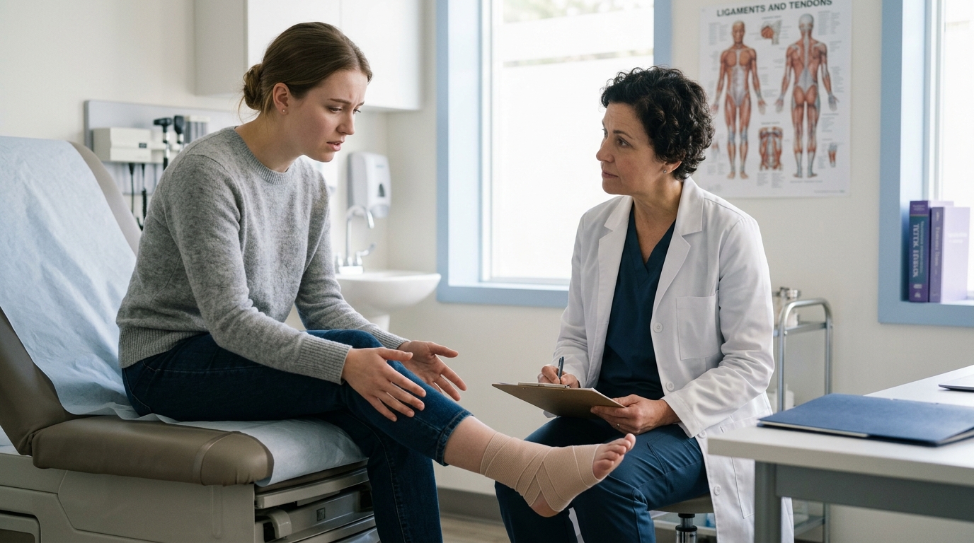
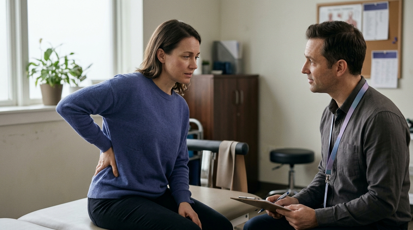
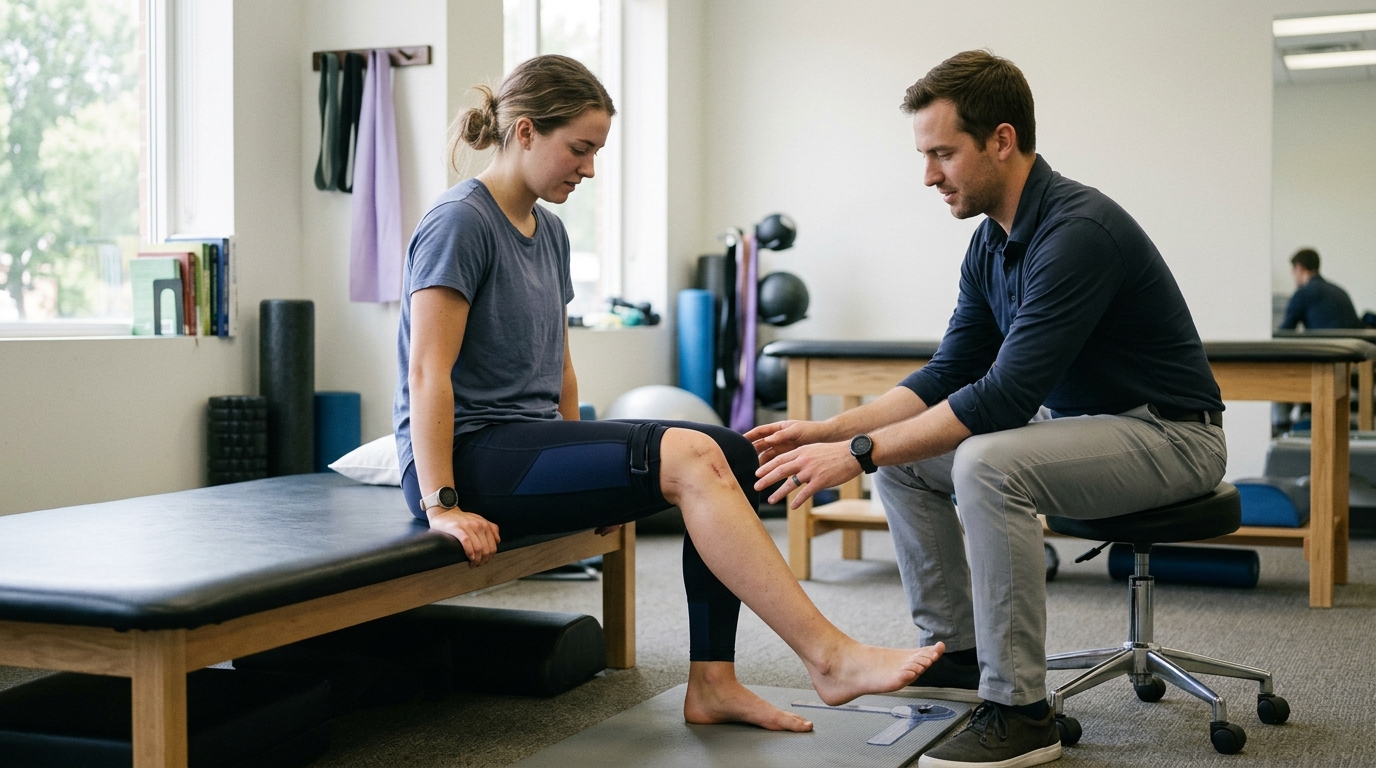
When you visit an urgent care center, emergency department, or clinic after an injury, the clinician does not automatically order X-rays or advanced scans for every sore joint. Instead, they use validated clinical decision rules and structured physical examinations to determine whether imaging is medically necessary.
One of the most widely researched clinical decision tools is the Ottawa Ankle and Foot Rules. Developed to reduce unnecessary radiography while maintaining high sensitivity for detecting fractures, this framework helps clinicians assess acute ankle and mid-foot injuries. Similar decision frameworks exist for the knee and cervical spine.
The Ottawa rules combine specific anatomical palpation points with a functional weight-bearing test. A clinician evaluates whether there is localized bone tenderness at exact landmarks along the lower leg and foot.
According to clinical guidelines, an ankle X-ray series is typically indicated only if there is pain in the malle
olar zone and any of the following findings:
Similarly, a mid-foot X-ray series is indicated if there is pain in the mid-foot zone and any of the following:
It is essential to understand that clinical decision rules are designed for trained medical professionals, not as self-diagnosis checklists for home use. Accurately locating the posterior edge of the lateral malleolus or the navicular bone requires practical anatomical knowledge. Pressing on swollen, inflamed soft tissues can easily be mistaken for direct bone tenderness by an untrained individual.
Furthermore, being able to limp through four painful steps does not rule out all serious soft tissue pathology, such as high ankle syndesmosis sprains or partial tendon avulsions. Clinicians integrate physical tests with a complete medical history, mechanism of injury evaluation, and joint stability testing. If you are uncertain about the severity of your injury, professional evaluation remains the safest course of action.
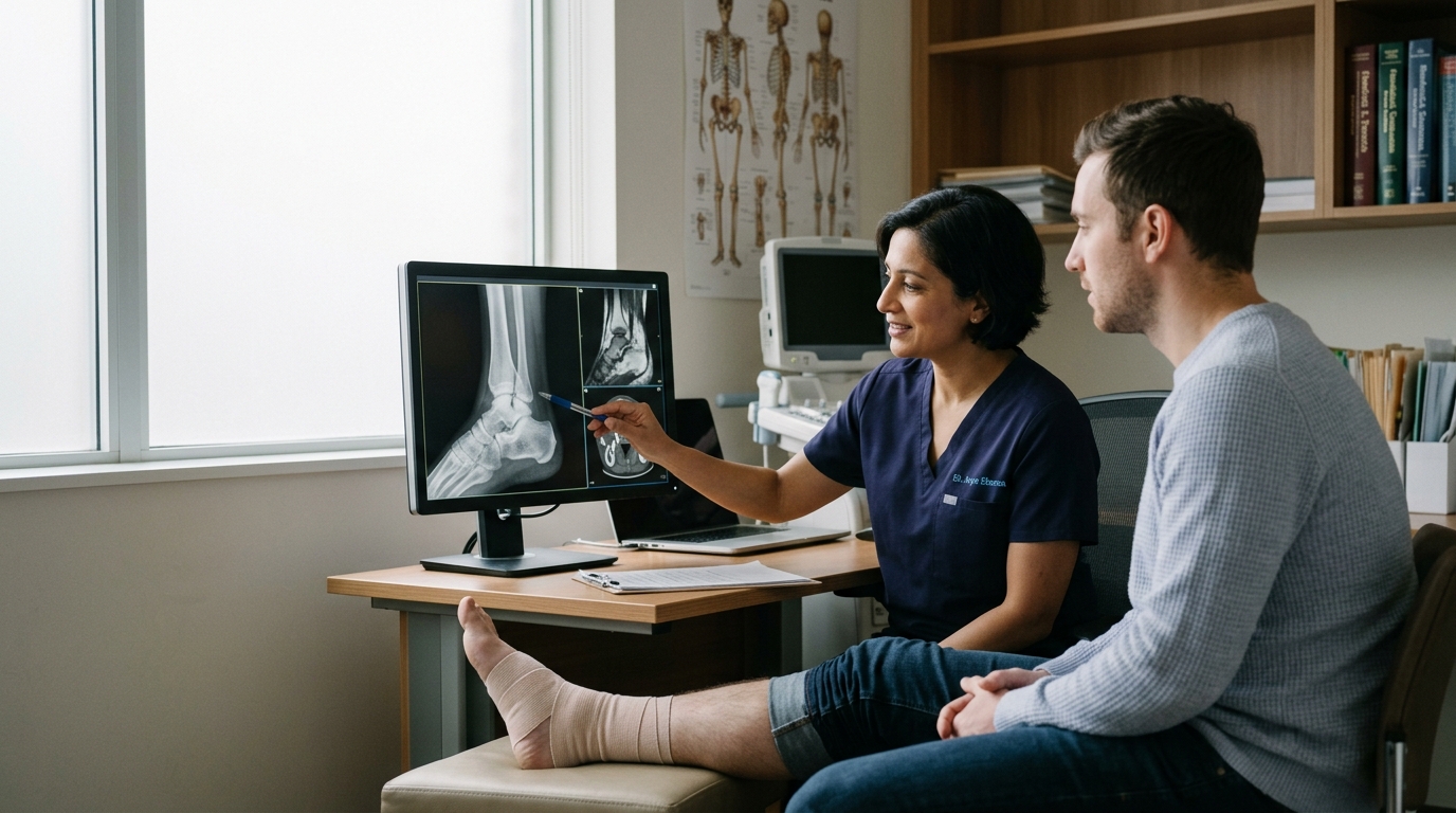
Standard radiography (X-rays) provides excellent visualization of dense bone structures, identifying fractures, bone chips, and joint subluxations. However, standard X-rays cannot visualize soft tissues such as ligaments, tendons, menisci, or cartilage.
If a bone fracture is ruled out on X-ray, but severe joint instability, locking, or complete loss of function persists, a clinician may consider advanced imaging:
Advanced imaging is rarely performed in the immediate first hours unless high-energy trauma, severe neurological deficits, or surgical emergencies are present. In many soft tissue injuries, clinicians prefer an initial period of protected load and clinical reassessment before deciding on an MRI.
The first 72 hours should be viewed as an active observation phase. Symptoms rarely stay completely static after an injury. Tracking the trajectory of swelling, pain, discoloration, and physical capacity helps you determine whether your body is stabilizing normally or if medical escalation is needed.
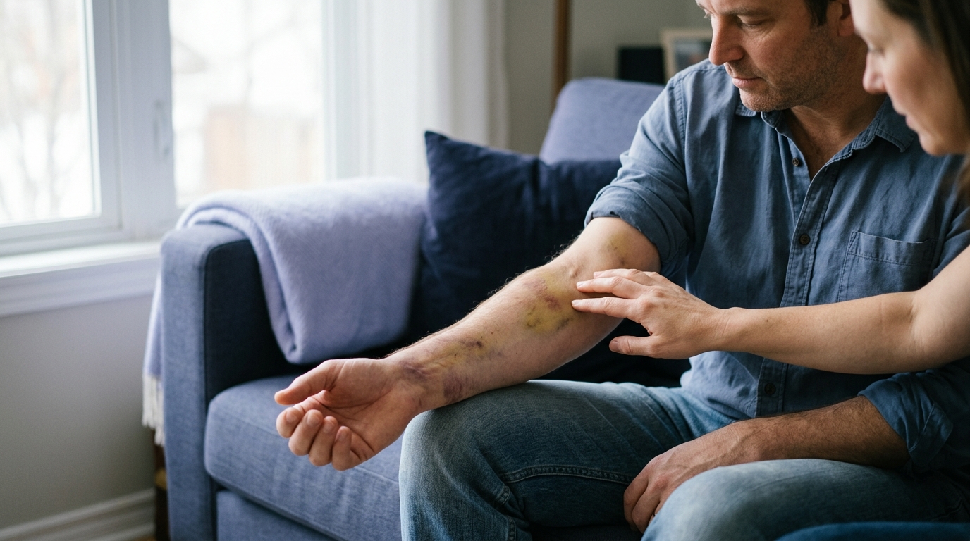
In a typical uncomplicated soft tissue injury, swelling and bruising often peak within the first 24 to 48 hours. Bruising may expand and change color from dark purple to reddish-blue as gravity pulls pooled blood downward through tissue planes. This downward migration of bruising into the toes or fingers is common and not necessarily a sign of worsening damage.
However, the intensity of sharp, resting pain should begin to level off or subtly decrease by day three. If pain levels steadily escalate despite resting, elevating, and protecting the limb, this is an abnormal trajectory. A joint that becomes progressively more swollen, hot to the touch, and intensely painful over 72 hours requires prompt reassessment.
During the first 72 hours, watch for emerging secondary complications that can arise after physical trauma:
A provincial clinical guideline for acute ankle and mid-foot injuries recommends a structured reassessment point if an injury does not improve. Even if an initial X-ray was not ordered, clinical follow-up is advised if significant pain or difficulty bearing weight persists after five to seven days.
This multi-day checkpoint allows acute swelling to subside, making physical examination and ligament stability tests easier and more accurate for the clinician. If your symptoms fail to show any meaningful functional progress as you move past the 72-hour window, schedule a visit with your doctor or physical therapist. Explore comprehensive strategies for ongoing recovery within our injury recovery and healing resources.
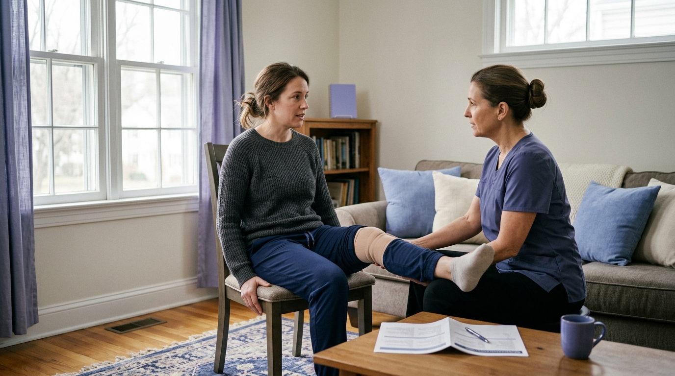
When you attend a medical appointment or urgent care clinic for an acute injury, asking focused questions helps you understand your diagnosis and recovery plan. Clear communication ensures that you leave with realistic expectations for daily activities.
Consider discussing these practical questions during your clinical evaluation:
A distinct popping sensation during a traumatic movement is an important clinical clue that warrants professional evaluation. While major ligament or tendon tears often cause rapid swelling, certain injuries may produce delayed or localized fluid accumulation. Joint stability cannot be verified by the absence of visible swelling alone. Protect the joint, avoid heavy weight-bearing, and schedule a clinical examination to test ligament integrity.
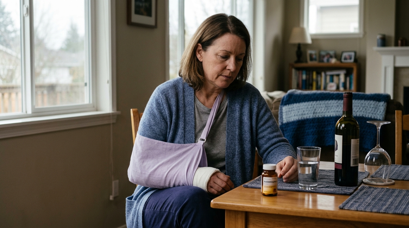
Over-the-counter medications can help manage acute discomfort, but they should be selected carefully based on your individual health profile. Acetaminophen helps reduce pain perception without significantly altering the inflammatory cascade. NSAIDs, such as ibuprofen or naproxen, reduce both pain and inflammation, though their widespread use in the immediate hours of soft tissue healing is debated. Always check with a pharmacist or physician before taking medications, especially if you have high blood pressure, kidney issues, stomach ulcers, or take prescription blood thinners.
Direct heat application should generally be avoided during the initial 48 to 72 hours after an acute injury. Heat causes vasodilation, which increases blood flow to the area and can worsen active bleeding, tissue swelling, and throbbing pain. Heat is more appropriately introduced in later subacute phases to relax tight muscle bellies and ease chronic stiffness once acute swelling has stabilized.
Head impacts in children must be handled with heightened caution. Children may not always verbalize subtle symptoms like dizziness or visual changes clearly. Watch closely for behavioral changes, excessive sleepiness, irritability, changes in nursing or eating habits, persistent crying, or vomiting. The CDC emphasizes that danger signs in children require immediate emergency medical care, and any suspected concussion warrants evaluation by a healthcare provider.
The first 72 hours after an injury are about safety, protection, and careful observation. Always address life-threatening hazards, responsiveness, breathing, and severe bleeding before attending to a painful limb. Recognize red flags, including head injury danger signs, visible bone deformities, and neurovascular changes like numbness or cold extremities, which demand immediate emergency care.
For suspected sprains and strains, focus on early joint protection, sensible elevation, and gentle, pain-free movement rather than aggressive testing. Avoid forcing painful joints through their range of motion, and do not rely on self-diagnosis tools to rule out structural fractures. If pain is severe, weight-bearing is impossible, or symptoms fail to improve over the first several days, seek a comprehensive evaluation from a qualified medical professional.

Use ReboundBody resources to understand common recovery stages, rehab terms, movement limits and strength rebuilding. Each guide is designed to make a complex comeback easier to understand.


Read practical guidance on injury recovery, rehabilitation, mobility and rebuilding strength as you work your way back to activity.
Explore Resources