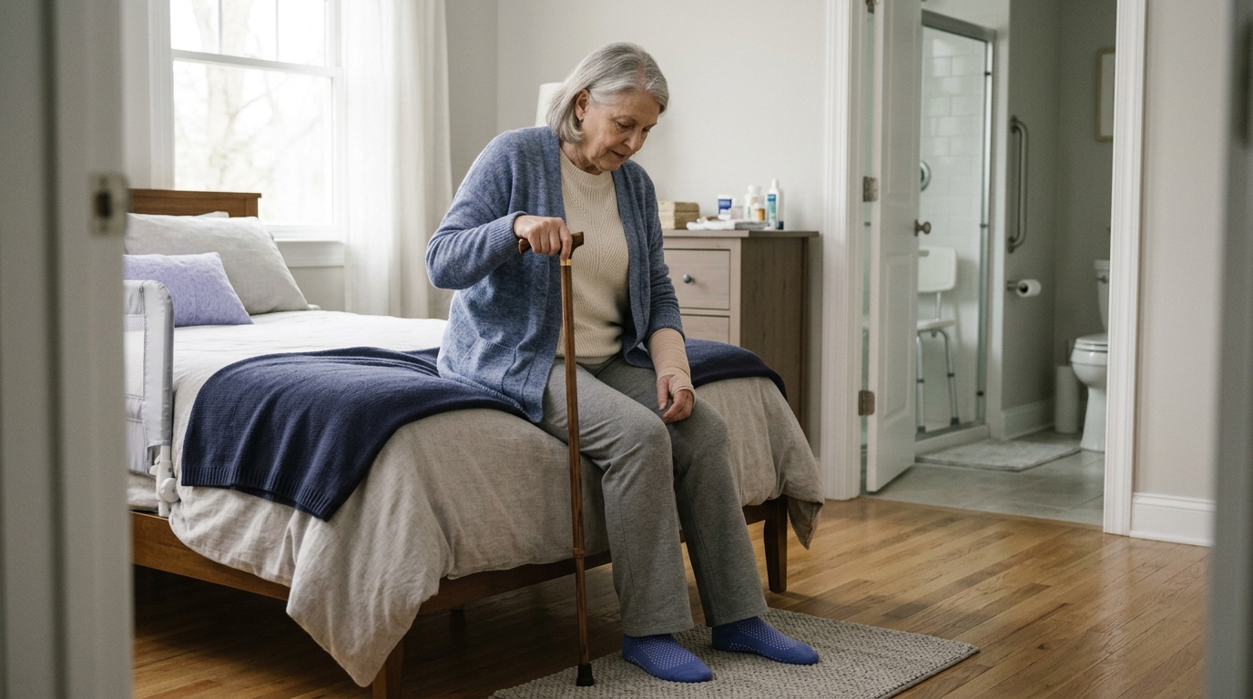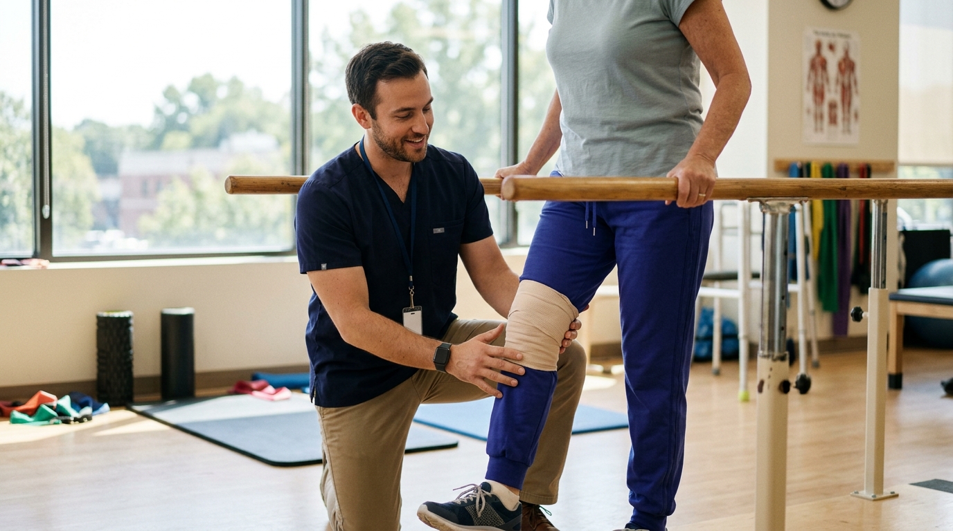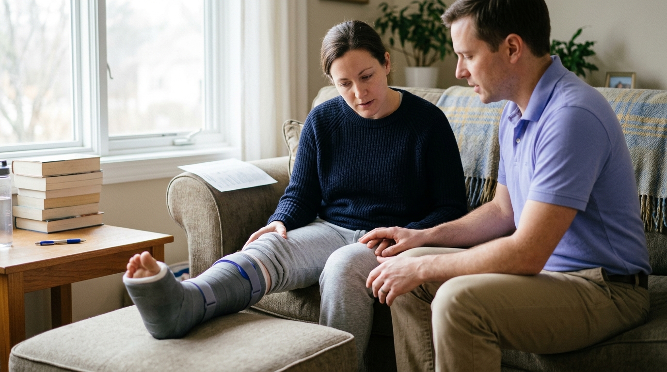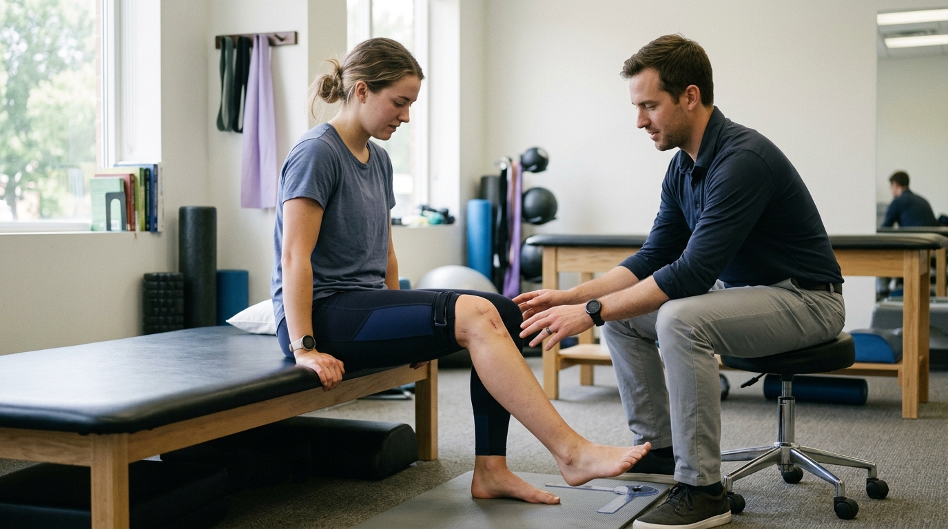

Noticing sudden fluid buildup around a fresh injury signals natural tissue repair that you can monitor at home using clear functional benchmarks.





A massive, puffy joint is often seen as definitive proof of a catastrophic injury, while minor swelling is assumed to mean everything is fine. Clinical research shows this common assumption is frequently incorrect. The physical size of swelling after an acute injury tells you surprisingly little about the actual structural damage or how long your recovery will take.
Swelling is a natural, protective biological response to tissue damage, but it is a clinical sign rather than a standalone diagnosis. It occurs when fluid, white blood cells, and proteins rush to an injured area to stabilize tissue and initiate healing. However, its overall volume does not accurately predict long-term recovery timelines. You can have a severe ligament tear with modest visible fluid, or a minor sprain with dramatic fluid accumulation. Because swelling does not tell the whole story on its own, monitoring the overall pattern of symptoms matters far more than obsessing over the exact size of a bump.
When you twist an ankle, strain a muscle, or take a direct blow, the body initiates an immediate inflammatory response. Capillaries and tiny blood vessels in the injured area dilate and become more permeable. This allows fluid, specialized proteins, and white blood cells to move out of the bloodstream and into the interstitial spaces between tissues. In clinical settings, this localized accumulation of excess fluid is called edema.
Edema serves an important biological purpose in the earliest hours after a setback. The localized fluid accumulation creates an internal splint that limits excessive joint motion. It also delivers the biological resources necessary to clear cellular debris and begin repairing damaged collagen fibers. While swelling can feel tight, uncomfortable, and restrictive, it is not an enemy that must be completely eliminated immediately. It is an active part of the natural healing cascade.
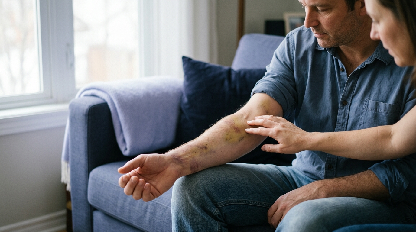
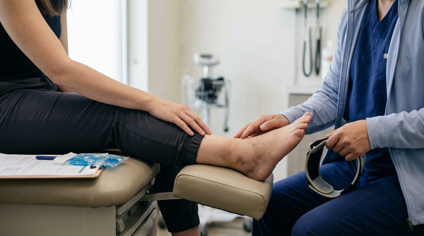
It helps to understand the difference between general swelling and bruising. Bruising, known clinically as ecchymosis, happens when small blood vessels break and leak red blood cells directly into the soft tissue beneath the skin. This leakage produces distinct black, blue, purple, or yellow discoloration. Swelling refers specifically to the expansion of tissue volume from fluid buildup. An injury can produce significant swelling without visible bruising, or widespread bruising with minimal fluid retention.
Because swelling is a non-specific response, it can accompany many different types of trauma. You will see swelling with mild ligament sprains, muscle strains, joint contusions, tendon irritations, and bone fractures. For this reason, seeing fluid build up around a joint does not reveal what specific tissue is injured. A comprehensive injury recovery and healing guide emphasizes that swelling must always be evaluated alongside physical function, joint stability, and localized pain patterns.
Clinical research challenges the idea that measuring swelling can tell you how well someone will recover. In a formal clinical study investigating recovery after acute lateral ankle sprains, researchers tracked objective physical measurements over several months. They measured ankle circumference directly across the malleoli and midfoot circumference between the outer ankle bone and the base of the second metatarsal. When the researchers analyzed long-term outcomes at seven months, these objective swelling measurements were not retained as reliable predictors of long-term recovery. A person with substantial initial swelling was just as likely to regain full function as someone with minimal swelling.
This finding is reinforced by clinical position statements from major athletic training organizations. A position statement from the National Athletic Trainers' Association noted that the magnitude of swelling does not correlate with self-reported function after an acute ankle sprain. An individual may experience substantial joint swelling while maintaining relatively low pain and decent mobility. Another person might have very little visible fluid retention but experience debilitating pain and an inability to bear any weight.
Evidence regarding acute management techniques also highlights the need for a nuanced perspective. A systematic review examining conservative treatments for soft-tissue injuries evaluated the comparative effects of temperature modalities on post-injury edema. The review cited an ankle sprain study where cold water immersion at 50 to 60 degrees Fahrenheit for 20 minutes across five days resulted in significantly less edema than heat therapy. Cold immersion resulted in an average fluid change of 3.3 milliliters, compared to 25.3 milliliters in the heat therapy group. While cold application helps manage acute discomfort and blunts excessive initial swelling, research shows it does not fundamentally speed up the biological timeline of tissue repair.
The evidence surrounding immobilization versus early movement is equally revealing. A systematic review evaluating ankle sprain management found that rigid immobilization for three weeks or longer was often less effective than functional treatment for reducing swelling and pain in the short and medium term. Keeping an injured limb completely motionless can actually slow down the clearance of fluid. Active muscle contractions act as a natural mechanical pump that moves stagnant lymph and venous blood out of the extremities. While the evidence quality in some early immobilization trials is considered low, modern rehabilitation protocols consistently favor protected, early functional movement over total prolonged casting for uncomplicated soft-tissue injuries.
To understand the broader science of tissue adaptation during rehabilitation, you can read our guide on recovery science and tissue remodeling.
Because a single measurement cannot tell you the severity of an injury, tracking symptoms over time is the best approach. You can monitor your recovery safely at home by using a practical three-part assessment framework focused on trend, function, and warning signs.
The first element to assess is the overall direction of the swelling over several days. You want to observe whether the fluid accumulation is decreasing, remaining stable, or actively expanding. In a typical uncomplicated sprain or contusion, swelling peaks within the first 24 to 48 hours and then gradually stabilizes before slowly declining. If swelling continues to expand rapidly after the initial two days, especially despite limb elevation and rest, this is an abnormal trend that requires clinical evaluation.
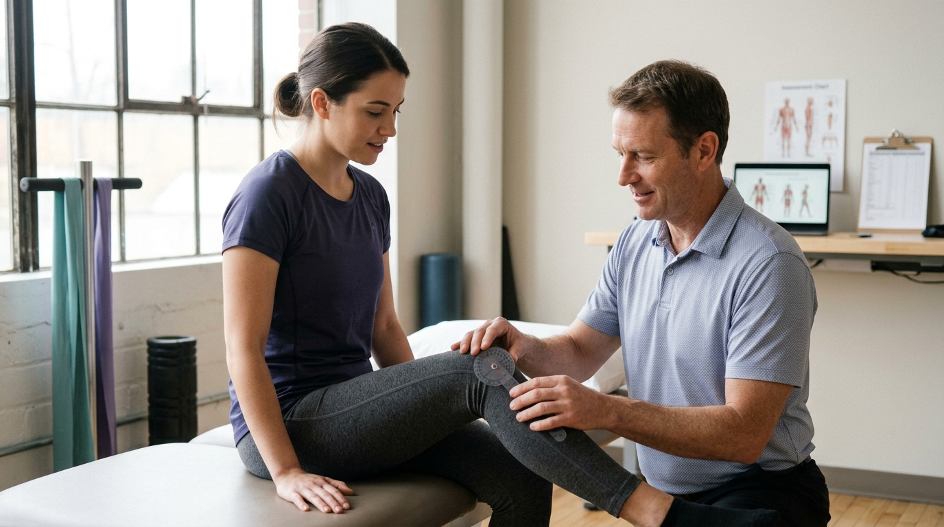
To track objective physical trends at home, you can use simple measurement methods:
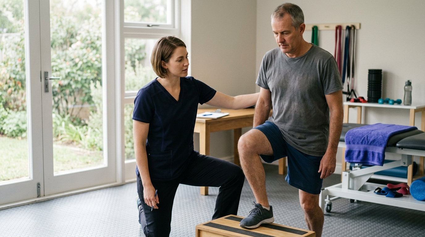
The second element of the framework is functional capacity. You need to assess how well you can actually use the injured body part for daily tasks. Functional ability is often a much better indicator of underlying joint integrity than visible puffiness.
When evaluating function, consider these practical questions:
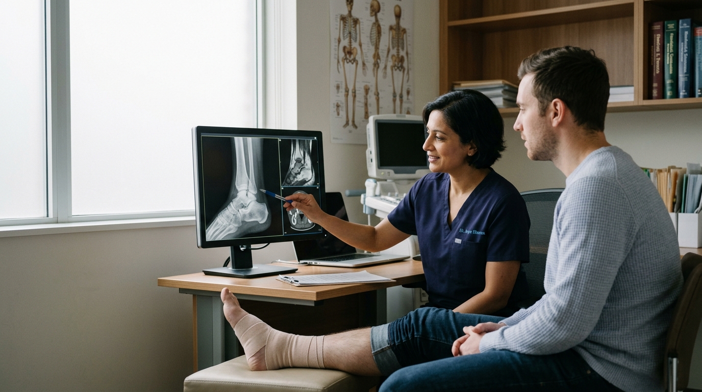
If you are unable to bear any weight on an injured lower limb immediately after the incident, clinical guidelines recommend a formal medical evaluation. Inability to take four steps often warrants an X-ray to check for a fracture.
The third component of the framework involves screening for urgent physiological warning signs. Swelling that occurs alongside nerve compression, compromised blood flow, or systemic infection represents an urgent medical situation.
You should monitor the affected limb for:
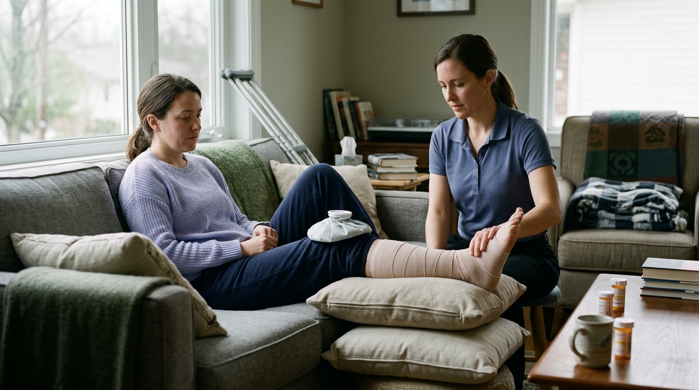
During the first 48 to 72 hours following a mild to moderate soft-tissue injury, conservative home care helps control excessive fluid accumulation and keeps you comfortable. Health organizations like the NHS recommend the PRICE protocol: Protection, Rest, Ice, Compression, and Elevation. Applying these steps correctly helps manage acute symptoms without interfering with the natural healing process.
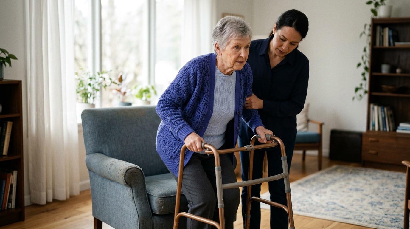
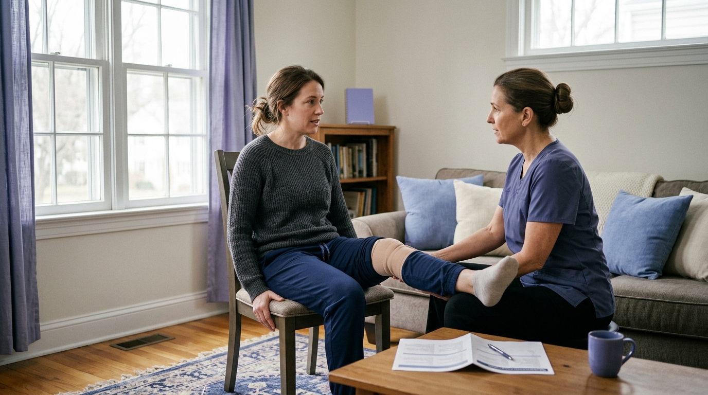
Protection involves shielding the injured tissue from further mechanical stress. Depending on the joint involved, this might mean wearing a supportive brace, using an elastic wrap, or temporarily using crutches.
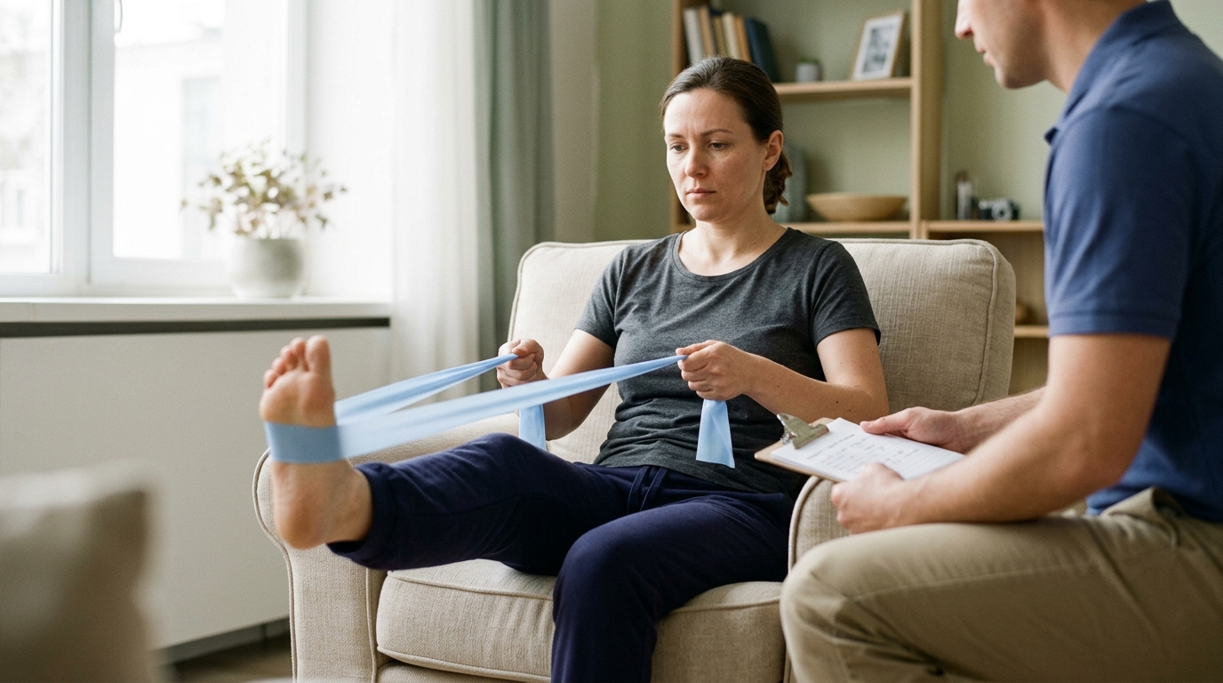
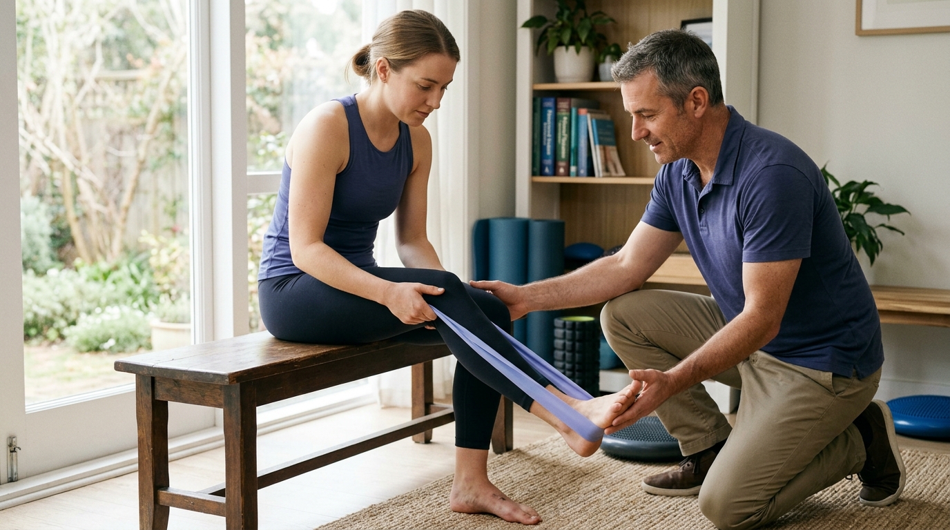
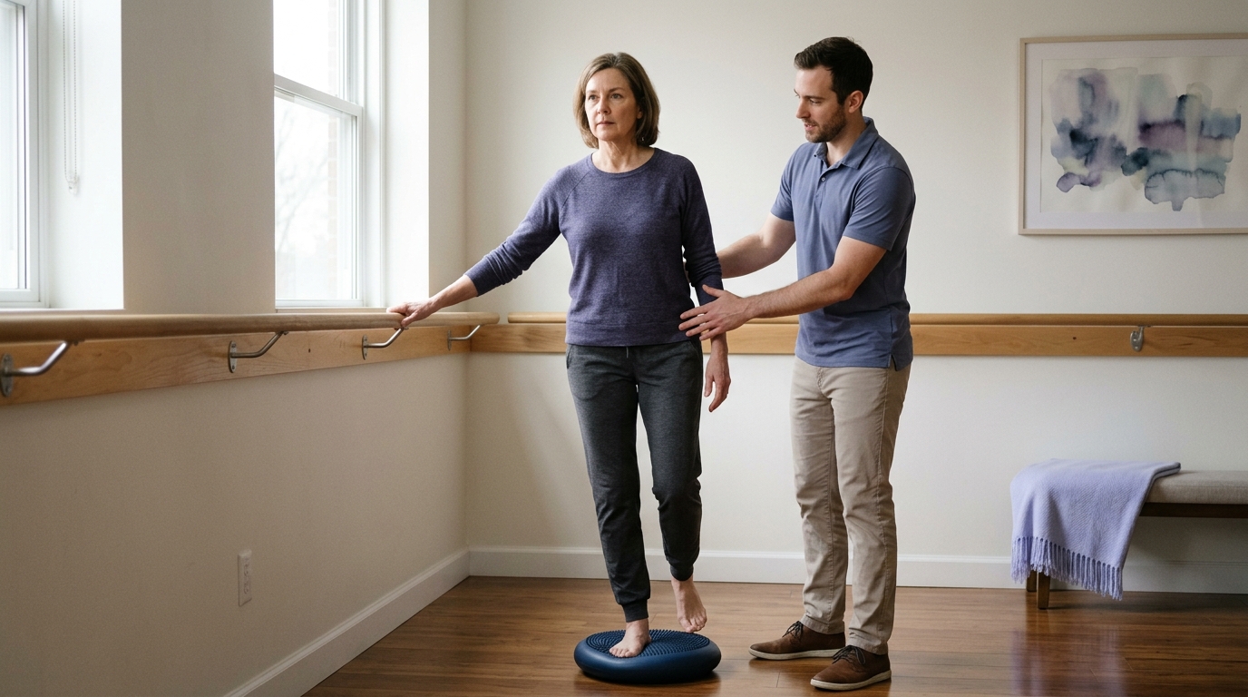
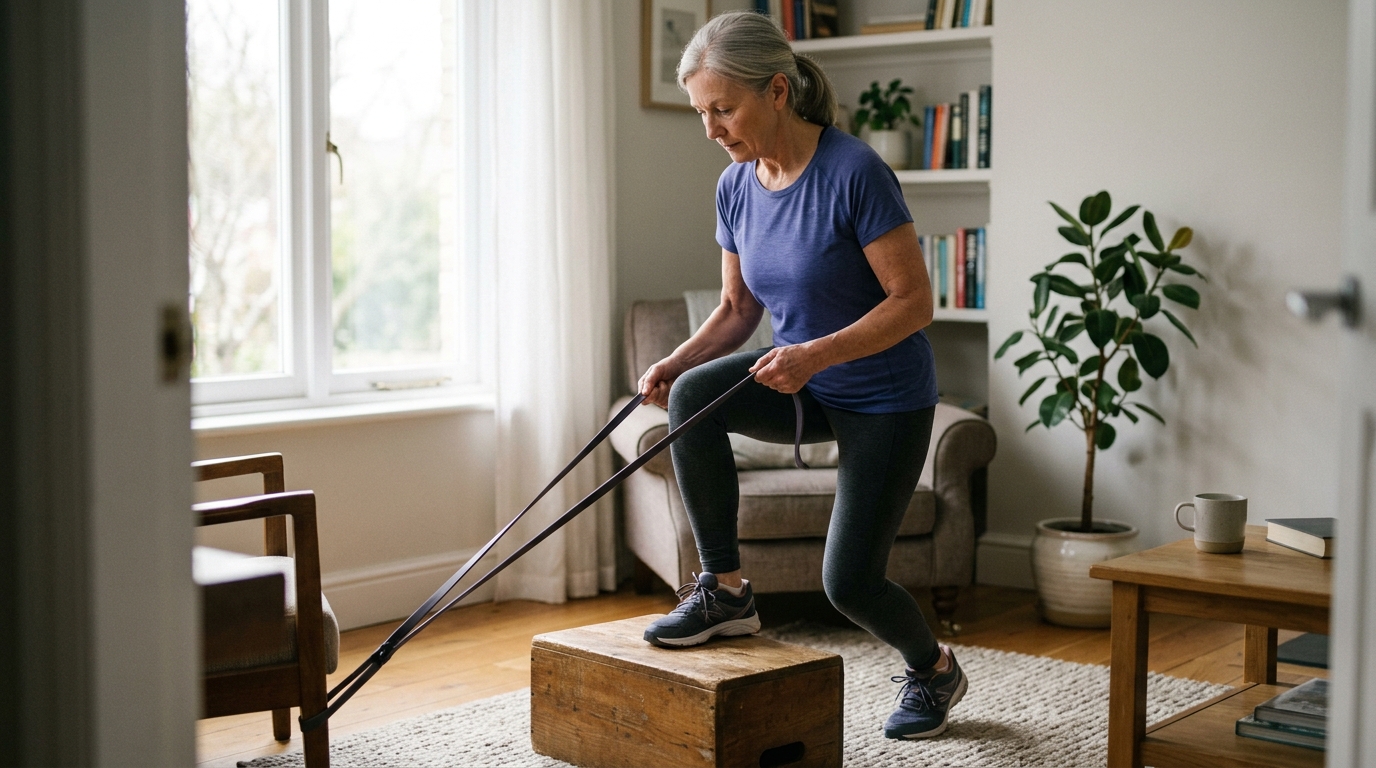
Rest during this early phase should be relative rather than absolute. You want to stop doing activities that cause sharp pain, such as running or heavy lifting. However, completely immobilizing a joint in bed for days is rarely helpful. Gentle, pain-free movements like light toe wiggling or seated ankle circles encourage fluid drainage through the lymphatic system without stressing repairing ligaments.
Ice helps numb acute pain and reduces excessive localized blood flow to the injured area. However, cold therapy must be applied safely to avoid skin irritation or tissue injury.
Apply an ice pack or a bag of frozen vegetables wrapped in a damp towel for 15 to 20 minutes every two to three hours during the first two days. Never place raw ice directly onto bare skin, as this can cause frostbite or superficial nerve injury. If you have peripheral vascular disease, diabetes, or poor sensation, consult a doctor before using cold therapy.
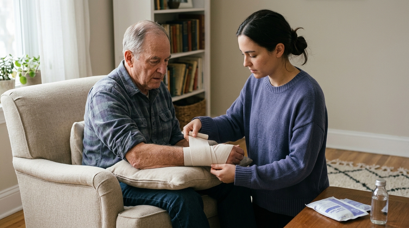
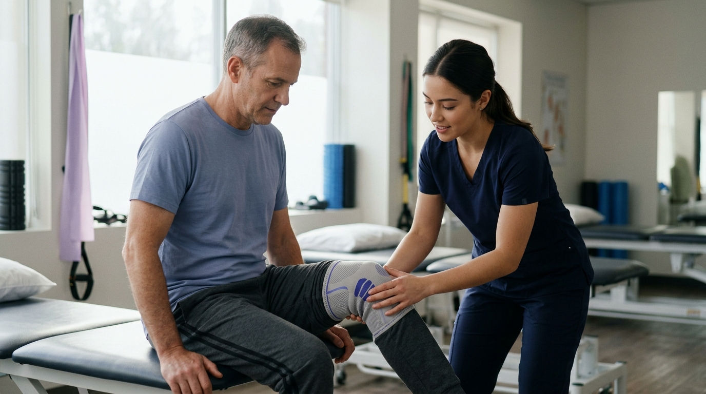
Compression provides external mechanical pressure that limits the accumulation of fluid in the interstitial spaces. An elastic bandage, such as an ACE wrap or a compressive sleeve, can provide comfortable support during the day.
When applying a compression wrap:
Elevation uses gravity to assist the passive drainage of excess fluid and venous blood back toward the central circulation. For elevation to be most effective, the injured joint should ideally be positioned above the level of your heart.
When resting on a couch or bed, prop your swollen leg or arm up on several firm pillows. Try to keep the limb elevated as much as reasonably possible during the first 48 hours. If you must sit at a desk, resting your foot on an elevated stool is better than letting it hang down, even if it is not fully above heart level.
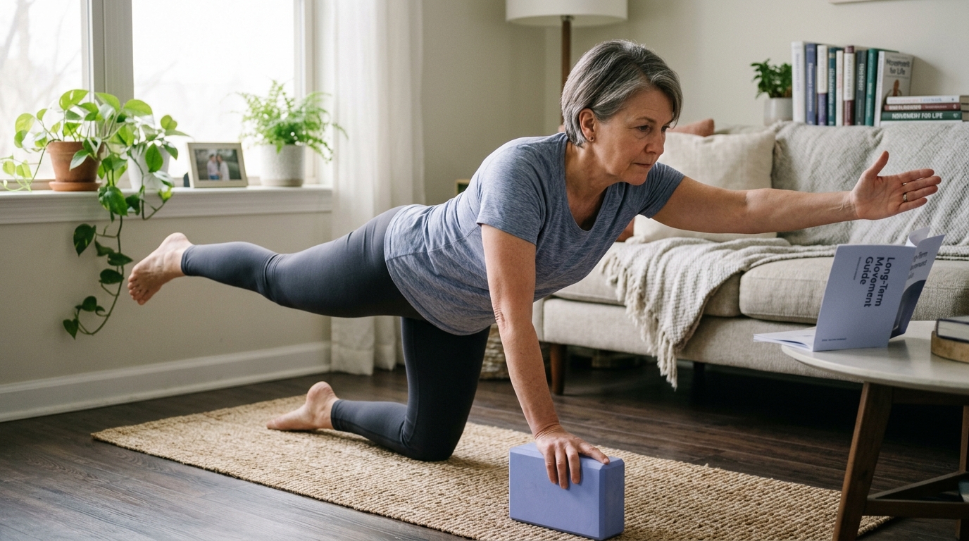
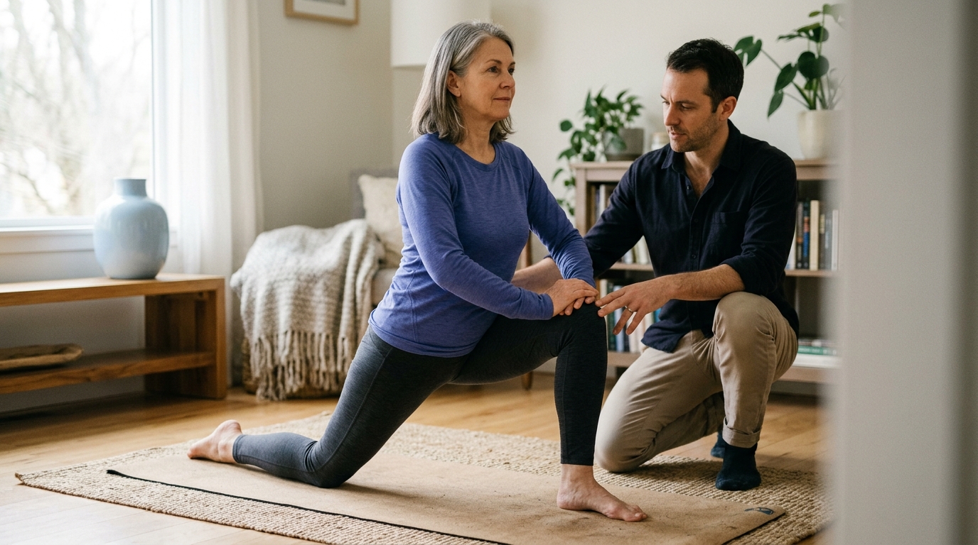
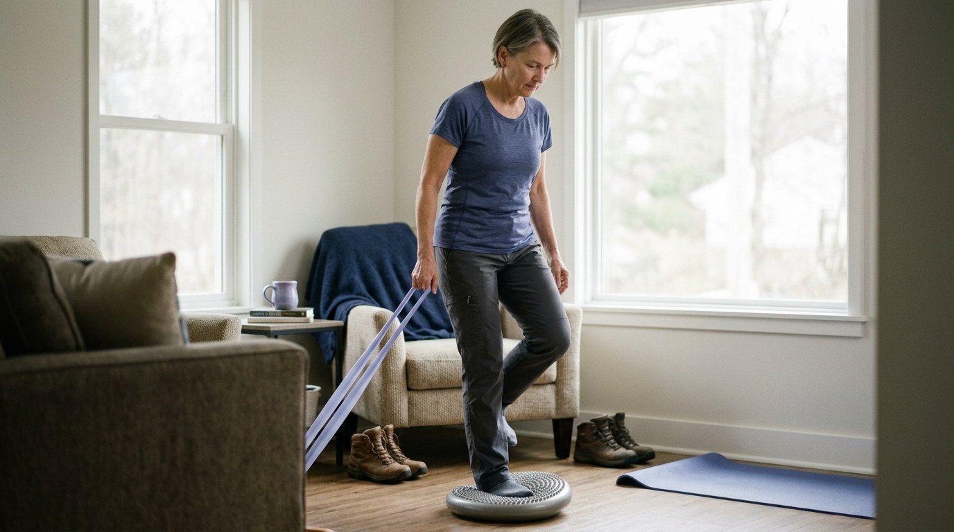
To learn more about transitioning from acute rest to safe physical activity, explore our guide on rehabilitation and mobility exercises.
Every person recovers from an injury at a different rate. The time it takes for swelling to resolve depends on several individual factors, including injury type, circulation, age, and activity habits.
Different bodily tissues have different blood supplies and cellular repair timelines. Highly vascular tissues, like skeletal muscle, often mount a vigorous early inflammatory response with significant bruising, but they clear metabolic waste relatively quickly. Conversely, intra-articular structures like the anterior cruciate ligament or meniscus have limited blood supplies. When these structures are damaged, swelling inside the joint capsule, known as an effusion, can linger for weeks or months.
Bone injuries also create distinct fluid patterns. A hairline stress fracture or non-displaced fracture triggers an extensive periosteal inflammatory response. This fluid buildup can persist until the initial bony callus stabilizes. If you have an injury that feels deep, achy, and stays puffy despite weeks of rest, structural bone involvement should be considered.
The physical location of an injury on your body significantly influences how fast swelling clears. Gravity naturally pulls fluid downward into the lowest extremities. An injured ankle or foot will almost always stay swollen longer than an injured wrist or shoulder.
Fluid clearance from the lower legs depends heavily on the venous muscle pump. When your calf muscles contract during walking, they compress deep veins and lymphatic channels, driving fluid upward past one-way valves. If you are forced to sit or stand still for long periods, fluid naturally settles in the lower leg and foot. Individuals with pre-existing venous insufficiency or varicose veins will clear post-injury fluid much more slowly.
As we get older, our blood vessels and lymphatic channels naturally become less elastic. The microcirculation responds more slowly to local trauma, and cellular clearance of inflammatory debris takes longer. Older adults often find that minor bumps and sprains leave lingering, low-grade puffiness long after the acute pain has disappeared.
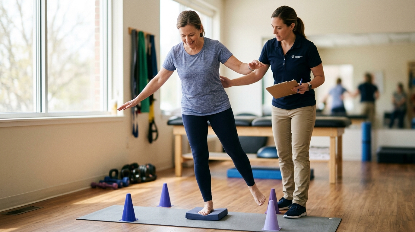
Age-related changes in collagen composition also influence how soft tissues handle mechanical stress during recovery. Tendons and ligaments lose some of their water-binding capacity and tensile elasticity over time. This makes progressive, gradual loading essential to avoid triggering recurrent inflammatory flare-ups.
The way you pace your daily activity directly influences fluid accumulation during recovery. Doing too much too soon is one of the most common reasons swelling flares back up weeks after an injury.
If you walk three miles on an ankle that is only ready for a gentle half-mile stroll, the overloaded tissues will produce a fresh wave of inflammatory fluid. The joint will feel stiff, tight, and warm the following morning. Recovery requires finding the middle ground between complete bed rest and premature athletic activity.
Your broader systemic health plays a quiet but powerful role in managing inflammation and fluid balance. Chronic poor sleep, elevated systemic stress, and high sodium intake can all encourage fluid retention and delay tissue repair.
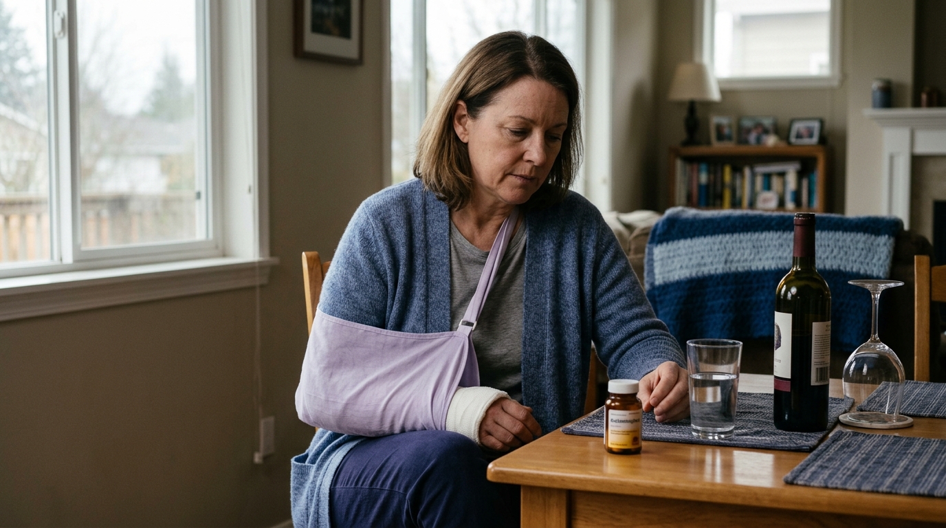
Hydration habits also matter. When the body is dehydrated, it tends to hold onto extracellular sodium and water, which can make localized swelling appear more pronounced. Maintaining balanced nutrition, prioritizing seven to nine hours of quality sleep, and avoiding alcohol during the acute healing phase provide the best physiological environment for recovery.
For a deeper look into how daily habits influence healing, see our resource on recovery nutrition and lifestyle strategies.
While most post-injury swelling is benign and resolves with basic conservative management, certain clinical signs indicate a more serious condition. It is critical to distinguish between normal recovery puffiness and urgent medical red flags.
Go to an emergency room or call emergency services immediately if you notice any of the following severe presentations:
Contact your primary care physician, an urgent care clinic, or a physical therapist promptly if you experience any of the following warning patterns:
Navigating the early days of an orthopedic setback is often confusing. Avoiding these common mistakes can help prevent unnecessary complications and support a smooth recovery.
A common myth is that a broken bone always prevents you from walking. In reality, non-displaced hairline fractures, avulsion fractures, and fibular stress fractures often allow people to bear partial weight despite structural damage. Never assume an injury is merely a simple sprain based solely on your ability to limp across a room. If you have severe focal bone pain, get a professional clinical evaluation.
Placing frozen gel packs or raw ice cubes directly against unprotected skin is a frequent mistake. Direct ice contact can damage superficial cutaneous nerves, cause cold-induced burns, and produce reactive vasodilation. Always place a damp cloth, thin towel, or elastic bandage between the cold source and your skin.
While taking it easy for the first 48 hours protects vulnerable tissues, extended bed rest can be counterproductive. Prolonged immobilization causes healthy muscle tissue to atrophy, stiffens joint capsules, and slows the natural clearance of lymphatic fluid. Once acute pain stabilizes, safe, pain-free active movement should be gradually reintroduced.
Soaking a newly injured, swollen joint in a hot bath or applying a high-temperature heating pad during the first two days is a common error. Heat causes peripheral blood vessels to dilate, which can increase localized swelling, bleeding, and throbbing pain. Save therapeutic heat for later stages of rehabilitation when managing chronic muscle stiffness.
Many people believe that once their visible swelling is gone, their injured ligament or tendon is fully healed. Conversely, others assume that because a joint still looks slightly puffy, they must avoid all physical activity. As clinical studies demonstrate, visible swelling does not correlate perfectly with tissue strength or functional recovery. Base your rehabilitation decisions on overall strength, mobility, joint stability, and professional clinical guidance.
The following examples show how the three-part monitoring framework can be applied to common everyday injury situations.
A 42-year-old recreational walker steps off a curb awkwardly, twisting the right ankle. Moderate swelling and localized tenderness develop over the outer ankle within three hours. The individual can still bear weight and walk across the room with a slight limp, and there is no numbness, coldness, or bone deformity.
A 55-year-old gardener twists a knee while pivoting on uneven grass. There is immediate, sharp discomfort, followed by significant joint tightness that gets progressively worse over the next 48 hours. Despite resting on the couch and elevating the leg with ice packs, the knee becomes noticeably tighter, warm to the touch, and increasingly difficult to bend past 45 degrees. The person cannot put full weight on the leg without sharp catching sensations.
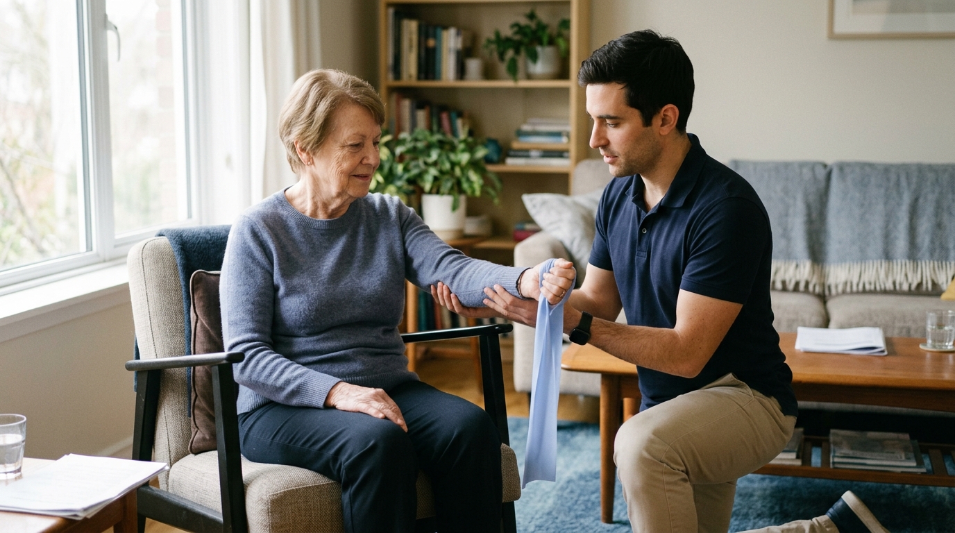
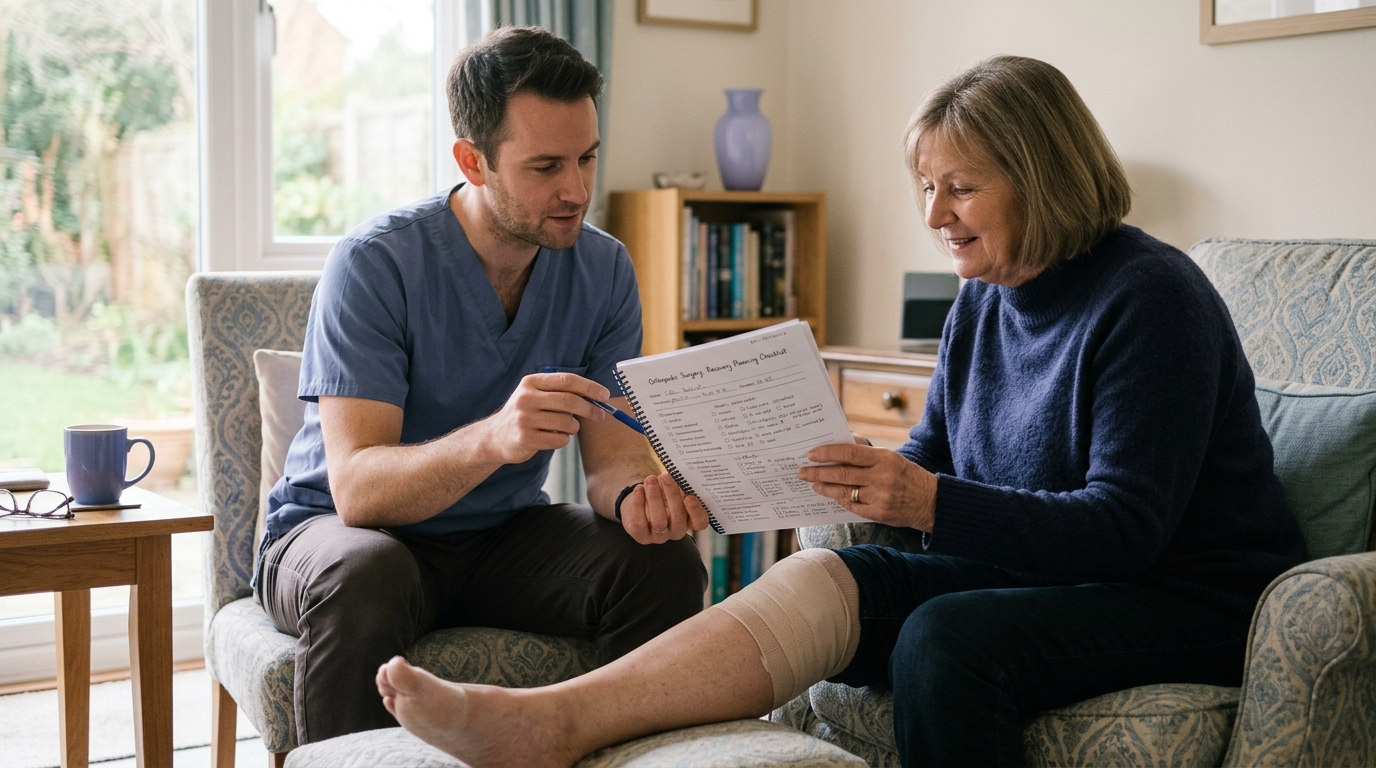
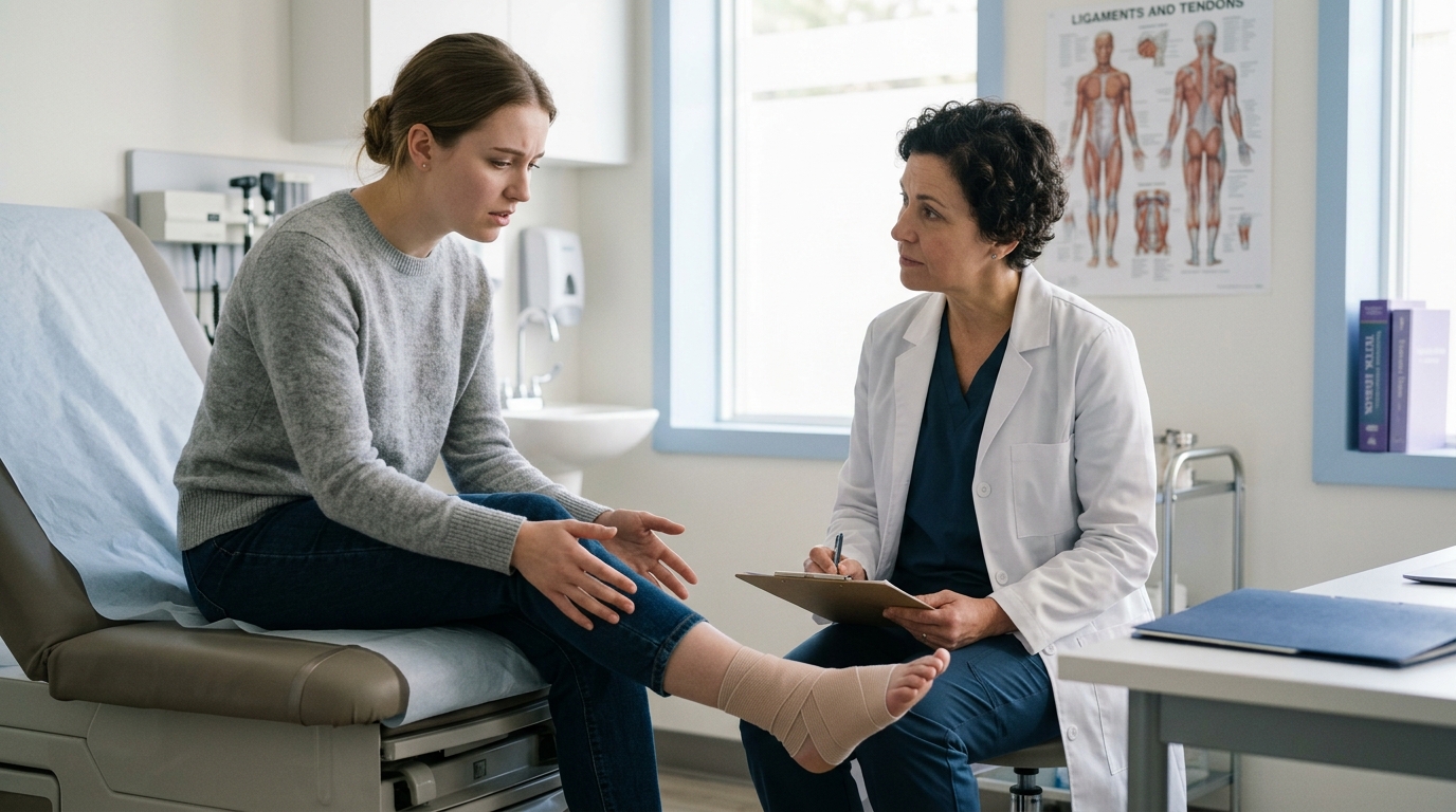
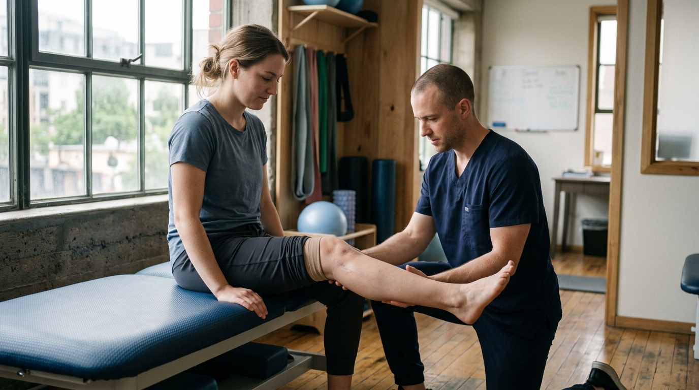
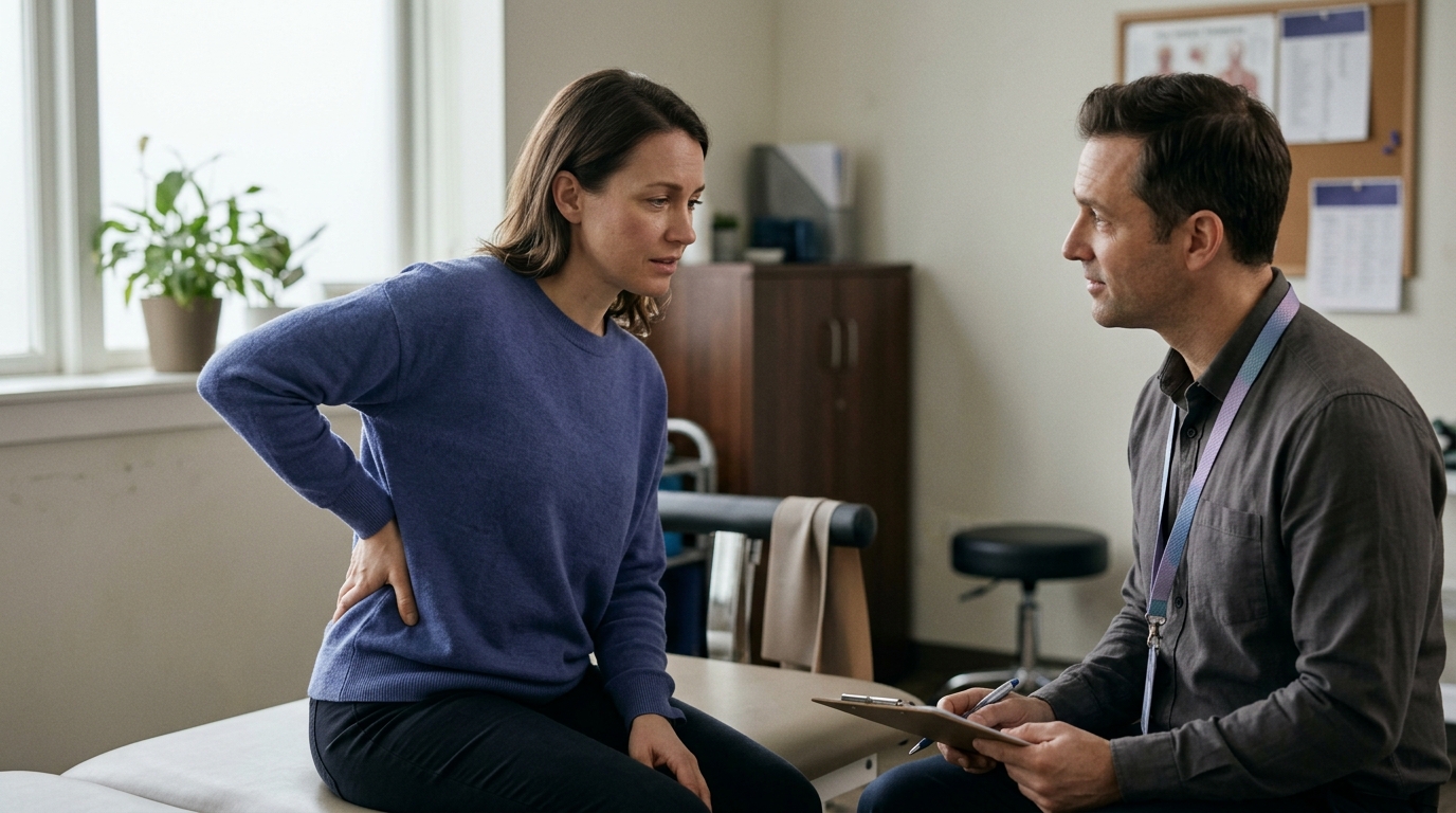
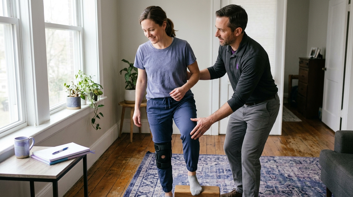
When you visit a physician, orthopedic specialist, or physical therapist for an injury evaluation, having clear, targeted questions helps you get the most out of your appointment.
Consider bringing these practical questions to your next clinical visit:
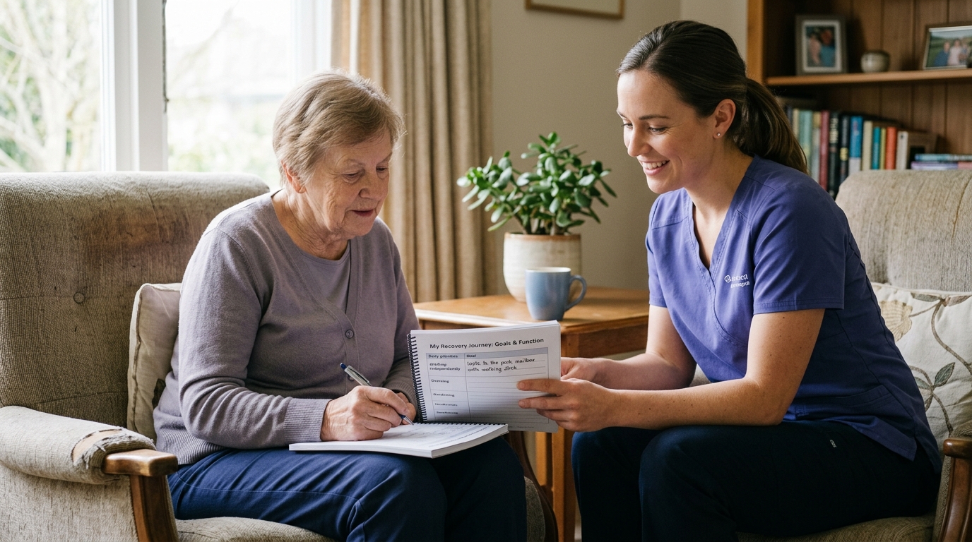
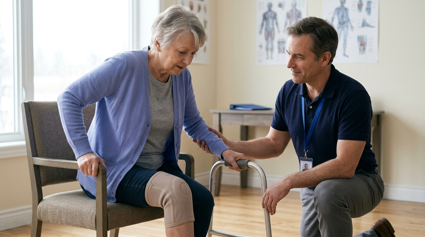
To learn more about working effectively with rehabilitation specialists, browse our articles on active aging and injury prevention.
Swelling is a natural, protective biological response to tissue damage, but its size alone cannot diagnose an injury or predict how long your recovery will take. Instead of focusing solely on the volume of fluid around a joint, monitor the broader pattern of your symptoms: the direction of the swelling trend, your functional ability to move and bear weight, and the presence of any neurological, circulatory, or systemic warning signs. Applying conservative home care during the first two to three days can help keep you comfortable, but worsening swelling, severe pain, inability to bear weight, or numbness should always prompt an evaluation by a qualified medical professional.
A patient, informed approach to monitoring swelling allows you to navigate the early stages of recovery safely and make confident decisions about when to manage an injury at home and when to seek professional care.

Use ReboundBody resources to understand common recovery stages, rehab terms, movement limits and strength rebuilding. Each guide is designed to make a complex comeback easier to understand.


Read practical guidance on injury recovery, rehabilitation, mobility and rebuilding strength as you work your way back to activity.
Explore Resources