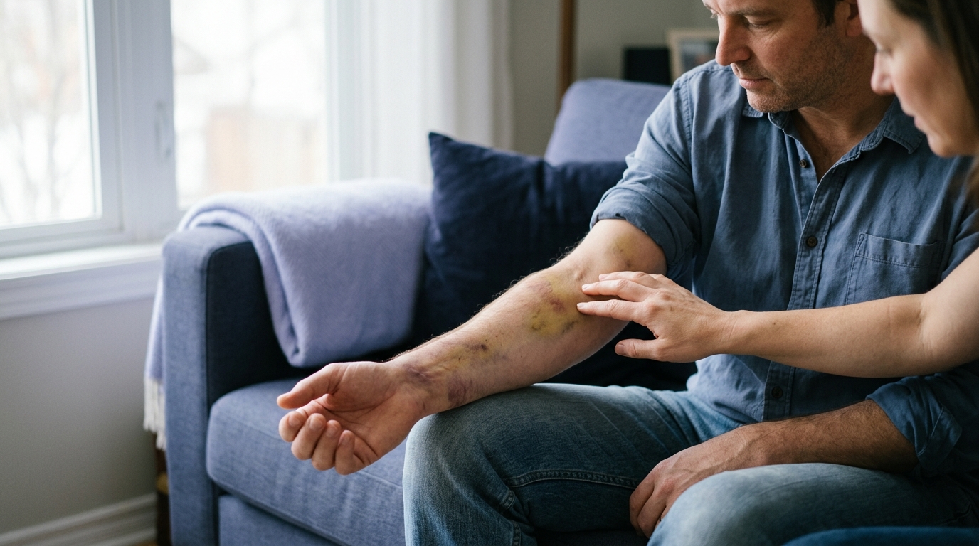

Watching a deep mark change color after a painful fall reveals how damaged tissues heal and why visible discoloration rarely reflects injury severity.





A bruise is visible proof that small blood vessels have leaked beneath intact skin. It is not a direct measure of how severely you have injured your muscles, ligaments, tendons, or bones. A deep joint injury can occur with almost no surface discoloration. At the same time, a minor bump can produce a broad, dark purple mark.
Looking at skin discoloration can give you a starting point for observation. However, skin color alone cannot confirm an injury grade or rule out a broken bone. It also cannot provide a dependable clock for when an injury occurred.
Understanding what a bruise can and cannot tell you helps you respond with realistic expectations. This guide explains the biology of bruising, breaks down clinical misconceptions, and outlines practical ways to monitor your physical recovery safely.
When blunt impact, sudden twisting, or excessive strain damages soft tissue, microscopic blood vessels tear open. Because the outer layer of skin remains unbroken, this escaping blood cannot exit the body. Instead, the fluid spreads into the surrounding interstitial spaces, subcutaneous fat, or muscle fibers.
Medical professionals use specific terms to describe this bleeding, though everyday language often mixes them up. Knowing these terms makes clinical conversations easier to follow.
Ecchymosis is the medical term for standard superficial bruising. It occurs when blood leaks into the upper layers of subcutaneous tissue and creates a flat patch of discoloration.
An area of ecchymosis does not form a palpable raised dome. It simply colors the skin as red blood cells escape damaged capillaries and settle between cells.
A contusion happens when a direct, blunt force crushes muscle tissue and local blood vessels against underlying bone. In casual speech, people use contusion and bruise interchangeably.
In clinical orthopedic medicine, a muscle contusion involves structural trauma to muscle fibers. The impact leads to localized bleeding, inflammation, and reduced muscle contraction.
A hematoma is a distinct, localized collection of blood pooling outside the vascular system within a confined space. Unlike flat ecchymosis, a hematoma often forms a palpable, raised lump or firm mass.
Hematomas can develop just beneath the skin. They can also develop deep inside thick muscle bellies or between fascial layers.
Intramuscular bleeding occurs when torn muscle fibers bleed directly within their enclosing sheath of connective tissue. Because the fascia holds the fluid tightly, this bleeding can create substantial internal pressure.
Deep bleeding may not produce immediate surface discoloration. The visible bruise might appear days later or remain hidden beneath thick tissue layers. For those seeking deeper insights into musculoskeletal health, injury recovery and healing resources provide extensive guidance on soft tissue trauma.
When fresh, oxygen-rich blood first leaks into tissue, the hemoglobin within red blood cells gives the skin a reddish or bright purple hue. Over the following days, your immune system begins cleaning up the site. Specialized cells called macrophages arrive to consume damaged red blood cells and break down the hemoglobin molecules.
This metabolic breakdown process causes a bruise to shift through several visual stages:
First, hemoglobin degrades into biliverdin, a bile pigment that creates dark green tones. Next, biliverdin converts into bilirubin, producing yellow and golden-brown shades. Finally, residual iron stores form hemosiderin, leaving faint brown patches before clearing completely.
People often assume you can pinpoint the exact age of an injury by its color. Forensic pathology and orthopedic research show that visual bruise color is completely unreliable for dating an injury.
Different areas of the same bruise can display distinct colors at the exact same moment. A single contusion might show purple in the center, green along the margins, and yellow around the outer perimeter.
Furthermore, the rate of chemical breakdown depends on local blood supply, tissue density, and individual metabolic activity. A dark purple mark could be twelve hours old or four days old. You cannot use color alone as a medical timeline.
Skin pigmentation significantly affects how bruise colors appear to the naked eye. In lighter skin tones, red, blue, and purple tones stand out clearly against the surrounding tissue.
In deeper, melanin-rich skin tones, initial red or blue tones may appear dark brown, deep purple, or black. Sometimes surface discoloration is faint or invisible, even when substantial tissue damage has occurred underneath.
Relying purely on visual color can lead to missed injuries in darker skin tones. Clinicians focus heavily on localized warmth, firmness, swelling, and tenderness for this exact reason.
While surface discoloration has clear limits, a bruise still provides valuable clinical clues when interpreted in the right context.
A visible bruise confirms that local forces were strong enough to disrupt capillaries or larger blood vessels. It verifies that physical trauma occurred in that anatomical region.
Even if initial pain was minor, the presence of discoloration proves that underlying tissues experienced mechanical stress. This observation can validate a person's experience of an unexpected collision or slip.
When discoloration is accompanied by a distinct, raised dome, it indicates a hematoma rather than simple ecchymosis. Identifying a hematoma is helpful because pooled fluid can irritate surrounding nerves and slow initial movement.
Tracking the size and firmness of this lump provides clear feedback on whether the body is successfully reabsorbing the blood.
Bruises often travel away from the original site of injury over several days. Observing where blood pools helps you understand how gravity and tissue pathways are managing internal swelling.
Seeing a bruise migrate does not mean your injury is actively spreading. It simply confirms that pooled fluid is following the path of least physical resistance.
One of the most common recovery misconceptions is equating the size of a bruise with the severity of tissue damage. A dramatic, dark purple patch can look frightening, but it does not automatically mean a severe muscle tear or structural failure.
The human body contains dense networks of fragile capillaries in the subcutaneous fat layers just below the skin. A direct, glancing blow can rupture many of these small vessels without damaging the underlying muscle or joint capsule.
Because fat tissue is loose, leaked blood can spread outward over a wide surface area. This creates a massive visual mark from an injury that involves minimal functional damage.
Major orthopedic injuries frequently occur with little to no visible surface bruising. The anterior cruciate ligament (ACL) inside the knee joint bleeds heavily when torn, but that blood remains trapped inside the joint capsule.
This internal joint bleeding causes rapid, tight swelling known as hemarthrosis. However, the surface of the skin over the knee may show zero purple discoloration.
Assuming an injury is minor because you see no bruise can lead people to walk on unstable joints prematurely. You can learn more about joint structures in our rehabilitation and mobility resources.
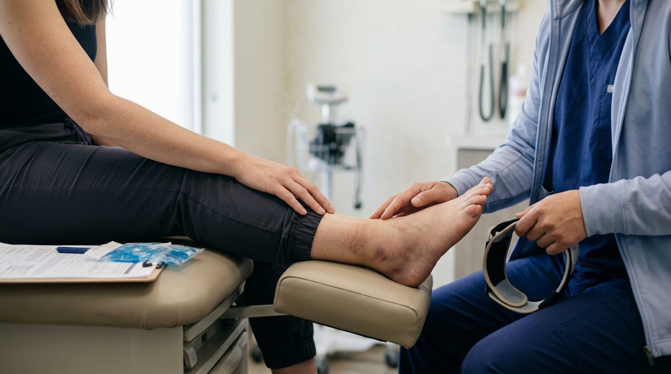
When clinicians evaluate acute injuries to the ankle or midfoot, they do not base their X-ray decisions on the darkness of a bruise. Instead, they rely on validated guidelines such as the Ottawa Ankle Rules.
Clinical guidelines look for specific functional markers:
A person with a non-displaced fracture might have minimal bruising. Another person with a mild lateral ligament stretch might develop widespread dark ecchymosis across the entire foot. Bruise appearance alone cannot determine whether bone is broken.
In sports medicine, clinicians classify muscle contusions by functional movement rather than visual size. The primary metric used to evaluate a crushed muscle belly is the loss of active range of motion:
A mild contusion on a thin area of the body can produce a very large surface bruise. A severe contusion deep in the thick quadriceps muscle might produce a small surface mark despite major functional limitation.
Blood is a liquid that responds to physical forces, structural boundaries, and gravity. Once blood leaves the vascular system, it does not remain locked in place. It migrates along tissue planes and settles where space allows.
When you spend hours sitting or standing, gravity pulls leaked blood downward toward the lowest anatomical points. This pooling effect is especially noticeable in the lower extremities.
If you twist your lateral ankle ligaments, the bleeding begins near the ankle bone. Over the next two to four days, the dark purple discoloration often migrates downward into the heel, arch, and toes.
Seeing your toes turn purple days after an ankle twist does not mean you have newly injured your foot. It simply means gravity has drawn the original pool of blood into the loose tissues of your lower foot.
Muscles are organized into anatomical compartments wrapped in tough sheets of connective tissue called fascia. Blood cannot easily penetrate through intact fascial barriers.
Instead, escaping fluid flows along the path of least resistance between muscle sheaths. In severe hamstring or calf muscle tears, extensive ecchymosis often emerges several inches below the actual tear site.
The visible bruise might show up behind the knee or near the Achilles tendon, far from the torn fibers. Relying on bruise location to find the exact source of damage can easily mislead you.
To evaluate an injury safely at home without relying on superficial color, you can use a simple observational framework. This approach combines four key areas of observation: Mark, Mechanism, Function, and Trend.
Look closely at the physical characteristics of the skin and underlying tissue.
Consider the exact physical force that caused the injury.
Direct impacts commonly cause contusions and large surface bruises. Twisting forces are more likely to damage internal ligaments, tendons, or cartilage without immediate surface discoloration.
Assess how well the injured body part can perform its normal daily tasks. Function is almost always more important than appearance.
If you have a massive purple bruise but can move freely with minimal pain, the injury is likely superficial. If you have no bruise but cannot bear weight or lift your arm, you need a professional medical assessment.
Observe how the injury changes over several hours and days.
A normal injury shows stable or gradually improving symptoms over forty-eight to seventy-two hours. An escalating symptom pattern requires prompt clinical evaluation, regardless of how small the surface bruise appears.
Two people can experience the exact same physical impact and display radically different degrees of bruising. Several biological, pharmacological, and lifestyle factors influence how easily blood vessels leak.
As we age, our skin and subcutaneous tissues undergo natural structural changes. The protective layer of subcutaneous fat thins out, leaving small blood vessels closer to the surface with less physical cushioning.
At the same time, the body produces less collagen and elastin, which makes capillary walls more fragile. Because of these changes, older adults frequently develop large, dark bruises from minor impacts that would not leave a mark on a younger person.
Many widely used medications reduce the body's ability to form blood clots or thin the blood. When capillaries experience minor stress, bleeding continues longer before a clot seals the leak.
Common substances that increase bruising include:
If you take any of these substances, you will likely develop larger, darker bruises from everyday bumps. Never discontinue prescribed medications on your own simply because of a bruise. Always consult your prescribing doctor first.
Unexplained or unusually extensive bruising can sometimes reflect internal health conditions rather than simple mechanical trauma. Disorders that affect blood platelets, liver function, or clotting protein synthesis can lead to spontaneous bleeding.
If bruises appear across your body without any known impact or fall, you should discuss this pattern with a physician. Understanding systemic recovery factors is a core focus of recovery science articles.
While most bruises resolve on their own, certain symptom combinations point to serious underlying complications that require immediate emergency evaluation.
Acute compartment syndrome is a limb-threatening medical emergency. It occurs when rapid bleeding and swelling build up immense pressure inside a rigid fascial compartment of the arm or leg.
This trapped pressure cuts off local blood supply and starves muscles and nerves of oxygen. If untreated, compartment syndrome can lead to permanent muscle damage or loss of the limb within hours.
Watch for the clinical warning signs of compartment syndrome:
A severe limb injury can progress to compartment syndrome with only a modest-looking surface bruise. Never wait for a bruise to look worse if you are experiencing severe, escalating pain and tightness.
You should seek prompt, same-day medical attention if your bruise is accompanied by any of the following clinical signs:
Managing a straightforward bruise involves supporting your body's natural cleanup processes without disrupting tissue repair.
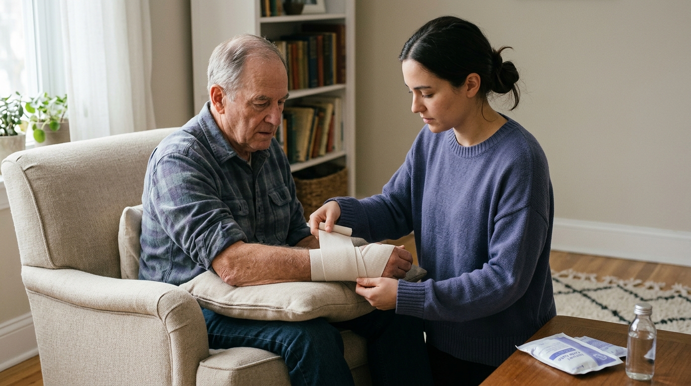
During the first 24 to 48 hours following a minor soft tissue injury, focus on comfort and symptom control:
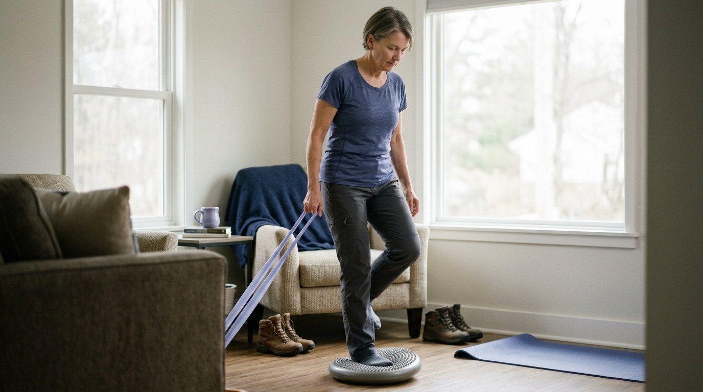
Once the initial 48-hour acute window passes, prolonged complete immobility can actually slow down recovery. Gentle, pain-free active movement stimulates lymphatic flow and encourages venous return.
Light muscle contractions help pump broken-down red blood cells and metabolic waste out of the tissue. Start with simple active range of motion exercises within a comfortable, pain-free boundary.
As tenderness subsides, you can gradually reintroduce normal walking, light resistance, and daily movement. For structured guidance on returning to movement safely, check out our mobility and movement resources.
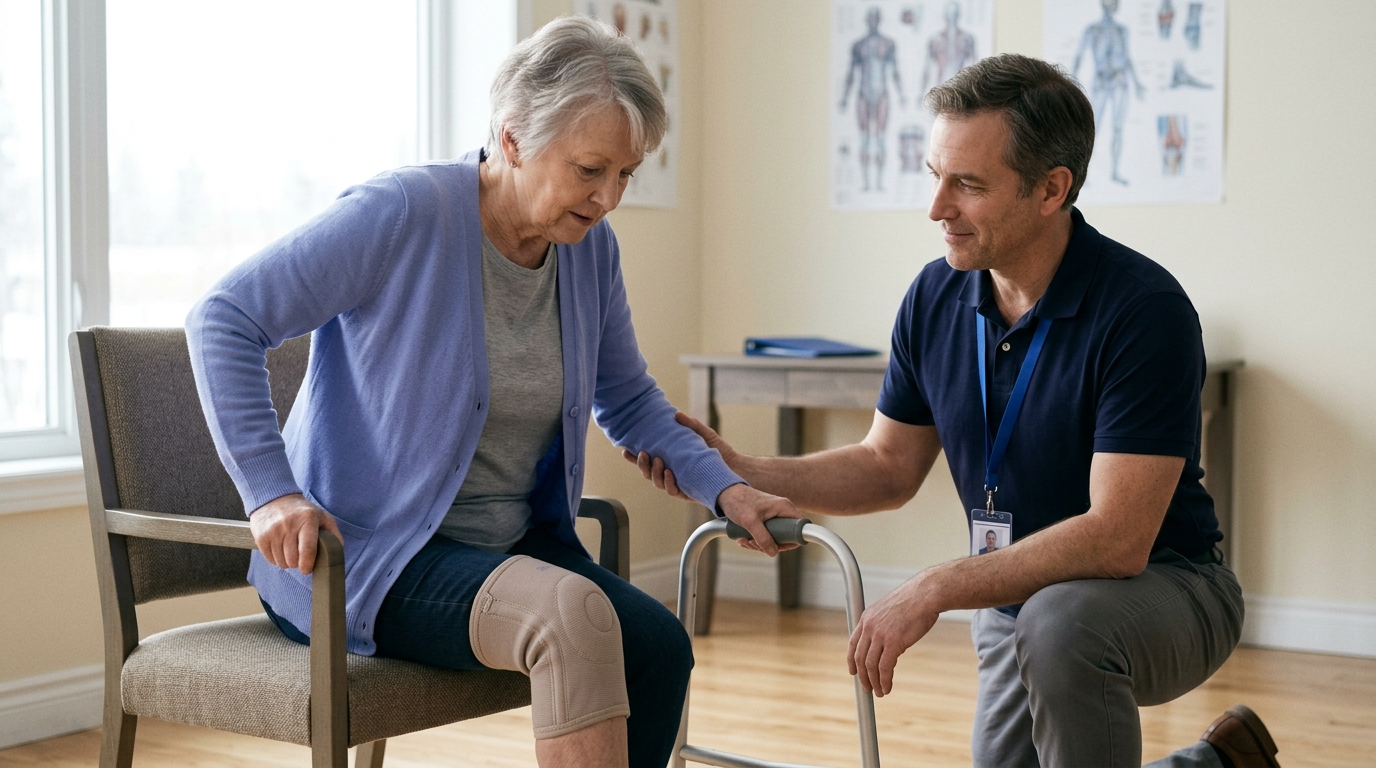
When you visit a doctor, physical therapist, or orthopedic specialist for an injury, asking targeted questions can help clarify your diagnosis.
Here are practical questions to bring to your clinical appointment:
Having these clear questions prepared ensures you leave your appointment with a realistic, actionable recovery plan. You can read more about structured clinical transitions in our injury recovery articles.
When blood pools densely in a confined tissue space, it forms a hematoma. As the body begins repairing the area, it deposits fibrin and organizes the pooled blood into a firm clot.
This clot can feel like a distinct, hard marble or knot beneath the skin. Over several weeks, the body slowly breaks down this clot and softens the tissue. If the lump becomes increasingly painful, red, or hot, you should have a clinician evaluate it.
You should avoid applying direct heat packs or soaking in hot baths during the first 48 hours after an acute injury. Heat causes local blood vessels to dilate and increases blood flow to the area.
Applying heat too early can restart internal bleeding and cause swelling to expand. Save gentle warmth for later stages of recovery when you want to loosen stiff muscles and stimulate circulation for tissue remodeling.
After a major contusion or large hematoma, large quantities of red blood cells are destroyed in the tissue. The iron contained in hemoglobin can be stored locally as a pigment called hemosiderin.
Hemosiderin staining can leave a light brown or tan discoloration in the skin that lasts for months. This residual staining is harmless and gradually fades as the skin cells turn over and clear the remaining iron.
Spontaneous bruising without a known impact can occur, particularly in older adults with fragile capillaries. However, frequent, widespread, or large unexplained bruises can also indicate medication side effects, vitamin deficiencies, or blood clotting disorders.
If you notice unexplained purple or blue patches appearing on your torso, back, or legs without any bump or fall, you should consult a primary care physician for blood tests.
A bruise is simply visible evidence that blood has escaped damaged microvessels beneath unbroken skin. It does not provide a dependable measure of injury severity, nor does it establish whether bones, ligaments, or deeper muscle fibers are intact.
Visual color progression cannot be used as an accurate clock to date an injury. Furthermore, gravity often pulls pooled blood into distant areas, causing discoloration far from the original site of trauma.
When evaluating a physical setback, look beyond surface color. Pay attention to how the injury happened, whether you can bear weight and move freely, and how your symptoms trend over time. If you experience extreme pain, tight swelling, numbness, or cannot use the limb, seek a prompt medical assessment.

Use ReboundBody resources to understand common recovery stages, rehab terms, movement limits and strength rebuilding. Each guide is designed to make a complex comeback easier to understand.


Read practical guidance on injury recovery, rehabilitation, mobility and rebuilding strength as you work your way back to activity.
Explore Resources