
Fracture surgery recovery involves biological bone healing stages and a structured rehabilitation plan to help patients safely restore mobility and strength.





Recovery after fracture surgery is not simply waiting for an incision to close or an implant to hold. It is a biological process where living bone tissue repairs itself under protected conditions, supported by a structured return to physical movement. Surgical fixation stabilizes broken bone fragments, but the surgery itself does not instantly mend the bone.
True recovery requires two parallel tracks working together. The first track is biological bone healing, which restores structural strength and continuity across the fracture site. The second track is physical rehabilitation, which rebuilds joint mobility, muscle strength, balance, and day-to-day functional independence.
Navigating this journey can feel confusing because progress rarely follows a straight line. Instructions that apply to one person, such as walking immediately after an ankle repair, may be unsafe for someone recovering from a complex knee or hip fracture.
This guide examines the biological phases of bone repair, the principles behind surgical stabilization, and the evidence regarding weight-bearing and exercise progression. It also outlines the individual factors that shape recovery timelines, providing clear information to help you understand your postoperative plan.
To understand postoperative guidelines, it helps to understand what happens inside the body after a broken bone is repaired. Fracture healing is a dynamic sequence of cellular events. Orthopedic science identifies three primary, overlapping phases of healing: inflammation, repair, and remodeling.
The inflammatory phase begins the instant a fracture occurs. Blood vessels tear, forming a localized hematoma around the fracture fragments. This blood clot releases signaling proteins that summon immune cells and specialized repair cells to clear damaged tissue. This early inflammation triggers the downstream cascade needed for new tissue growth.
The reparative phase follows over the subsequent weeks. During this period, the body forms new blood vessels and develops a soft callus composed of fibrous tissue and cartilage. As mineral deposition progresses, osteoblasts convert this soft framework into a hard callus made of woven bone. This hard callus acts as a biological bridge across the structural gap.
The final phase is bone remodeling, which can continue for many months or years. During remodeling, bone cells reshape the bulky hard callus along natural lines of mechanical stress. Woven bone is gradually replaced by organized, dense lamellar bone, restoring the internal canal and original architecture.
Surgeons also classify healing into direct or indirect pathways based on how the bone is stabilized:
Surgical implants, whether plates, screws, intramedullary nails, or external frames, provide mechanical stability and maintain anatomical alignment. The AO Foundation established four core principles that guide fracture fixation: restoring anatomy, providing appropriate stability, preserving local blood supply, and enabling early mobilization.
Fixation protects the biology of the healing bone, but the hardware is not a replacement for biological repair. Understanding these basic principles helps clarify why your care team creates specific movement boundaries during your surgical recovery and comeback.
It is common to assume that once an X-ray shows bone union, your recovery is complete. In clinical practice, bone healing and functional recovery are distinct milestones. Bone healing refers specifically to the structural continuity and mechanical strength of the skeleton.
Functional recovery encompasses the return of joint mobility, muscle power, neuromuscular control, balance, and confidence in daily tasks. The bone may be solid, but the surrounding muscles, ligaments, and tendons often require extended rehabilitation to recover from surgery and immobilization.
When a limb is protected after a fracture, surrounding muscles lose volume and strength. Joint capsules tighten, and the nervous system adapts to altered movement patterns. Even after a bone has achieved solid biological repair, a person may still limp, experience stiffness, or struggle with stairs.
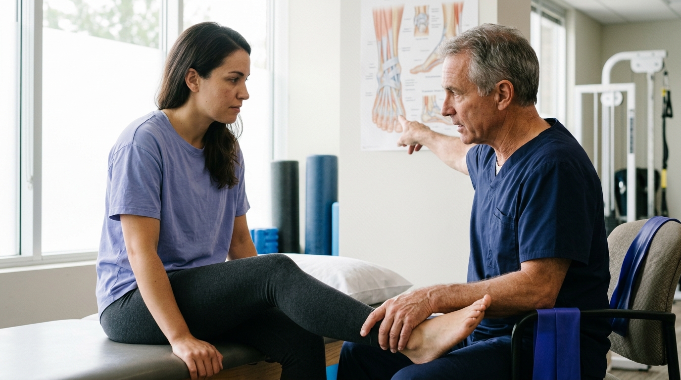
Rehabilitation bridges the gap between a healed bone and a restored body. Clinical literature identifies five primary objectives in post-fracture physical therapy:
A comprehensive program balances tissue protection with progressive exercise. Attempting high-demand activities before muscle capacity is restored can place abnormal stress on adjacent joints. A structured approach to surgery and rehab ensures that physical capacity advances alongside skeletal healing.
One of the most confusing aspects of fracture recovery is the wide variation in weight-bearing instructions. One person might be encouraged to put weight on a broken ankle within days, while another with a broken tibia must remain non-weight-bearing for months. These differences reflect the distinct mechanical demands and stability profiles of different fractures and surgical constructs.
Weight-bearing orders are medical prescriptions calculated to manage mechanical strain at the repair site. Excessive strain can loosen implants or disrupt the forming callus, while total lack of mechanical stimulation can slow bone remodeling. The goal is finding the right mechanical environment for each specific injury.
Clinical guidance illustrates these distinct paths:
There is also an important distinction between permission to bear weight and the physical ability to do so. A surgeon may grant permission for full weight-bearing based on X-ray stability, but pain, muscle weakness, and balance deficits may require crutches or a walker. Following your surgeon's specific weight-bearing orders is essential, because protocols cannot be safely transferred between different injuries.
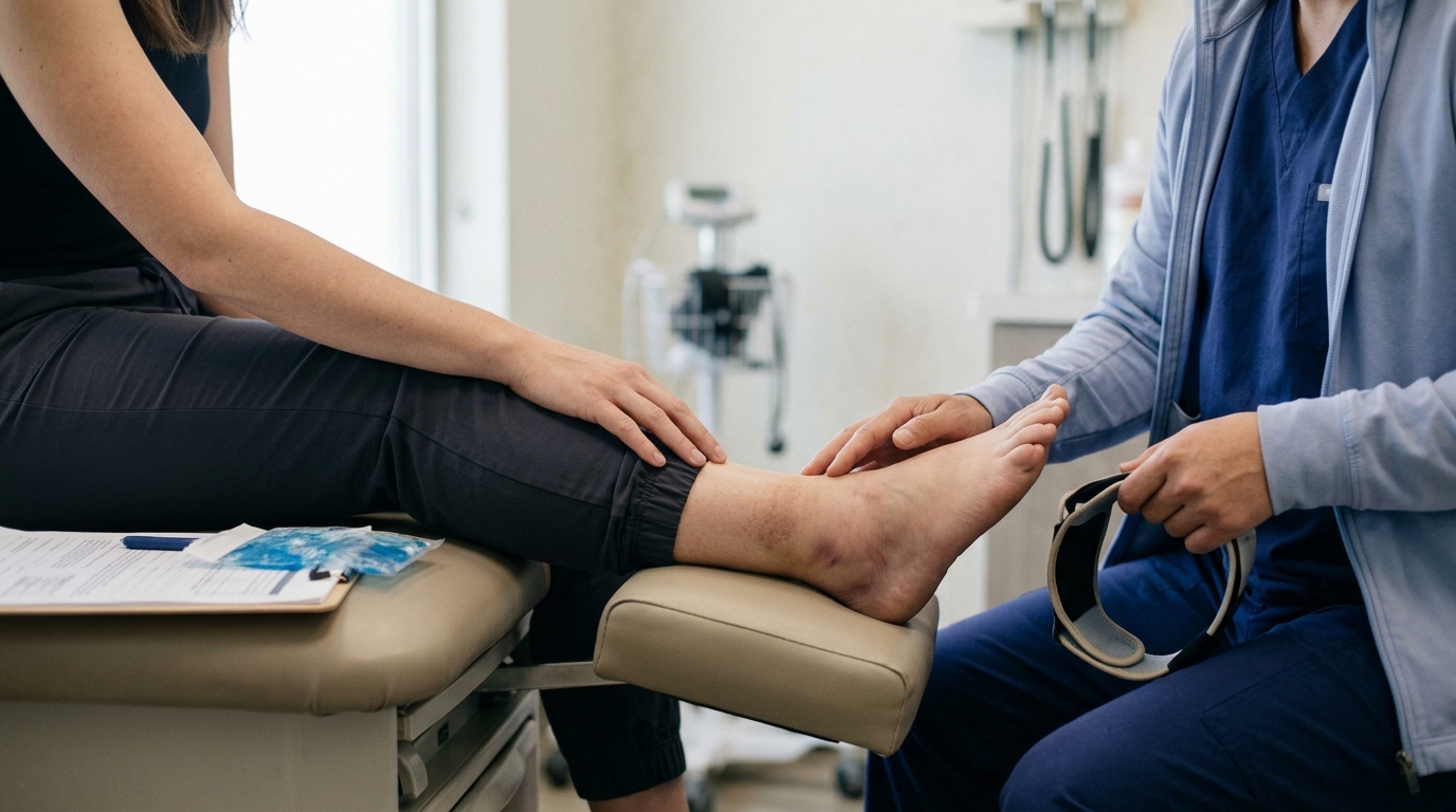
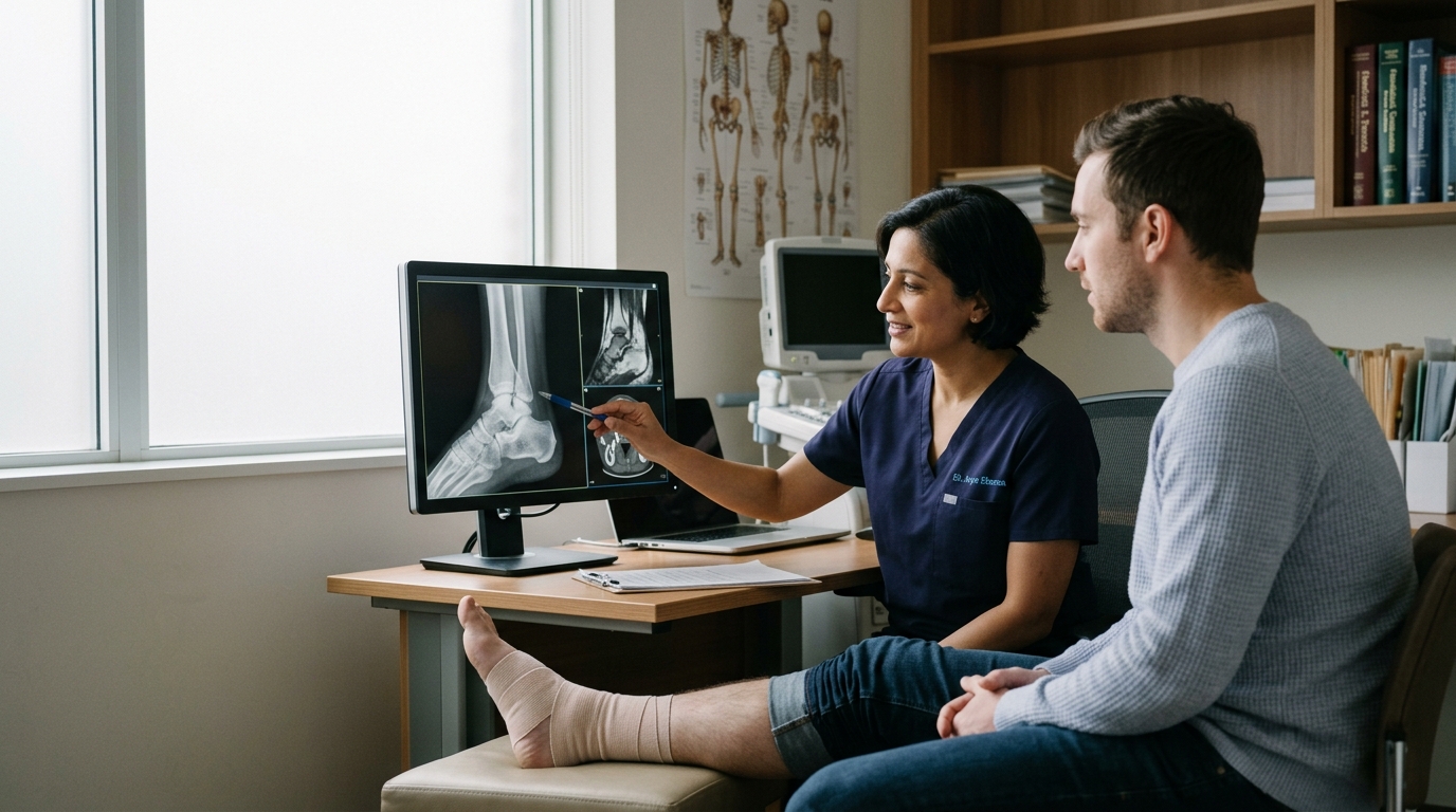
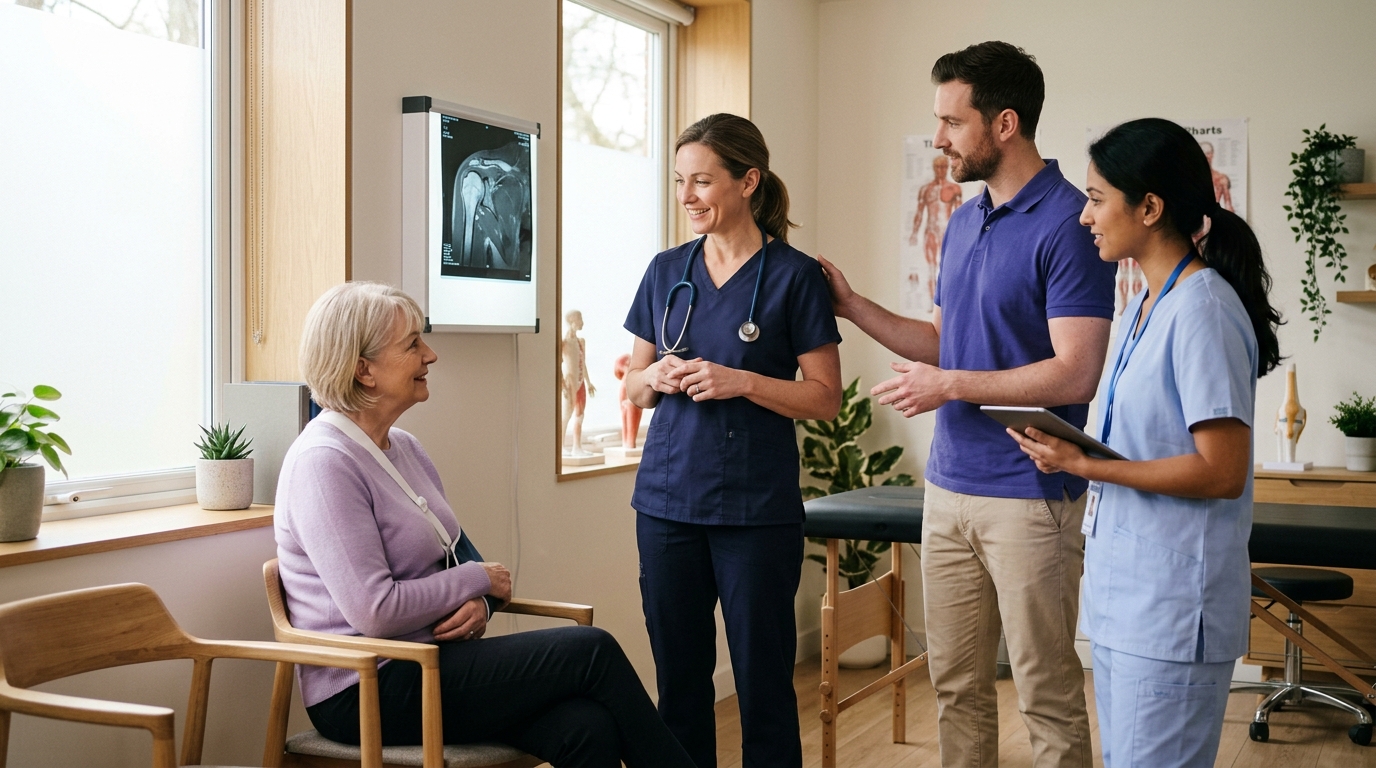
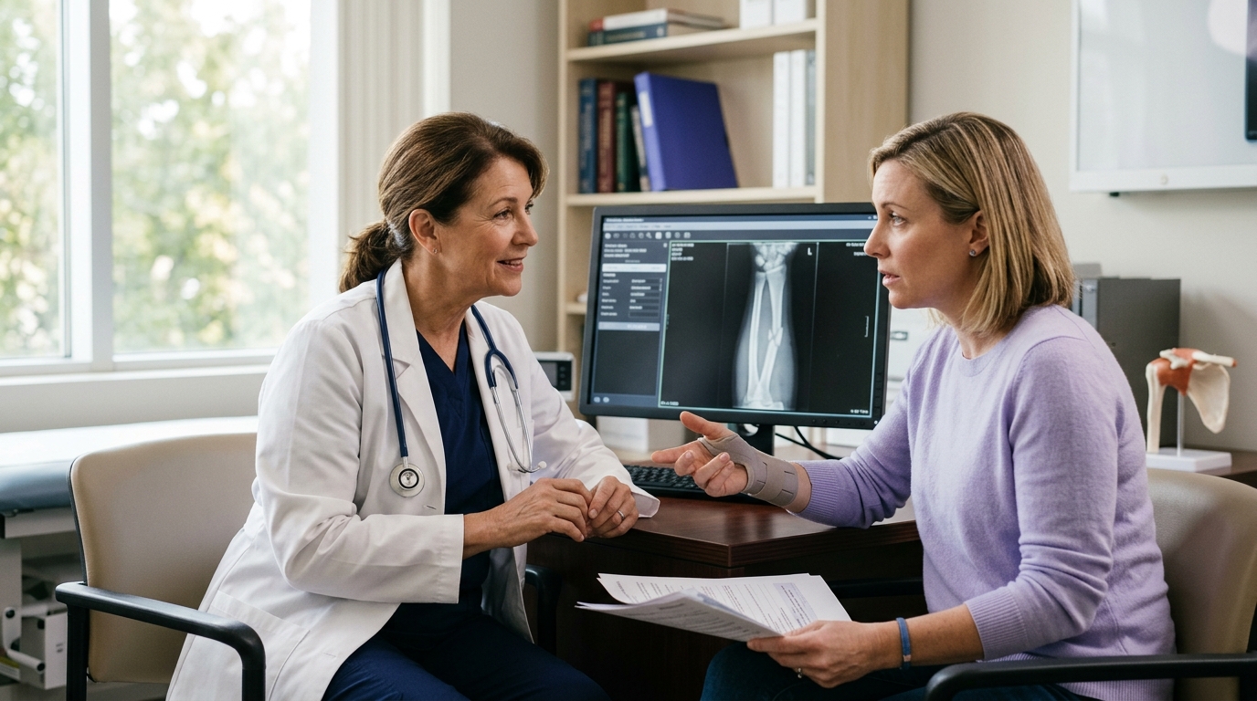
Determining whether a fracture is fully healed is a clinical judgment rather than an automatic calculation. There is no single universal test that confirms bone healing. Instead, orthopedic surgeons combine clinical examinations with radiographic imaging to evaluate progress over time.
Research reviews evaluating fracture healing criteria have identified significant variation across clinical studies. In a systematic evaluation of fracture repair criteria, researchers noted over a dozen distinct clinical benchmarks. Clinicians evaluate a patient's healing trajectory by looking at several complementary factors.
Clinical criteria focus on physical symptoms and functional capacity. Common clinical signs of healing include:
Radiographic criteria rely on serial X-ray views to monitor bone remodeling. Key radiographic markers include:
For specific injuries, such as tibial shaft fractures treated with an intramedullary nail, clinicians may use structured tools like the Radiographic Union Score for Tibia (RUST). The RUST system scores the presence of callus and the visibility of fracture lines across four distinct bone cortices seen on standard X-rays. While helpful, scoring tools are structured aids rather than standalone clearance tests.
Clinical union and radiographic union often progress at slightly different speeds. A patient may feel comfortable walking weeks before an X-ray shows complete disappearance of the fracture line. Conversely, early callus may appear on an image while the site remains tender to physical touch. Evaluating both clinical and radiographic findings ensures safe clearance for physical demands.
Healing timelines are broad estimates rather than rigid schedules. Two people with the same type of fracture may heal at different rates due to biological, mechanical, and lifestyle variables. Understanding these factors provides valuable context for your recovery trajectory.
Injury characteristics play a fundamental role in healing speed. High-energy trauma, such as a motor vehicle collision, causes extensive damage to both the bone and surrounding soft tissues. The periosteum and adjacent muscles supply the blood vessels that deliver oxygen, nutrients, and repair cells to the fracture site.
Open fractures, where the broken bone pierces the skin, carry higher risks of delayed healing and infection due to compromised local vascularity. Highly fragmented (comminuted) fractures require more biological bridge-building than simple, clean breaks.
Patient health and systemic biology also shape bone repair:
Tobacco and nicotine use represent significant, modifiable risk factors for bone healing complications. A comprehensive meta-analysis evaluating over 8,000 adult fracture and fusion cases found that smokers had roughly 2.2 times the risk of delayed union or nonunion compared to non-smokers. Furthermore, among patients whose bones successfully united, the healing process took an average of 27.7 days longer in smokers.
A separate meta-analysis on non-pathological fractures reported an odds ratio of 2.50 for nonunion among smokers. Nicotine causes peripheral vasoconstriction, reduces tissue oxygenation, and inhibits osteoblast activity.
These variables represent cumulative risk factors rather than guaranteed outcomes. Having a health condition or experiencing a severe injury does not mean a fracture will fail to heal. Discussing your overall health profile with your surgical team helps them customize your rehabilitation timeline and monitoring plan.
When a fracture does not heal within the expected timeframe, clinicians evaluate the condition for delayed union or nonunion. While these terms can cause anxiety, they represent distinct clinical states with clear management paths.
Delayed union means that a fracture is healing more slowly than anticipated for that specific bone, injury pattern, and patient. It does not mean the fracture will never heal. Delayed union is an indicator that biological repair is lagging, often requiring continued immobilization, modified weight-bearing, nutritional optimization, or temporary activity adjustments. Research reviews generally identify delayed union when healing progress stalls between roughly three and six months post-injury.
Nonunion represents a formal cessation of the bone repair process. The biological mechanisms driving repair have halted, leaving a persistent structural gap. A widely referenced regulatory definition from the Food and Drug Administration (FDA) characterizes nonunion as a fracture that is at least nine months old and has shown no radiographic signs of healing progression for three consecutive months.
In clinical practice, surgeons do not wait an arbitrary nine months to intervene if healing has stalled. If serial X-rays over several months show no callus formation, or if a patient experiences persistent pain and instability, the care team will investigate earlier.
Nonunions are generally classified into two mechanical and biological categories:
Understanding the difference between delayed union and nonunion helps keep recovery milestones in perspective. Slower progress is a signal for careful clinical review and targeted adjustments rather than an irreversible setback.
Rehabilitation following fracture surgery is goal-oriented rather than governed by a rigid calendar. Progression depends on tissue healing, pain levels, joint range of motion, and muscle control. A structured physical therapy program moves through four sequential stages, matching mechanical loading to the bone's biological capacity.
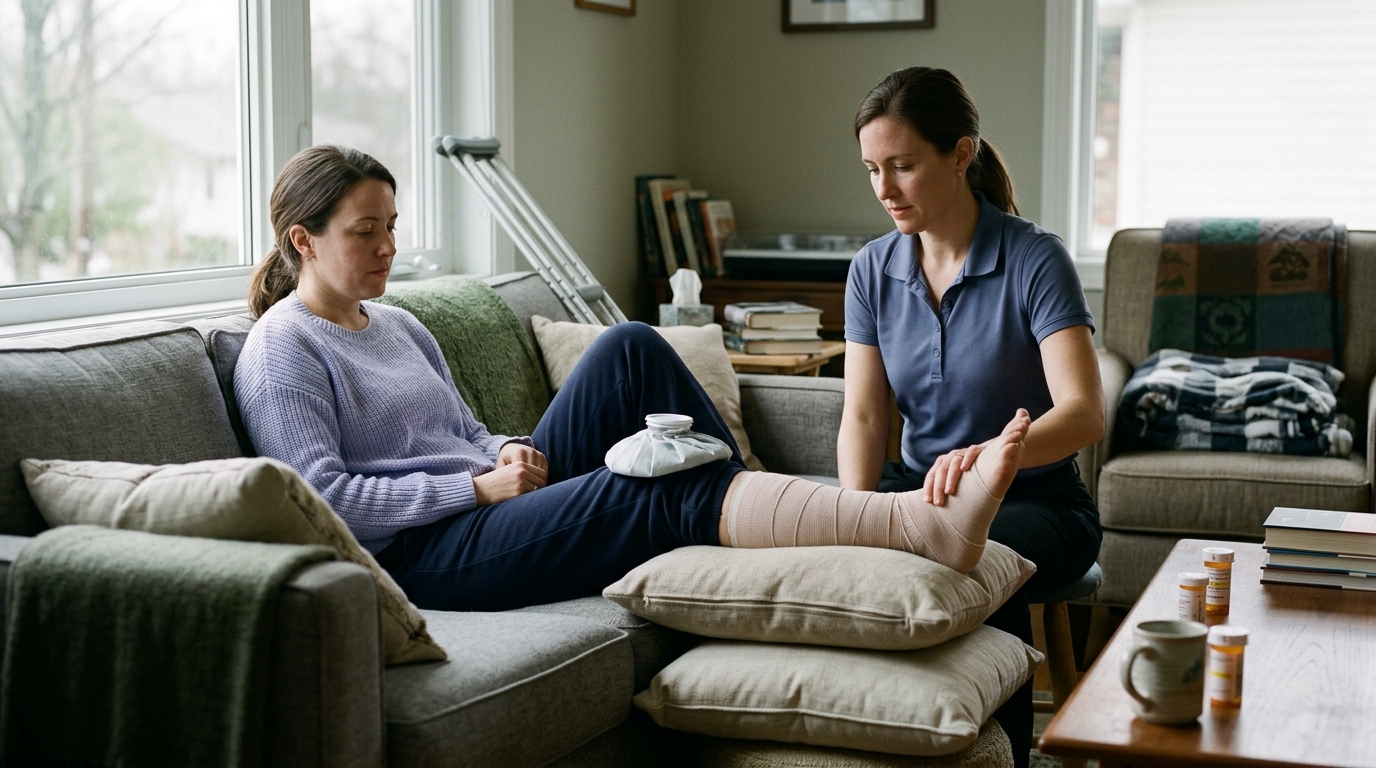
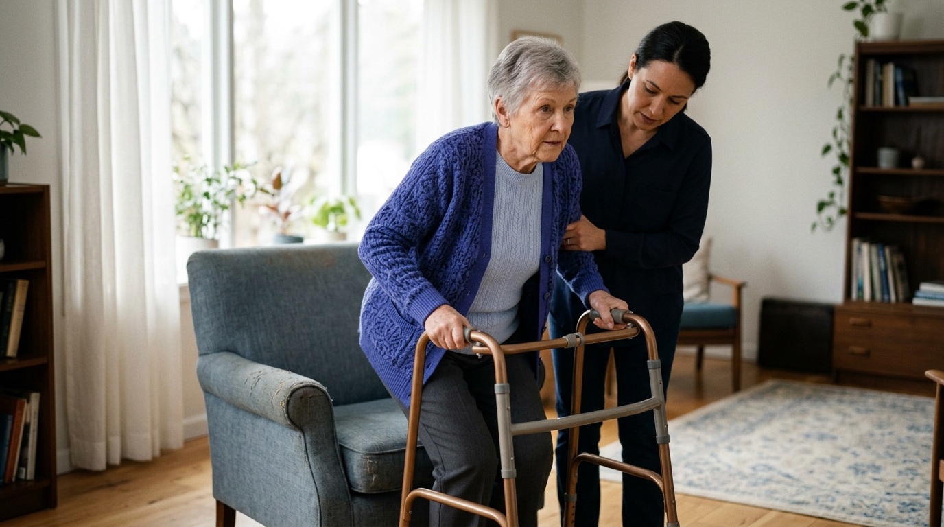
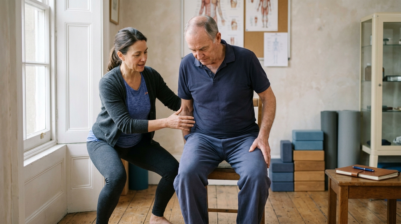
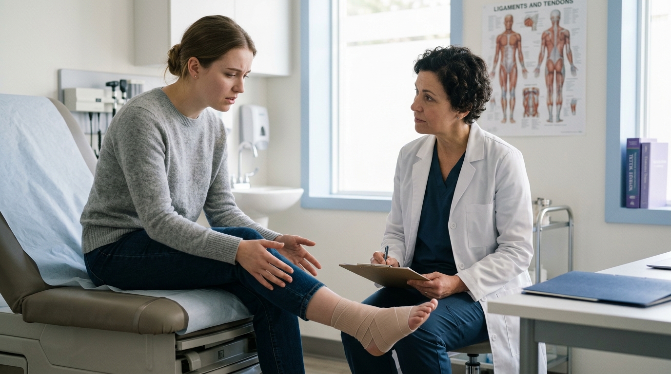
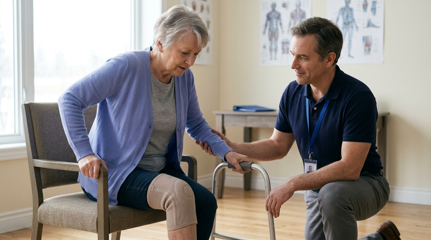
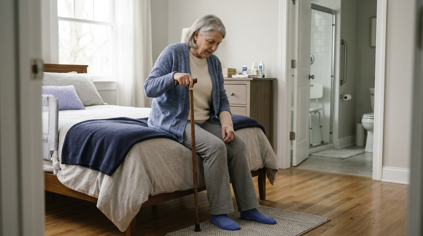
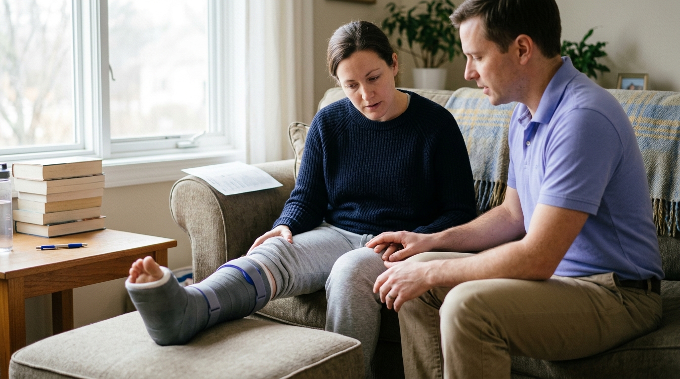
The primary goal of the initial postoperative weeks is protecting the surgical construct while keeping you safe and independent. Therapy focuses on managing acute pain, reducing local swelling, and mastering mobility aids like crutches, walkers, or knee scooters.
During this phase, patients learn safe techniques for bed mobility, standing transfers, and navigating stairs without violating weight-bearing restrictions. Gentle movements of non-immobilized adjacent joints, such as moving the toes and hips during ankle recovery, are encouraged to maintain circulation.
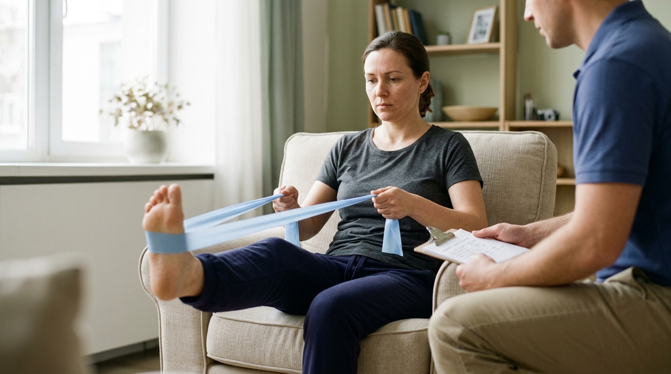
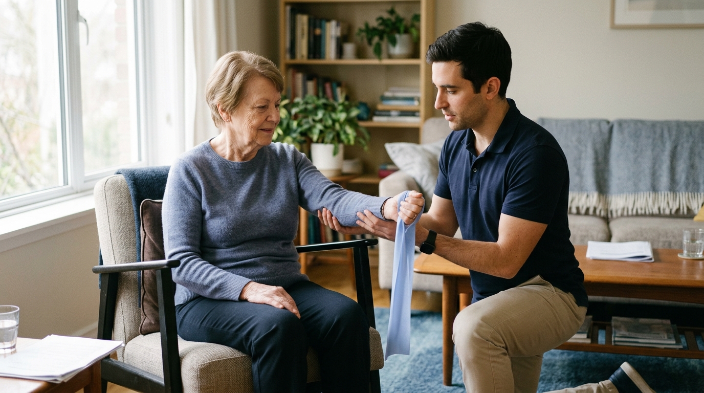
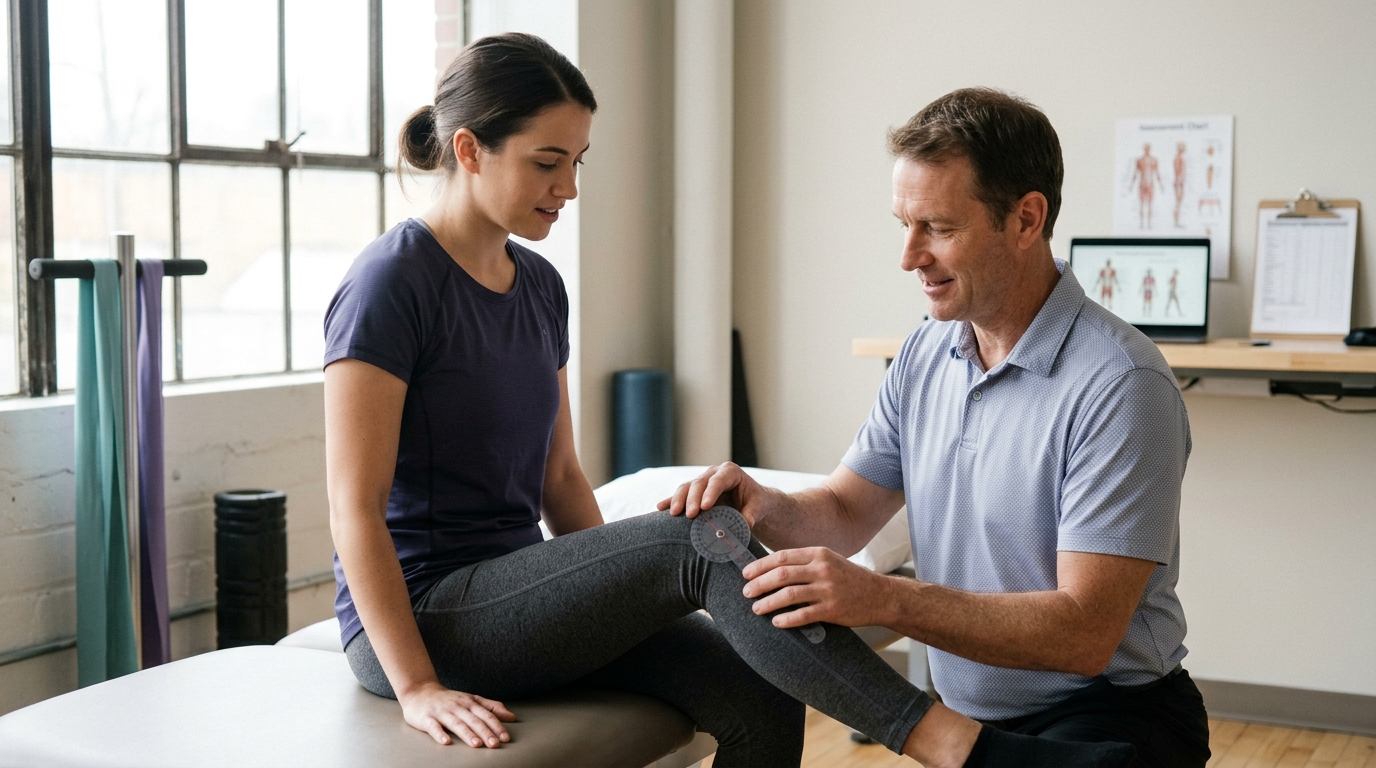
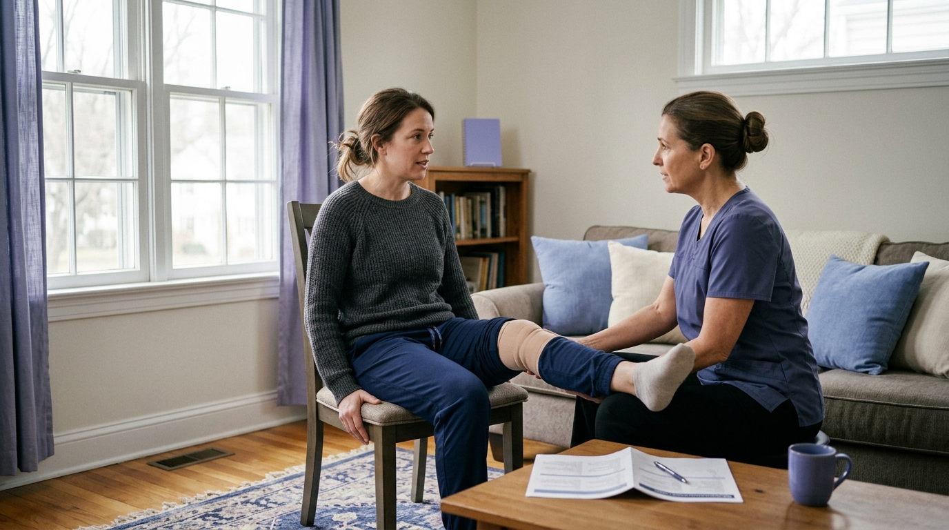
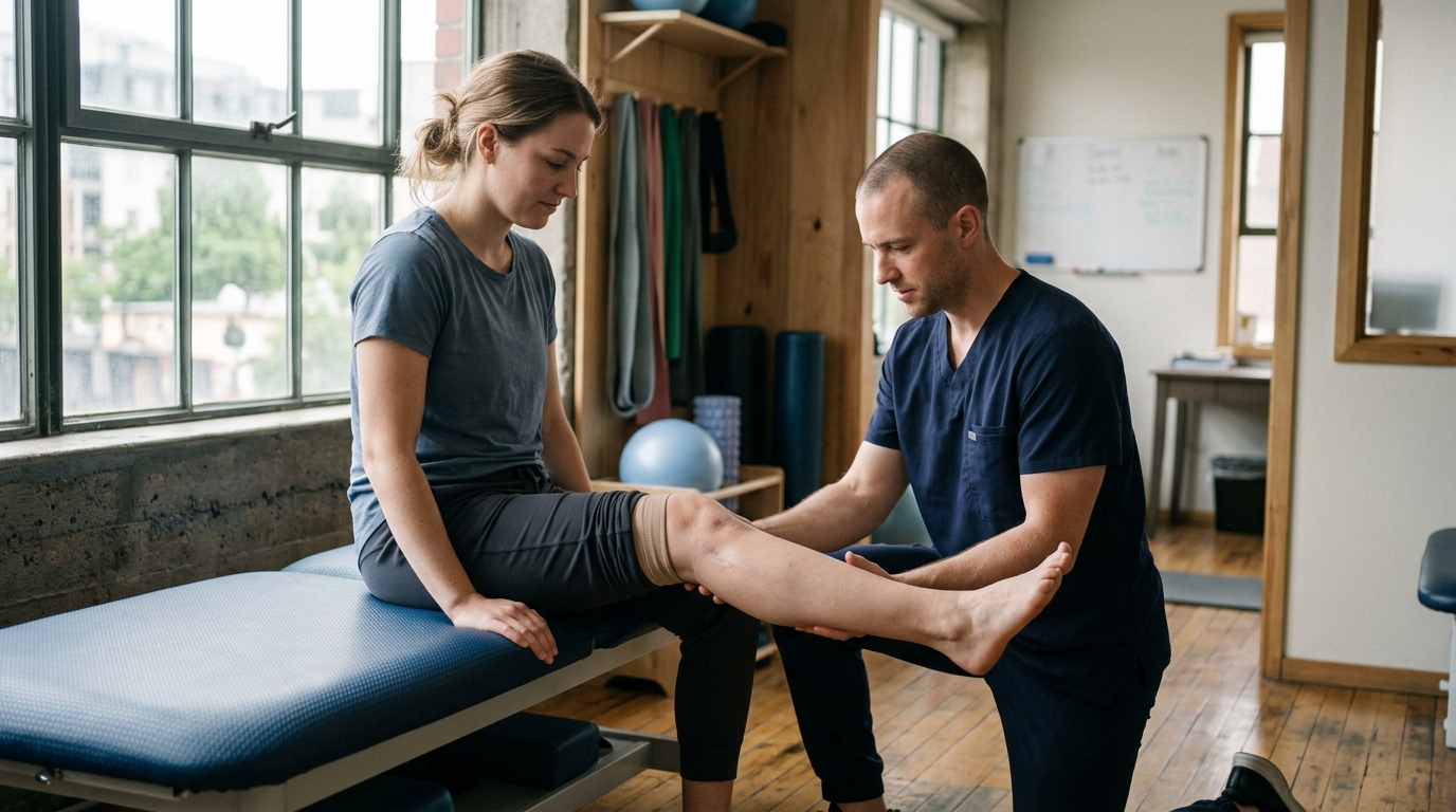
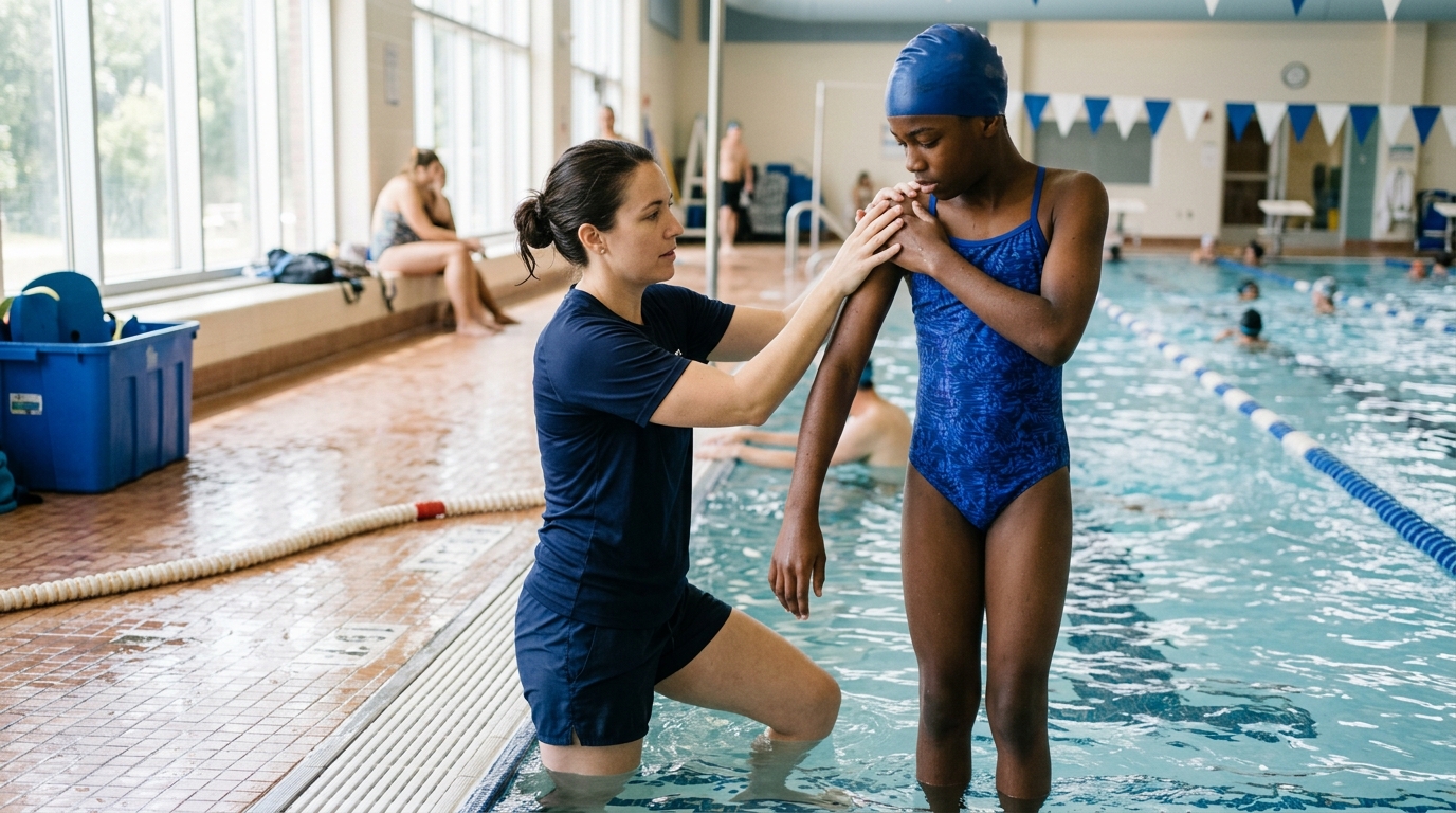
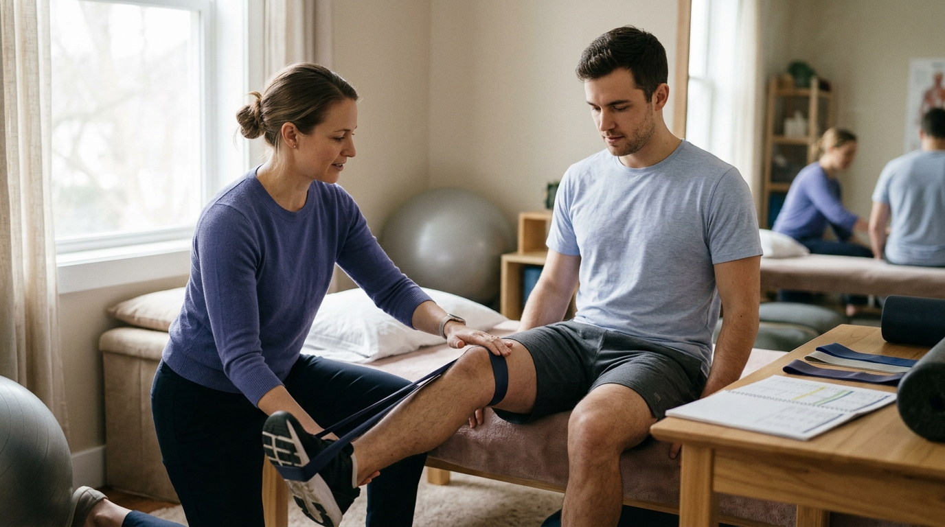
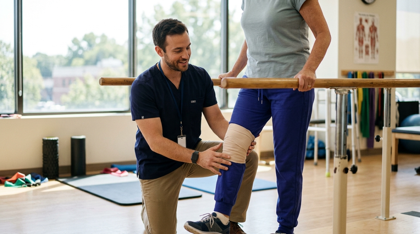
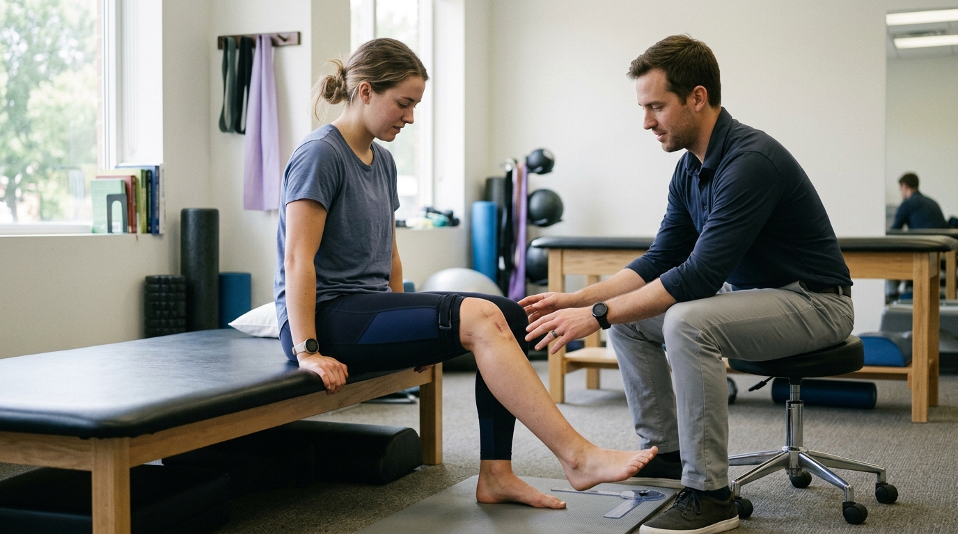
Once acute surgical swelling subsides and initial tissue healing occurs, rehabilitation introduces targeted range-of-motion exercises. Depending on surgical stability, this may involve passive movement guided by a therapist, active-assisted exercises, or self-directed mobility routines.
Muscles that cross the fracture site often experience neural inhibition after trauma. Gentle isometric exercises, which contract muscles without moving the healing joint, help re-engage dormant muscle fibers without creating shear strain across the fracture. Exploring principles of rehabilitation, mobility, and movement during this stage helps prevent long-term joint stiffness.
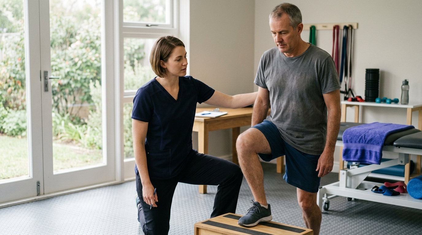
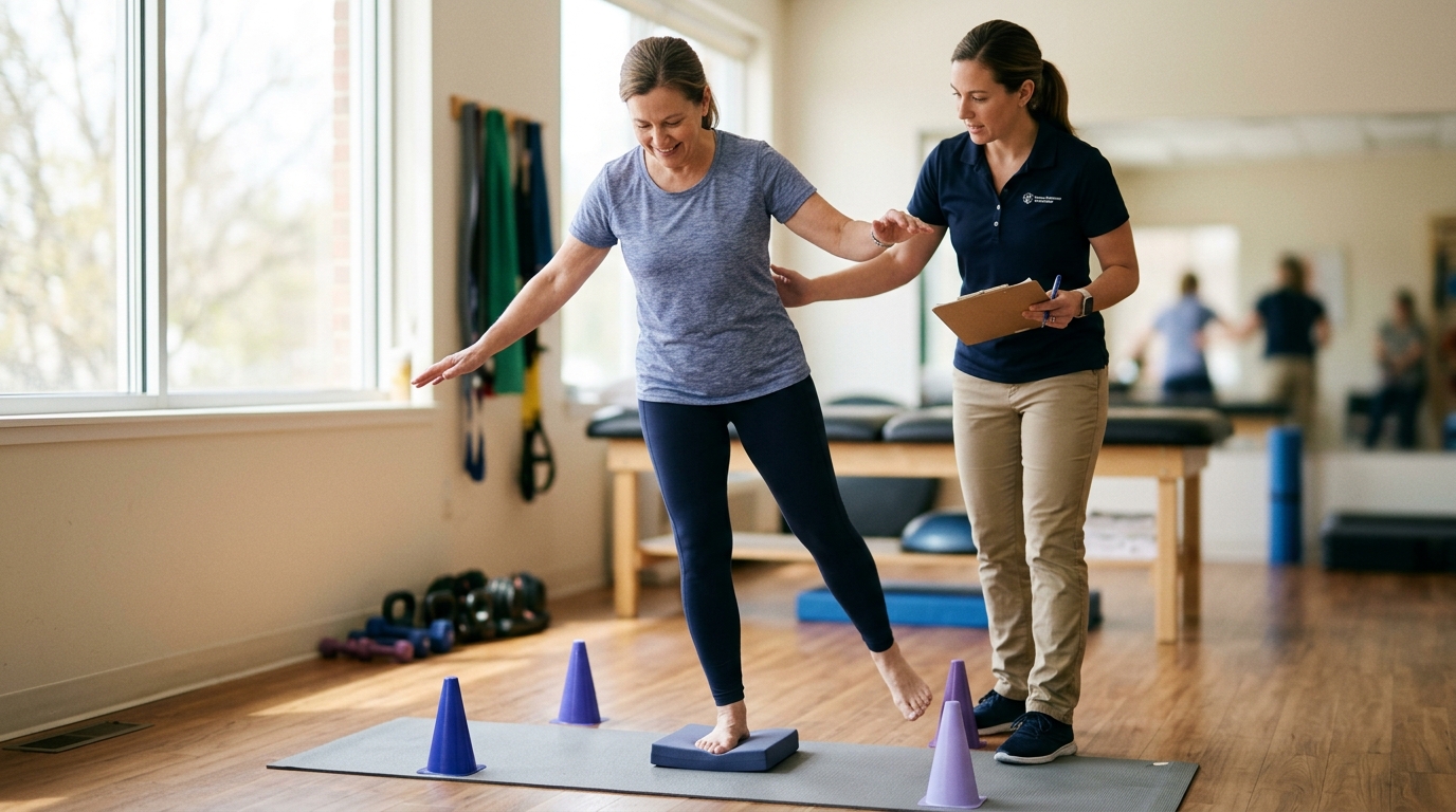
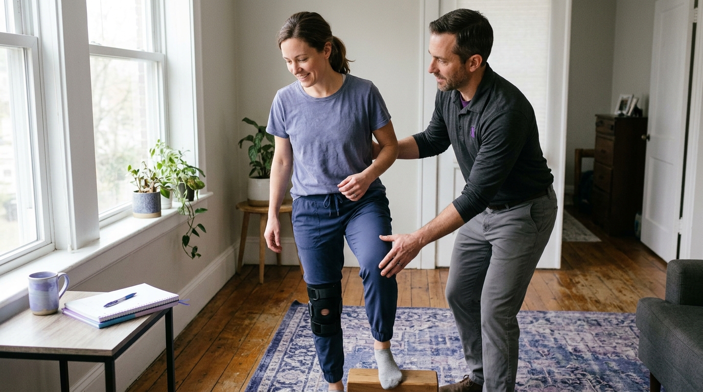
When clinical evaluation and follow-up imaging confirm adequate callus formation, the care team approves progressive mechanical loading. Weight-bearing is advanced systematically, moving from partial loading toward full weight-bearing based on comfort and stability.
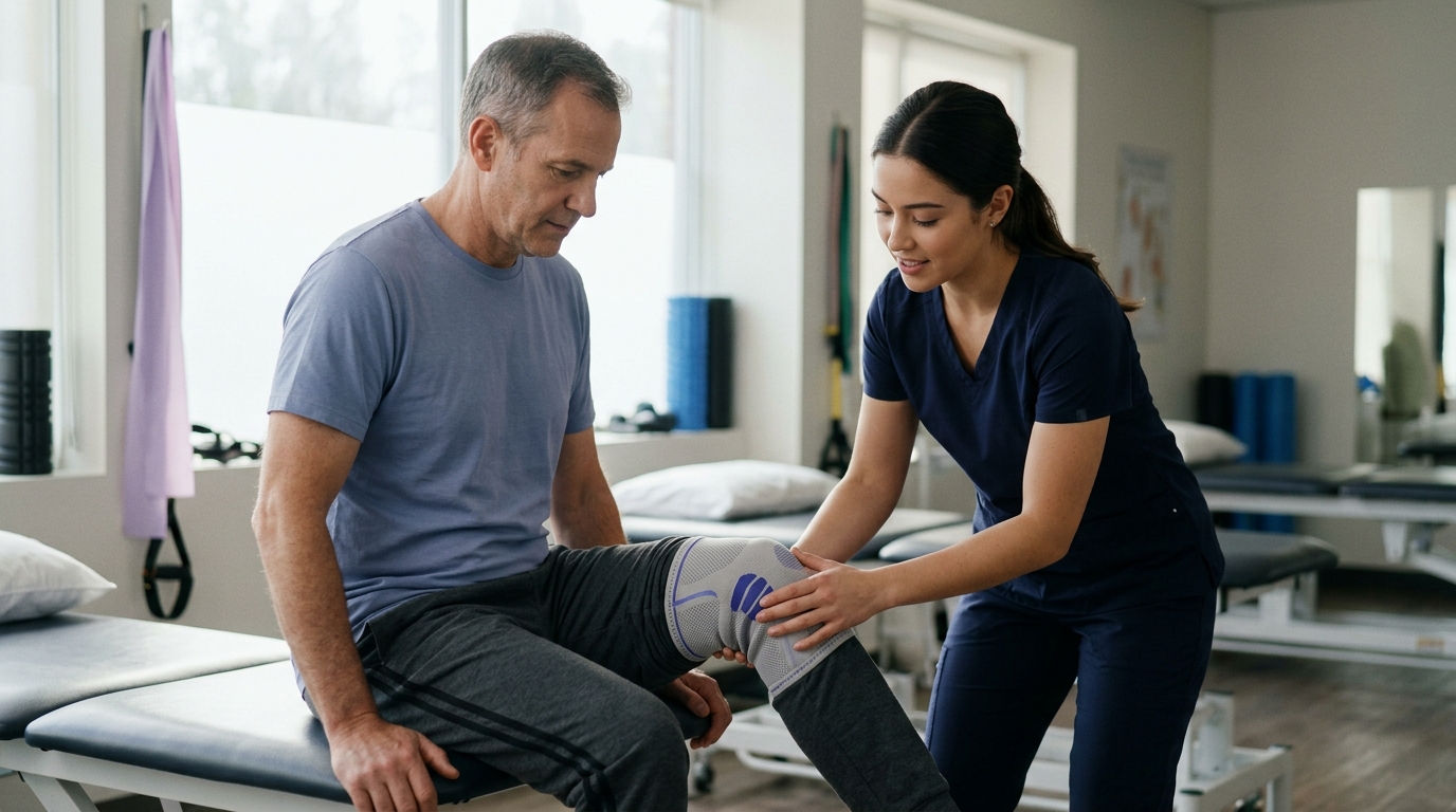
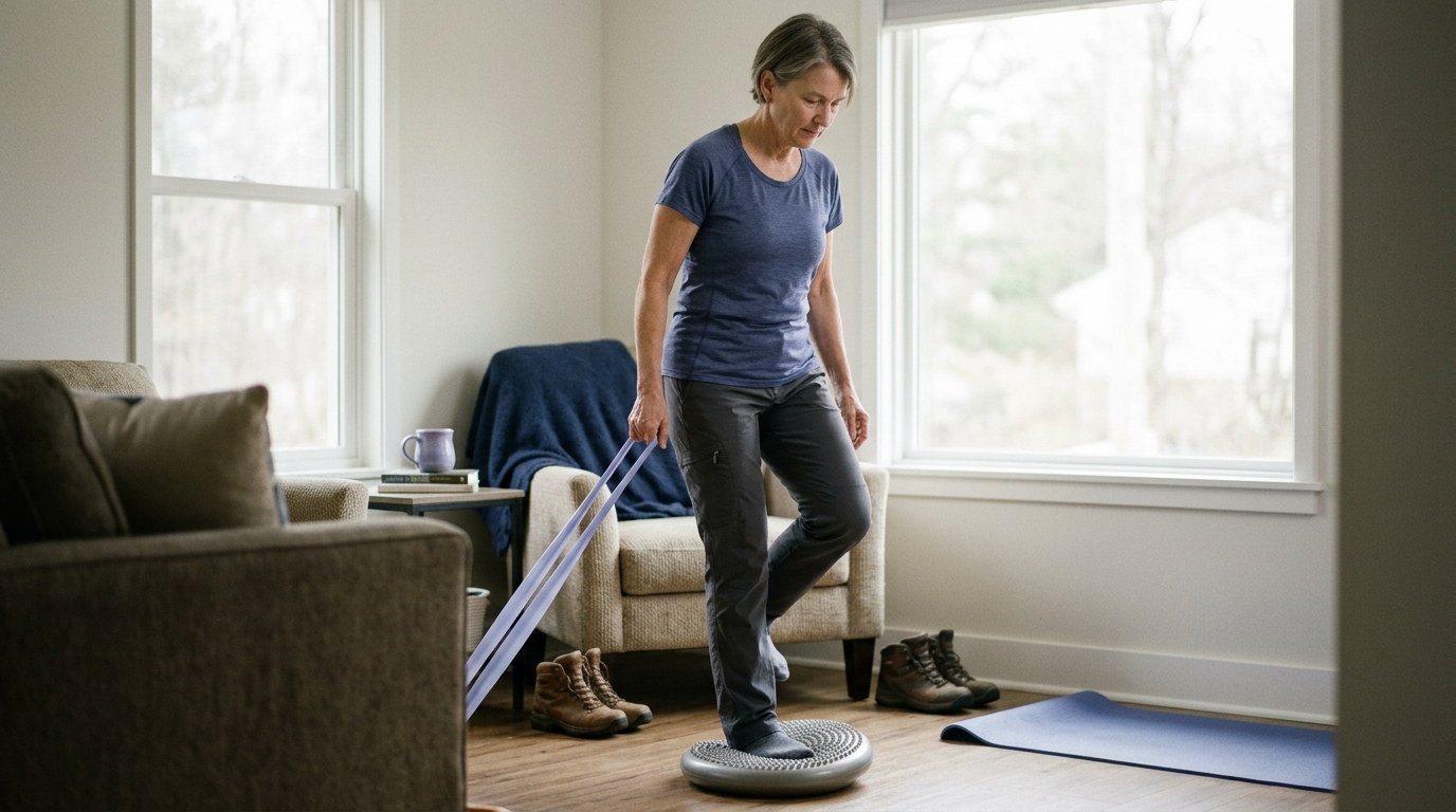
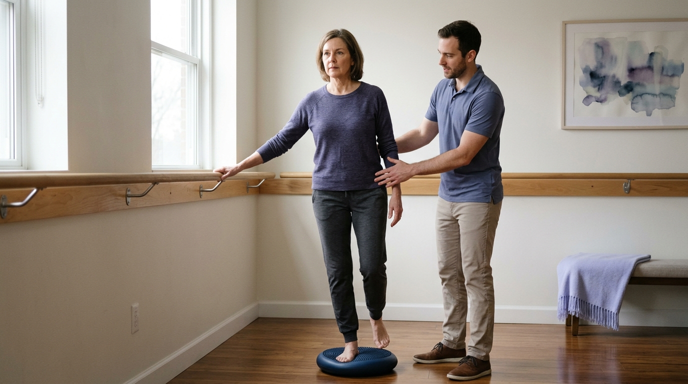
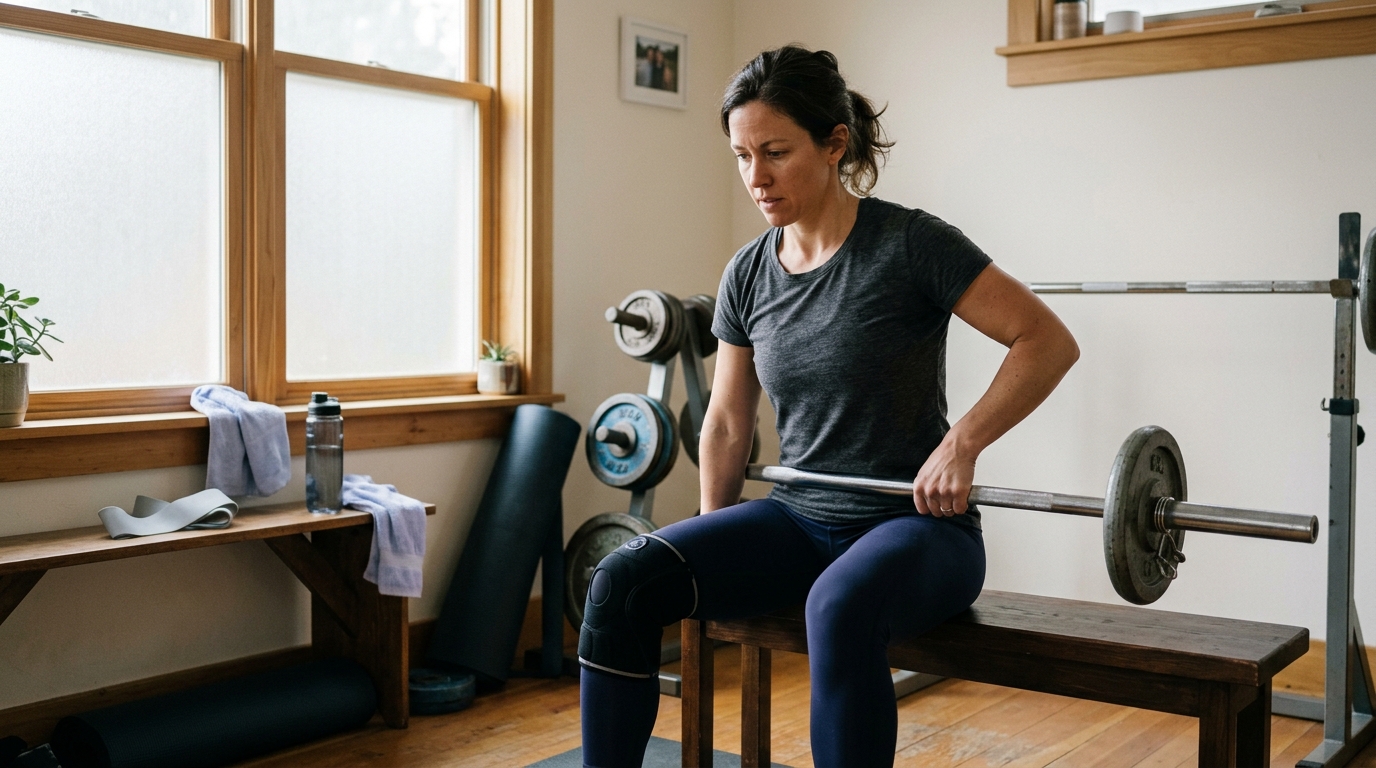
Strength training progresses from low-resistance, closed-chain exercises to higher-demand movements. Gait retraining is a central focus during this stage. Therapists work to eliminate compensatory limping, balance discrepancies, and postural asymmetries that developed during periods of limb protection.
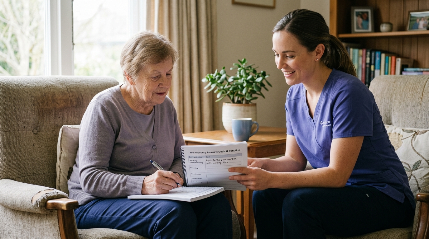
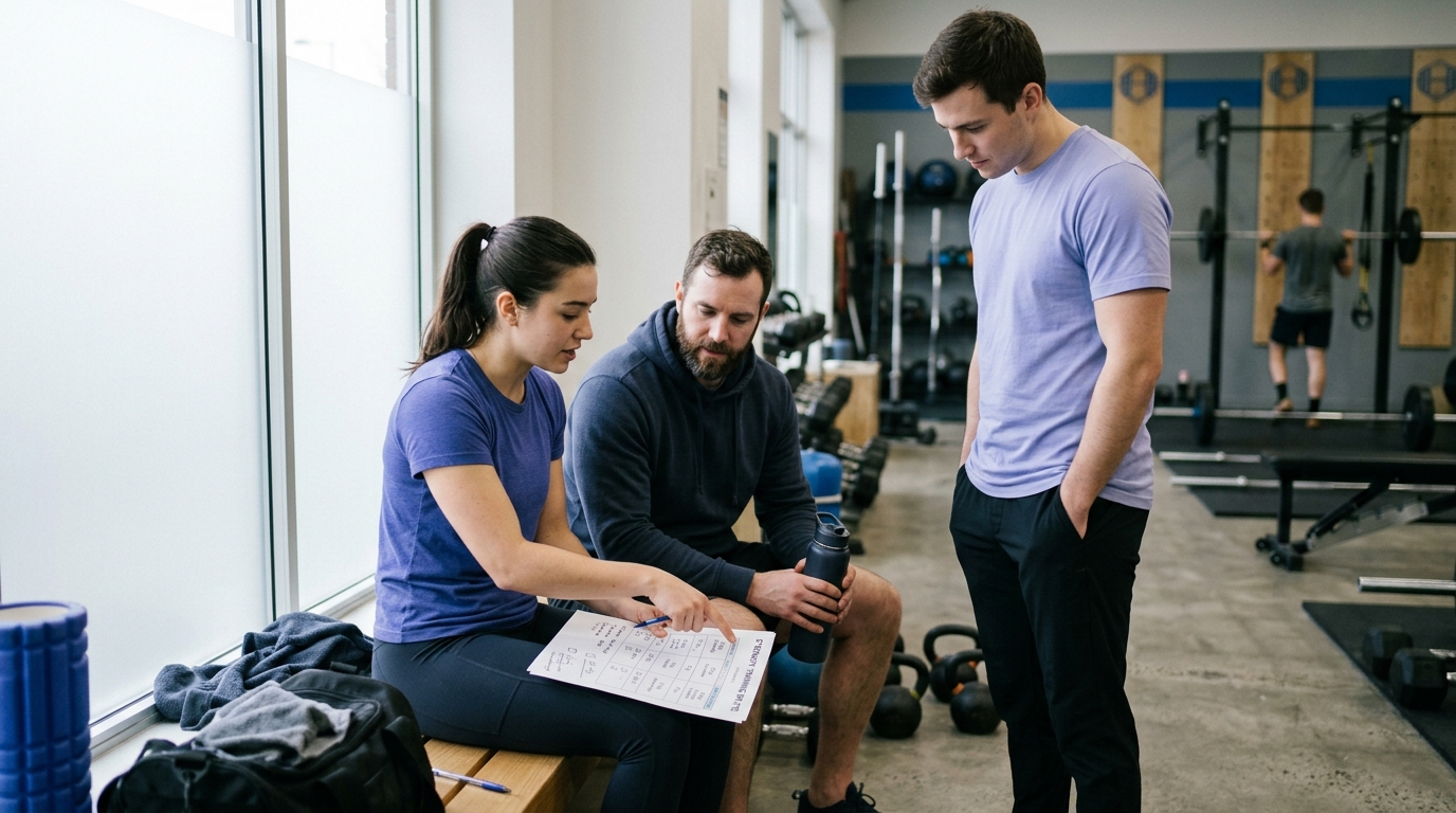
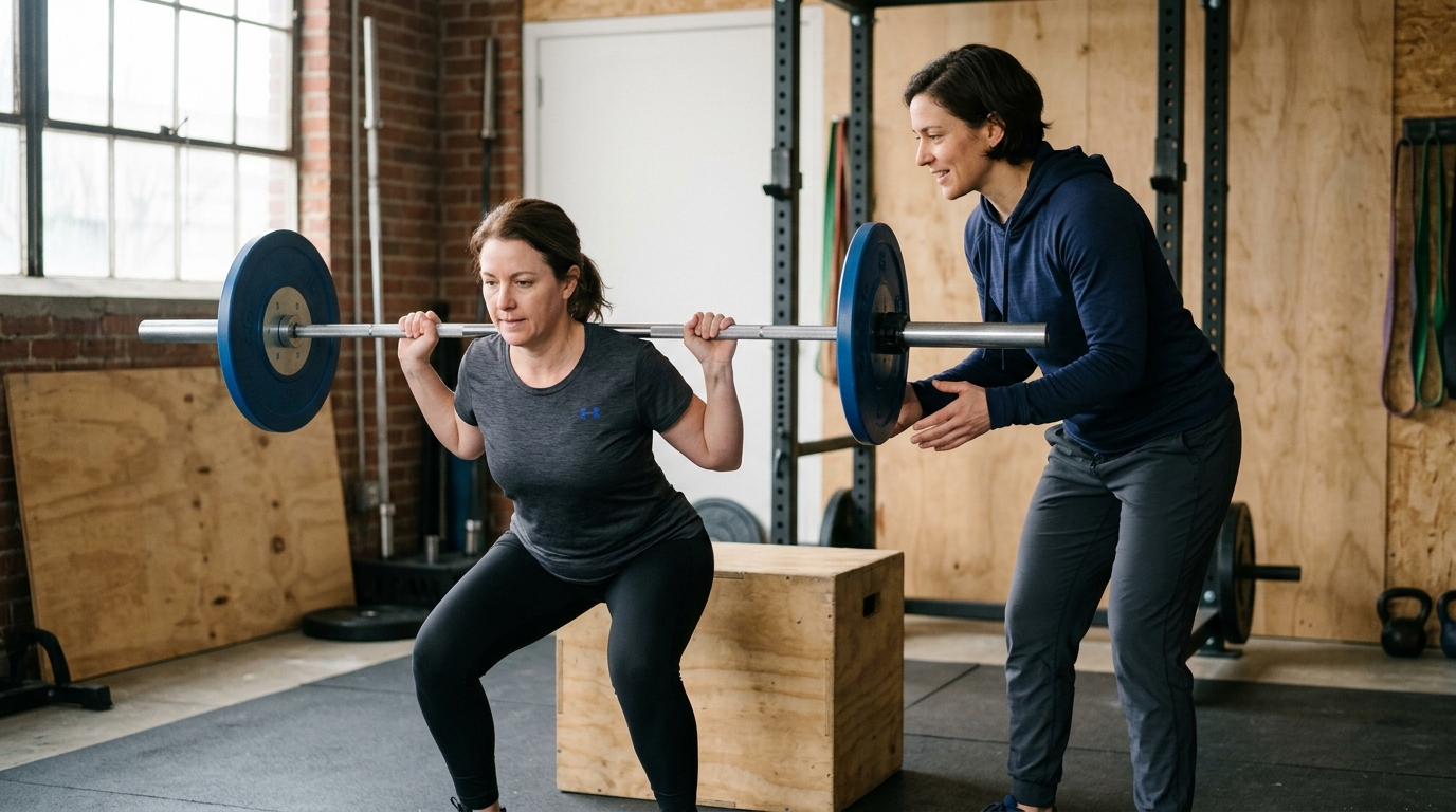
The final phase prepares you for everyday independence, occupational tasks, and recreational activities. Training shifts toward functional movement patterns, multi-directional balance, coordination, and muscular endurance.
Returning to higher-impact activities, such as running or sports, represents a separate clinical decision from basic daily walking. Clearance for impact loading requires solid radiographic union, full joint range of motion, symmetrical limb strength, and successful completion of functional movement assessments. Understanding the underlying recovery science ensures that you transition into high-demand recreation safely.
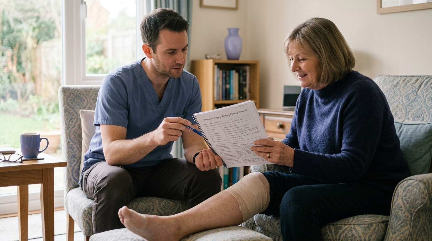
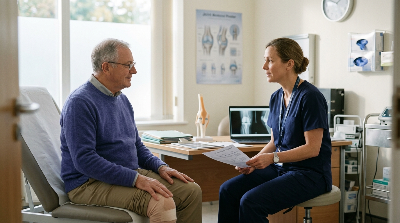
Because fracture patterns and surgical constructs vary, open communication with your care team ensures you understand your individualized boundaries. Bringing specific questions to follow-up visits helps clarify your expectations and keeps your
rehabilitation on track.
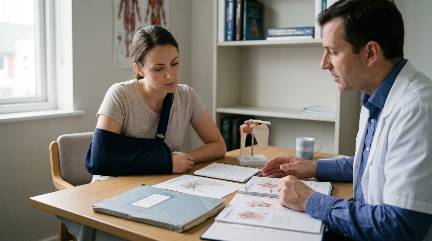
Consider discussing these practical questions at your next appointment:
Engaging proactively with your physical therapist and orthopedic surgeon creates a collaborative environment. Clear communication helps you progress with confidence while respecting your body's biological repair timeline.
Most modern orthopedic implants are made of titanium alloys or high-grade stainless steel. These materials are biocompatible and rarely trigger standard security metal detectors, although high-sensitivity scanners at airports may occasionally detect them.
Routine hardware removal is generally not necessary unless the implants cause localized tendon irritation, skin prominence, or persistent discomfort after bone healing is complete. If hardware removal is ever considered, surgeons typically wait until the fracture is fully consolidated and remodeled to prevent refracture.
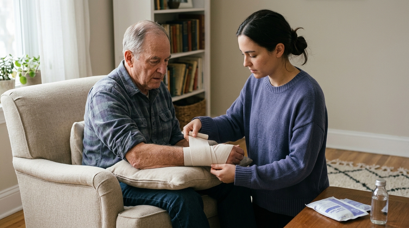
Post-traumatic and postoperative swelling frequently lasts longer than bone union itself. Surgical repair disrupts local lymphatic vessels and microcirculation, while periods of limb protection reduce the natural pumping action of contracting muscles.
As you increase daily standing, walking, and physical loading, fluid naturally pools in dependent extremities due to gravity. This residual swelling gradually improves as vascular networks regenerate, muscle pump efficiency is restored through mobility and movement, and tissue remodeling progresses.
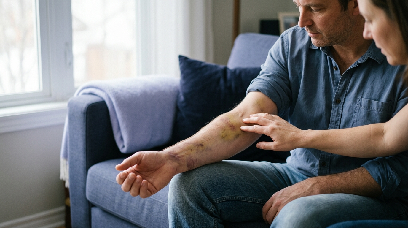
While mild soreness, stiffness, and fluctuating swelling are standard parts of recovery, certain warning signs require immediate medical attention:
Adjunctive therapies, such as capacitive coupling or pulsed electromagnetic field bone stimulators, are sometimes prescribed for nonunions or high-risk fractures to encourage cellular activity. Similarly, targeted nutrition supports the metabolic building blocks required for collagen synthesis and mineralization.
However, neither stimulators nor dietary supplements replace progressive mechanical loading and physical rehabilitation. Dynamic muscle contractions and guided weight-bearing provide the essential mechanical signals needed to restore bone density, joint mobility, muscle power, and neuromuscular coordination.
Surgical fixation provides the mechanical framework for recovery, but the living biology of bone repair determines the timeline. Healing unfolds through overlapping stages of inflammation, callus formation, and remodeling. Surgical hardware stabilizes fragments in correct alignment, creating the protected biological environment needed for new bone to form.
Bone healing and functional recovery are complementary milestones. While an X-ray demonstrates structural continuity, physical rehabilitation restores joint mobility, muscle strength, balance, and daily independence.
Weight-bearing restrictions and rehabilitation timelines are individualized prescriptions based on your fracture pattern, surgical stability, and personal health profile. By working closely with your surgical and physical therapy team, you can protect your repair and steadily rebuild functional capacity.

Use ReboundBody resources to understand common recovery stages, rehab terms, movement limits and strength rebuilding. Each guide is designed to make a complex comeback easier to understand.


Read practical guidance on injury recovery, rehabilitation, mobility and rebuilding strength as you work your way back to activity.
Explore Resources