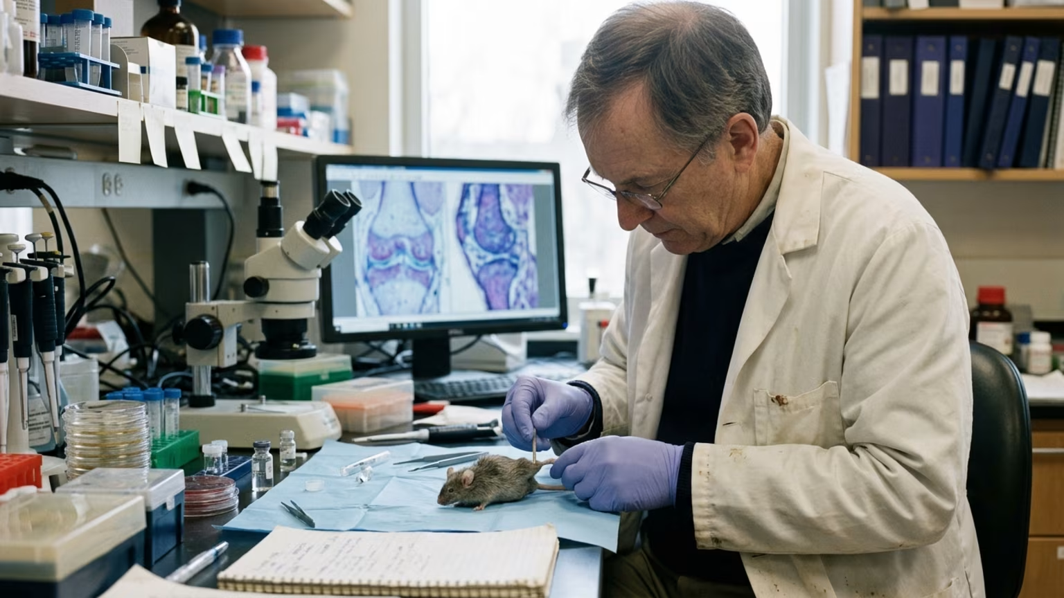

A 2026 Stanford study found that blocking the enzyme 15-PGDH promoted cartilage regeneration in older mice. Learn why this research requires human trials.





Navigating physical recovery often involves interpreting complex clinical research that appears in mainstream news. In 2026, a highly discussed study published in the journal Science introduced new findings regarding joint tissue. A Stanford Medicine-led team reported that blocking the enzyme 15-PGDH promoted cartilage regeneration in older mice. The researchers detailed how this experimental approach affected joint tissue in a strict laboratory setting. This research offers an interesting look into the biology of tissue repair and cellular function.
The formal study is titled “Inhibition of 15-hydroxy prostaglandin dehydrogenase promotes cartilage regeneration.” It appeared in volume 391 of the widely respected science publication. The specific findings and laboratory data can be located in issue 6789 on page 1053. The entire research initiative was guided by senior authors Helen Blau and Nidhi Bhutani. Helen Blau is a Stanford professor of microbiology and immunology. Nidhi Bhutani serves as an associate professor of orthopaedic surgery.
Lead authors Mamta Singla and Yu Xin Wang also contributed heavily to the published laboratory findings. During the testing phase, researchers used a small-molecule 15-PGDH inhibitor. This specific inhibitor was delivered either by injection into the abdomen or directly into the knee. The team then carefully observed the physical changes in the joint structure over time. Tracking these changes allowed the researchers to measure the direct physical impact of the inhibitor.
The Stanford researchers reported thicker cartilage across the joint surface in the treated older mice. The regenerated tissue was specifically described by the team as hyaline articular cartilage. This is the smooth cartilage needed for healthy and pain-free joint movement. These structural changes mark an important observation in the ongoing study of joint biology. Understanding how tissues respond to chemical changes helps researchers design better future treatments.
Before investigating joint health directly, the Stanford research group studied how 15-PGDH levels affect other physical tissues. Their prior work had linked 15-PGDH inhibition to increased muscle mass and endurance in old mice. This earlier research laid the necessary foundation for examining how the same enzyme interacts with aging joints. It demonstrated a biological pathway that the research team wanted to evaluate further in a different bodily system. Finding overlapping mechanisms in different tissues is a common step in medical research.
Researchers describe 15-PGDH as an enzyme that breaks down prostaglandin E2 within the body. They refer to this specific enzyme as a gerozyme. A gerozyme is simply defined as an enzyme that becomes more abundant as physical bodies age. Understanding this natural aging mechanism is vital for individuals focusing on active aging and prevention strategies. Managing expectations about how our bodies change requires accurate information about these cellular processes.
In mouse knee cartilage, the team found that 15-PGDH levels roughly doubled with age. This significant increase suggested the enzyme might play a direct role in joint health deterioration over time. The previous success in muscle tissue prompted the team to apply a similar inhibitory approach to joint cartilage. They wanted to see if artificially lowering the enzyme level could alter the joint environment favorably. Testing this theory required careful observation and controlled animal models.
The 2026 study tested the inhibitor on a separate mouse model of knee injury to gather functional data. In this specific animal model, the inhibitor was given twice a week for four weeks after the initial injury occurred. Untreated control mice developed osteoarthritis within four weeks of their injury. By contrast, treated mice were substantially less likely to develop the degenerative condition. Comparing these two groups provided researchers with clear data on the inhibitor's effectiveness in mice.
The physical outcomes for the treated mice were notably different from the untreated control group. Treated mice moved more normally during the four-week observation period. They also placed more weight on the paw attached to the injured leg. These functional improvements aligned closely with the positive structural changes observed in the joint tissue. Measuring functional movement is a critical step in evaluating any joint-related treatment.
The laboratory outcomes prompted strong reactions from the primary study authors. Nidhi Bhutani said the cartilage regeneration in aged mice took the team by surprise. She formally called the effect remarkable for the broader field of joint research. Bhutani also stated that existing cartilage cells changed their gene-expression patterns during the study. These cellular changes indicate a fundamental shift in how the tissue operates under the influence of the inhibitor.
Helen Blau described the laboratory findings as a new way of regenerating adult tissue. She noted that the approach holds significant clinical promise for future scientific study. Blau highlighted that the cartilage regeneration occurred through changes in existing cartilage cells rather than through stem cells. This specific distinction provides a new perspective on how different musculoskeletal tissues heal naturally over time.
It is important to clearly identify the subjects tested in this specific medical study before drawing conclusions. The reported cartilage-regeneration findings come entirely from mice and laboratory-treated human tissue. The researchers did successfully test human cartilage removed during total knee replacement surgery. However, the human-tissue experiment was conducted strictly outside the body in a controlled laboratory environment. The study absolutely did not test cartilage regeneration in treated living patients.
During the laboratory test, human tissue was exposed to the enzyme inhibitor for exactly one week. After this brief exposure, the treated tissue showed fewer cells expressing 15-PGDH. The samples also demonstrated lower activity of genes associated with cartilage degradation and fibrocartilage. Researchers reported early signs of articular-cartilage production in the treated laboratory samples. Measuring these microscopic changes helps scientists understand the fundamental cellular mechanics of joint tissues.
While these cellular changes are interesting, they do not show what would happen inside a living human joint. The study did not test how human bodies would process the inhibitor systemically. It also did not report any human pain outcomes or mobility improvements following treatment. The findings strictly represent early laboratory results rather than a finished clinical therapy ready for patient use.
For active adults managing joint stiffness, understanding early scientific research can provide helpful educational context. However, the headline-level promise of cartilage regeneration is not a current treatment option for orthopedic patients. The study does not provide a proven recovery protocol for human rehabilitation or injury management. Its specific mouse injury schedule is strictly an experimental regimen designed for laboratory testing.
The schedule of two treatments per week for four weeks is not clinical guidance for human self-treatment. Readers should view these laboratory findings as foundational science rather than a reason to change a clinical rehabilitation plan. The study reports no patient results showing reduced pain or restored mobility in human subjects. It also does not show avoidance of joint replacement surgery for people living with advanced arthritis.
You should strictly continue following your established knee rehabilitation and movement protocols. Maintain consistent communication with your medical provider regarding your daily functional progress and any symptom changes. Incorporating realistic expectations into your daily routine remains absolutely critical for long-term joint health. Discarding proven therapies for experimental concepts often leads to physical setbacks and unnecessary frustration.
If you are currently learning about protecting knees after injury, this research reinforces the vast complexity of joint biology. While medical science advances steadily, your daily focus should remain on supervised strength rebuilding and mobility work. Following a clear physical therapy path is the safest and most reliable way to rebuild physical capacity safely.
Significant clinical questions remain completely unanswered regarding how this treatment might work in living humans. Formal clinical trials specifically testing cartilage regeneration are still needed before any patient applications can begin. These comprehensive trials will determine whether the biological approach is safe and effective for actual joint patients. Until those human trials are completed, the medical community cannot recommend this approach for joint recovery.
Helen Blau said that Phase 1 trials of a 15-PGDH inhibitor for muscle weakness had shown safety and activity in healthy volunteers. This optimistic statement refers to drug development for a different physical indication entirely. Safety findings in healthy volunteers for general muscle weakness do not prove that cartilage treatment is safe. People with severe osteoarthritis or complex joint injuries present entirely different biological challenges that require specific testing.
The current research does not report human surgery outcomes or long-term safety data for joint patients. Until clinical trials focus strictly on cartilage regeneration in living patients, the practical application remains entirely uncertain. Those preparing for imminent surgery should still rely on established knee replacement recovery timelines and procedures.
Medical guidance is consistently based on years of documented human outcome data and rigorous safety testing. Experimental laboratory findings offer hope for the future but do not replace current medical standards of care. The global medical community will carefully monitor future clinical trials to see if these laboratory results eventually translate to humans. Maintaining patience while science progresses ensures that you protect your physical health with proven methods.
While new laboratory research highlights exciting future possibilities, your physical recovery requires actionable steps you can take right now. Focus on the proven rehabilitation methods that support your joints through consistent and carefully measured physical movement. Maintain clear and honest communication with your orthopedic team to ensure your plan fits your current capabilities perfectly. Your most effective action today is to complete your prescribed physical therapy exercises and report your functional progress to your clinician.
Misinterpreting early laboratory studies on cartilage regeneration as immediate medical treatments can cause recovering adults to abandon proven physical therapy routines. ReboundBody addresses the confusion about recovery timelines and what normal variation can look like, providing clear context so you can navigate rehabilitation milestones safely. We translate complex clinical updates into objective educational material without making unrealistic recovery promises.

Follow ReboundBody for practical articles on injury recovery, rehabilitation, mobility and rebuilding strength. New posts turn research into clear ideas for the road back to activity after injury or surgery.






Read practical guidance on injury recovery, rehabilitation, mobility and rebuilding strength as you work your way back to activity.
Explore Resources