
Ankle rehabilitation relies on integrated mobility and balance training rather than passive rest to restore walking mechanics and prevent recurring injuries.





Ankle mobility and lower-leg strength are not simply about having loose joints or large calf muscles. Instead, they represent the coordinated mechanical capacity that allows your foot and lower leg to absorb shock, roll forward smoothly, balance on uneven ground, and propel your body forward. At the same time, an ankle issue is not a single, universal condition. A sudden ligament sprain, persistent joint stiffness after a fracture, tendon irritation, or gradual age-related weakness each place unique demands on the body and require different recovery approaches.
This guide provides a comprehensive examination of how the ankle and lower leg function during everyday movement. We will examine the mechanics of the walking cycle, review what clinical research says about recovery and training, outline practical steps to rebuild functional capacity, and highlight critical warning signs that warrant medical evaluation.
To understand how the ankle functions, it helps to establish what ankle capacity is and what it is not. Ankle function is not merely a single flexibility measurement taken while sitting on an examination table. It is the dynamic ability of the lower leg, foot, and surrounding nervous system to manage load while you are upright.
When you walk, step off a curb, or climb a hill, your ankle must alternate between being a mobile shock absorber and a stiff lever for propulsion. If mobility is restricted, your body must find that motion elsewhere, often placing extra stress on the foot, knee, or hip. If strength or coordination is lacking, your body may struggle to generate forward momentum or maintain steady balance on shifting surfaces.
The primary motions of the ankle are dorsiflexion and plantarflexion. Dorsiflexion occurs when the foot moves upward toward the shin, or when the shin moves forward over the planted foot. Plantarflexion occurs when the foot points downward away from the shin, such as when pushing off the ground or standing on your toes.
Functional movement requires an adequate balance of both directions. Having excessive range of motion without muscular control can leave the joint feeling unstable. Conversely, having high strength without sufficient movement can make walking feel rigid and tiring. Successful everyday movement depends on matching your physical capacity to the demands of your daily tasks.
Walking may feel automatic, but it requires precise timing and coordination across multiple joints. In biomechanics, the period from the initial contact of one foot to the next contact of that same foot is called the gait cycle. In typical adult walking, roughly 60 percent of this cycle is spent in the stance phase, where the foot is on the ground. The remaining 40 percent is spent in the swing phase, where the foot travels through the air to prepare for the next step.
During the stance phase, the foot and ankle progress through three distinct pivot points, commonly referred to as rockers. Each rocker serves a specific mechanical purpose to maintain smooth forward momentum.
The heel rocker begins the moment your heel contacts the ground. At this instant, the ankle is slightly plantarflexed or in a neutral position. As your body weight shifts onto the leg, the foot pivots around the heel and lowers toward the floor.
The muscles along the front of the shin, particularly the tibialis anterior, work eccentrically during this phase. Eccentric muscle action means the muscle lengthens while producing force, acting like a brake. This controlled lowering prevents the foot from slapping hard against the ground and helps absorb initial impact forces.
Once the entire foot is flat on the ground, the ankle rocker takes over. During this stage, the foot remains firmly planted while your body weight rolls forward, causing the lower leg to travel over the foot.
This forward movement of the shin requires progressive ankle dorsiflexion. As the shin moves forward, the calf muscles (the gastrocnemius and soleus) control the rate of forward tilt. This action stabilizes the lower leg, protects the knee from collapsing forward, and stores elastic energy in the Achilles tendon for the upcoming push-off.
The forefoot rocker occurs at terminal stance as your body moves ahead of the planted foot. The heel begins to rise off the floor, and the pivot point shifts to the metatarsal heads across the ball of the foot.
During this stage, the ankle transitions rapidly into plantarflexion. The calf complex contracts forcefully to generate upward and forward propulsion. Research into ankle biomechanics indicates that the plantarflexor muscles can provide up to 50 percent of the positive mechanical work produced in a single walking stride. This powerful push-off drives the body forward into the next step.
Once the toes leave the ground, the leg enters the swing phase. The ankle must immediately dorsiflex to lift the toes away from the floor. This motion provides adequate foot clearance, reducing the risk of catching a toe on the ground or tripping over small surface irregularities.
If you are exploring practical ways to restore comfortable movement patterns, reading our mobility and movement articles can provide helpful context on how different lower-body joints work together during daily locomotion.
People often use the terms mobility, strength, and balance interchangeably when talking about their ankles. However, these represent distinct physical capacities that rely on different biological structures and neural pathways. Understanding these differences helps clarify why an exercise program targeting only one component may not resolve a movement difficulty.
Flexibility generally refers to the passive length of a muscle or the available range of motion when the limb is unloaded. For example, pulling your toes toward your nose with a strap while sitting down tests non-weight-bearing flexibility.
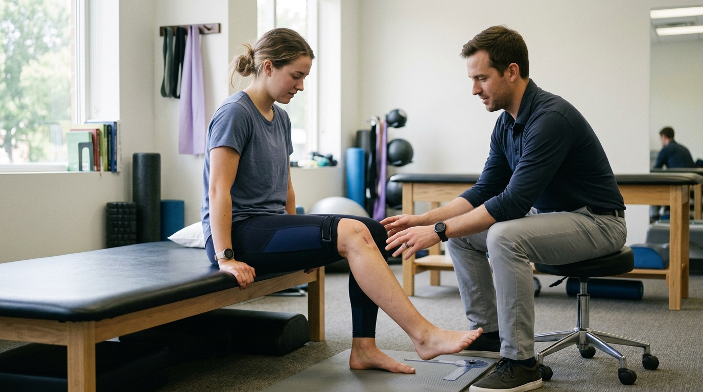
Weight-bearing mobility, on the other hand, describes how far the joint can move while carrying your body weight. A common clinical measurement is the weight-bearing lunge test. In this test, a person places their foot on the floor and lunges their knee forward toward a wall without letting their heel lift.
Clinical research shows that weight-bearing mobility tests often provide a more practical reflection of daily movement than non-weight-bearing tests. During walking, stair climbing, and squatting, the joint experiences compression and active muscular tension. A person might show acceptable passive motion on an examination table yet experience restricted movement when attempting a full squat or stepping down from a high curb.
Strength refers to the amount of force your muscles can produce to move a joint or resist external loads. In the lower leg, the calf complex must produce substantial force to support your body weight and propel you forward.
The two main muscles responsible for plantarflexion are the gastrocnemius and the soleus:
Muscles on the outside of the lower leg, known as the peroneals, and muscles along the inside and back, such as the tibialis posterior, provide lateral stability. They control side-to-side tilting of the foot and support the arch under load.
Balance is not simply a matter of muscle size. It relies heavily on sensorimotor control, which is the nervous system's ability to take in sensory information, process it, and execute rapid muscular corrections.
Specialized sensory receptors in the ligaments, joint capsules, tendons, and muscles of the ankle provide continuous feedback about joint position and movement. This internal sense is known as proprioception. When you step on an uneven patch of grass, these receptors instantly detect the tilt and signal your muscles to contract, preventing your ankle from rolling over.
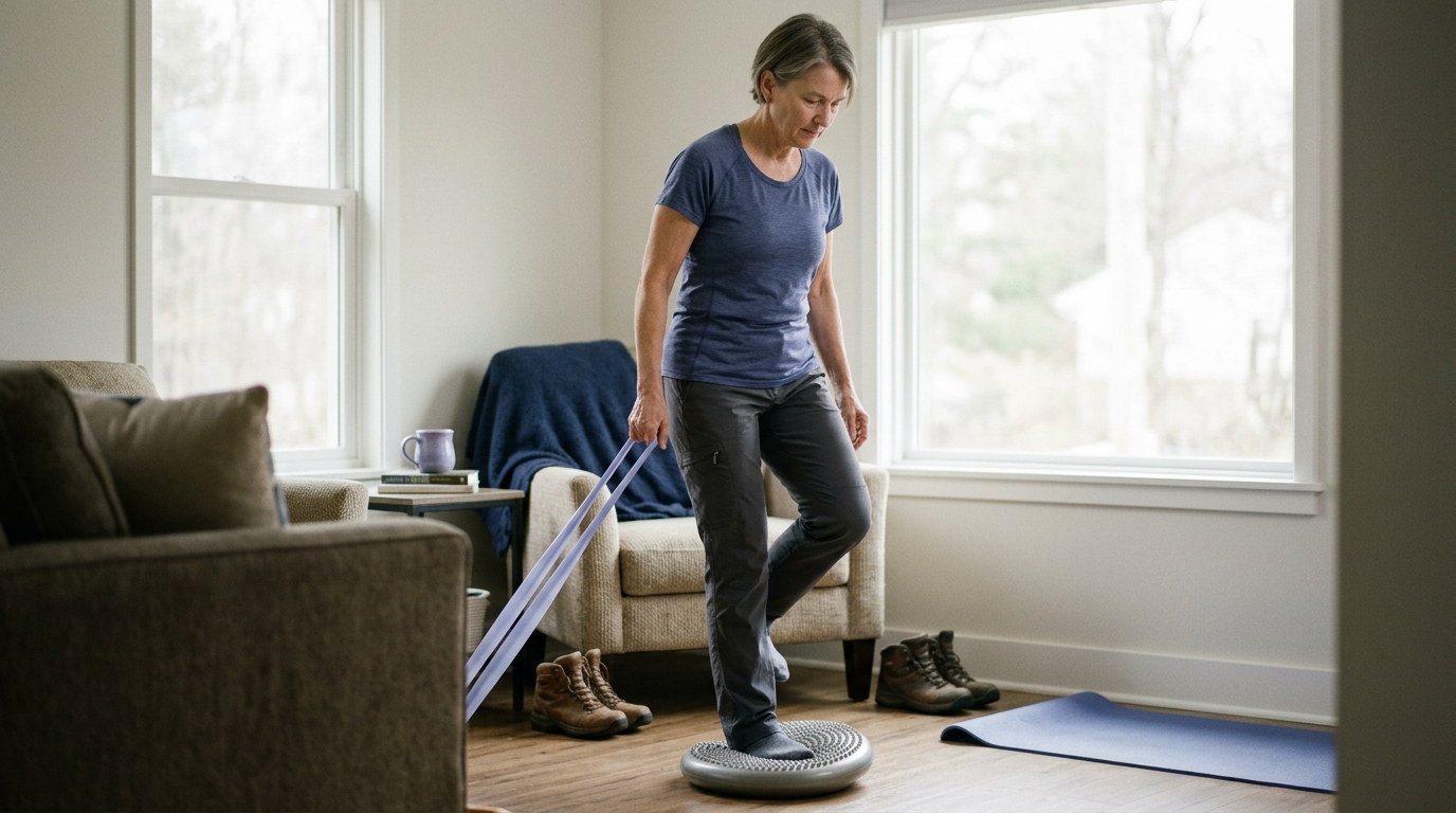
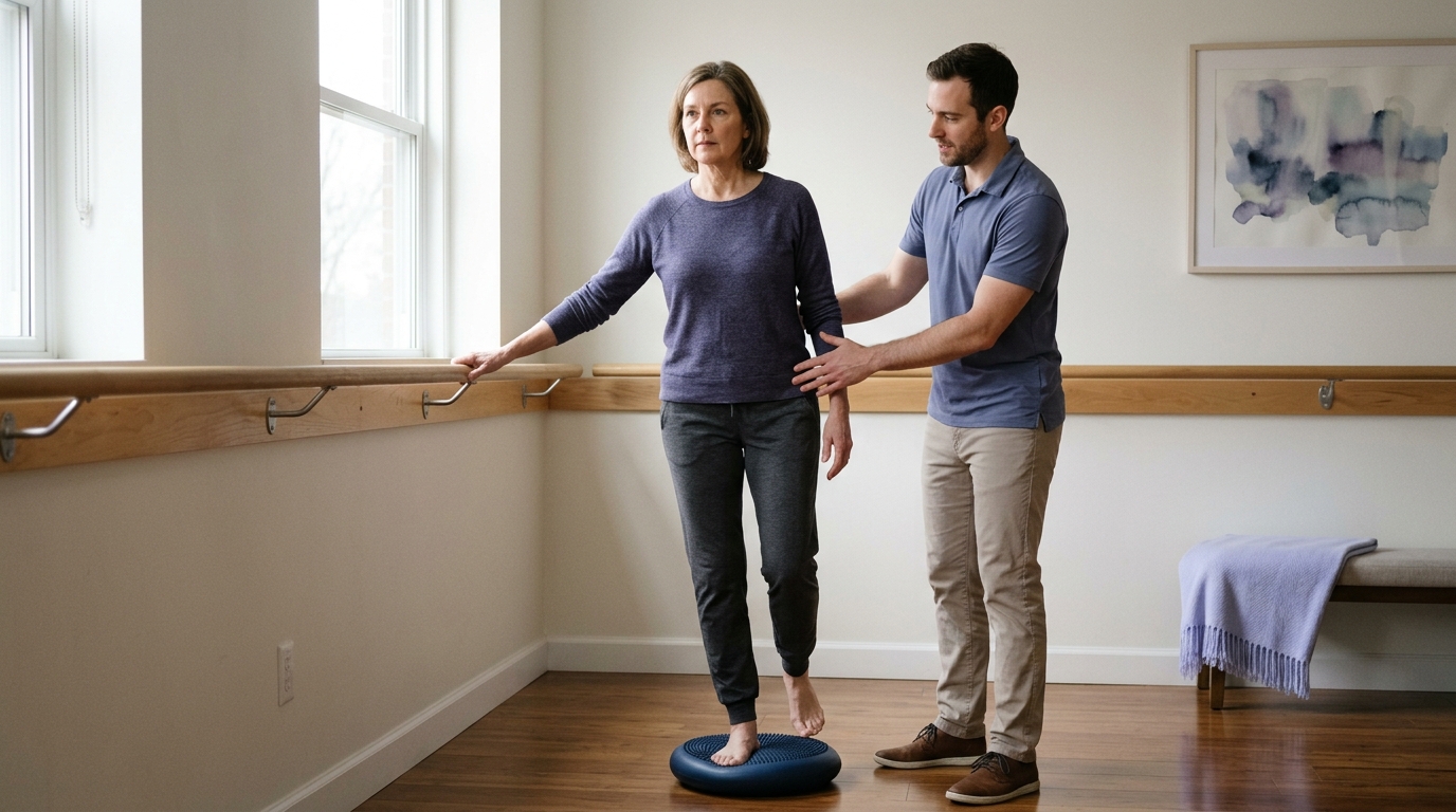
After an ankle injury, these sensory pathways can become disrupted. Even after ligament tissue heals and basic muscle strength returns, impaired proprioception can leave a person with persistent feelings of instability or frequent giving-way. Rebuilding sensorimotor control requires deliberate balance and coordination exercises, not just stretching or resistance training.
Clinical practice guidelines and peer-reviewed studies offer valuable clarity regarding what works in ankle assessment and rehabilitation. The 2021 clinical practice guideline published in the Journal of Orthopaedic Sports Physical Therapy provides an extensive, evidence-based synthesis for lateral ankle ligament sprains and chronic ankle instability.
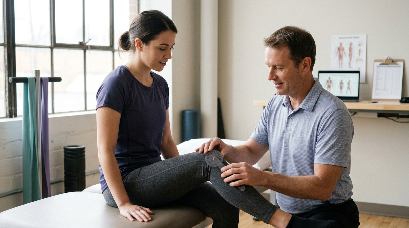
The guideline emphasizes that physical examination should include objective, reproducible measurements. For evaluating dorsiflexion, the guideline specifically recommends using the weight-bearing lunge test.
This test has been shown to be reliable and practical for identifying movement limitations. However, the guideline also notes that dorsiflexion measurements can vary based on individual technique and anatomy. Furthermore, because a person may have bilateral restrictions, comparing the injured ankle only to the uninjured side can sometimes be misleading. Clinicians are encouraged to interpret mobility scores alongside gait quality, pain levels, and specific daily limitations.
For acute lateral ankle sprains, modern clinical evidence strongly supports a functional treatment approach over prolonged cast immobilization. The guideline recommends:
For severe sprains with substantial tissue damage, short-term immobilization in a semi-rigid brace or below-knee cast may be indicated, but this should generally be limited to a maximum of 10 days. Studies show that functional treatment combining early protected weight bearing with exercise leads to a faster return to work and daily activities than extended immobilization.
The evidence supporting balance and neuromuscular training after ankle injury is robust. In data synthesized by the clinical practice guideline, balance training was associated with a 6.5 percent absolute risk reduction and a 46 percent relative risk reduction in future ankle sprains.
Meta-analyses highlighted in the guideline demonstrate that exercise-based rehabilitation significantly lowers the odds of recurrent lateral ankle sprains (odds ratio 0.59). When compared with standard usual care, structured exercise interventions reduced reinjury odds at 7 to 12 months post-injury (odds ratio 0.53).
These findings indicate that completing a dedicated course of balance and coordination exercises is one of the most effective strategies for preventing recurrent instability. If you want to explore broader therapeutic principles, our collection of rehabilitation, mobility and movement resources outlines how progressive movement training supports long-term recovery.
Maintaining ankle capacity is equally vital for active older adults. As we age, changes in muscle mass and tendon stiffness can alter how we walk and balance.
A systematic review and meta-analysis examining foot-and-ankle exercise programs in older adults found that structured interventions consistently improved balance performance and ankle flexibility. However, the review noted that existing evidence was insufficient to confirm definitive improvements in standalone muscle strength or broad functional scores.
A separate 2025 systematic review highlighted that toe-strengthening exercises performed three times per week contributed meaningfully to balance improvements. While these studies show clear benefits for balance and joint motion, they also remind us that exercise programs must be matched to realistic, specific goals rather than assumed to solve every mobility challenge.
No two ankle problems follow the exact same timeline or require the identical set of exercises. Multiple factors influence how quickly tissues heal, how much strength is needed, and how movement must be adapted.
The term ankle sprain covers a wide variety of clinical presentations:
Guideline recommendations for a lateral ligament sprain should not be applied blindly to tendon tears, bone fractures, or joint impingement. Each diagnosis involves different structural constraints.
The physical demands placed on the ankle change dramatically depending on the terrain. Walking on a flat, smooth sidewalk requires moderate ankle dorsiflexion and predictable muscle recruitment. Walking uphill or across uneven trails demands substantially more joint motion and muscle power.
In a biomechanical study comparing level walking to uphill walking at a 9-degree incline in older adults, researchers documented significant shifts in mechanical demand:
These figures illustrate why an individual might walk comfortably around their home but experience fatigue, calf tightness, or anterior ankle discomfort when walking up hills. The incline forces the ankle into deeper dorsiflexion while simultaneously demanding far greater propulsive force from the calf muscles.
As adults age, the way the body distributes mechanical work during walking can change. Studies examining joint kinetics show that older adults often produce lower peak plantarflexor torque during walking compared to younger individuals.
In one comparative study, older adults demonstrated an average plantarflexor strength of 0.87 Newton-meters per kilogram, compared to 1.39 Newton-meters per kilogram in younger adults. This reduction in ankle strength was directly associated with lower peak positive ankle power during walking.
To compensate for reduced ankle push-off power, the nervous system often redistributes mechanical work toward the hip joints. The hip flexors and extensors perform a greater proportion of the work required to advance the leg. Recognizing this redistribution helps explain why rehabilitation for walking and balance should consider the entire lower extremity, including hip strength and trunk control, rather than focusing entirely on the foot.
For individuals interested in maintaining independence and capacity over time, our articles on active aging and injury prevention provide evidence-based insights into lower-body conditioning.
A well-designed rehabilitation progression builds capacity step by step. Rather than following a rigid calendar, a task-based progression advances as your symptoms stabilize, your movement improves, and your tolerance for load increases.
The initial stage focuses on protecting healing tissues while preventing the negative effects of complete immobilization.
As swelling and resting pain subside, the focus shifts toward restoring functional joint motion and reintroducing light resistance.
Once basic walking is comfortable and standing calf raises are pain-free, the emphasis moves toward building robust strength and restoring sensorimotor control.
If your goals include returning to recreational sports or demanding fitness routines, our resources on strength and return to sport discuss how to safely bridge the gap between basic rehab and high-demand activities.
The final stage prepares the ankle for the specific environments, speeds, and terrains you encounter in daily life or recreation.
Do not rely on a single milestone to determine complete recovery. A comprehensive check of functional readiness should include:
Navigating ankle recovery can be confusing due to conflicting advice found online. Avoiding common misconceptions helps ensure a smoother, safer rehabilitation process.
It is easy to assume that if some ankle flexibility is good, maximum flexibility must be better. However, the true objective is functional, usable mobility that matches your daily activities.
Forcing an ankle into extreme dorsiflexion, especially when a joint capsule is inflamed or structurally blocked, can lead to anterior impingement and persistent joint irritation. Mobility exercises should feel like a muscular stretch in the calf rather than a sharp, bony block or pinch in the front of the ankle.
Passing a single test, such as being able to stand on one foot for 20 seconds, does not mean the ankle is prepared for all physical demands. Balance on a flat floor does not replicate the dynamic forces of walking down a steep, rocky trail or quickly reacting to a stumble. Readiness should be judged across a broad battery of movement tests, strength benchmarks, and real-world task tolerances.
Timelines reported in medical literature vary widely. For a mild lateral ankle sprain, return to basic participation can range from a few days to three weeks. For more severe sprains or high-demand physical occupations, recovery commonly extends to six to eight weeks or longer.
Healing is a biological process influenced by age, tissue health, sleep, and overall physical condition. Advancing your activity level based on functional milestones rather than an arbitrary calendar date reduces the risk of setbacks.
While the calf complex is essential for propulsion, functional ankle rehabilitation requires far more than just calf raises. Restoring side-to-side stability from the peroneal muscles, rebuilding arch support from the deep foot musculature, and retraining sensory feedback through balance drills are equally vital for long-term joint health.
Comparing your injured ankle to your uninjured side is standard practice, but it has limitations. Research shows that individuals with chronic ankle instability or previous lower-limb injuries often have bilateral sensorimotor deficits. Your opposite leg provides helpful reference data, but it should not be viewed as an infallible benchmark of optimal function.
While many mild ankle strains and sprains improve steadily with conservative management, certain signs and symptoms require prompt professional evaluation. Distinguishing routine soreness from potential structural injury is critical for safe recovery.
According to emergency care guidelines from major health organizations such as the NHS, you should seek urgent medical evaluation if you experience any of the following:
In clinical settings, healthcare providers use a validated decision framework known as the Ottawa Ankle Rules to determine whether an acute injury requires diagnostic X-rays. These rules assess:
Clinical studies report that the Ottawa Ankle Rules have a sensitivity between 92 and 100 percent for detecting significant fractures. If these specific criteria are met, an X-ray is strongly recommended to rule out broken bones before proceeding with active exercise.
If an ankle injury fails to improve after several weeks of conservative care, or if you experience recurrent giving-way, a comprehensive reassessment is warranted. A healthcare provider can investigate alternative or coexisting conditions, such as:
For additional guidance on navigating clinical care pathways and understanding diagnostic processes, you can review our injury recovery and healing resources.
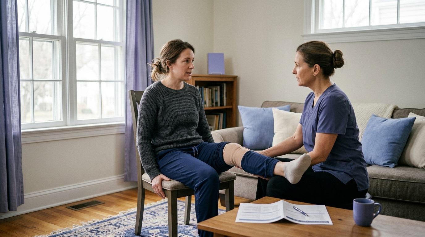
Collaborating with a physical therapist, orthopedic specialist, or primary care physician ensures your rehabilitation program matches your exact diagnosis. Here are practical questions you can bring to your next appointment:
The ankle is a dynamic, load-bearing joint system that plays a central role in shock absorption, forward propulsion, and standing balance. Successful ankle function depends on a combination of weight-bearing mobility to let the body roll forward over the foot, plantarflexor muscle strength to drive push-off, and sensorimotor control to adapt to uneven terrain.
Recovery from an ankle setback is not a universal process, nor does it follow a rigid calendar. While high-quality evidence supports early protected weight bearing, functional bracing, and progressive balance training for common lateral sprains, other conditions require tailored protection and medical oversight. By building physical capacity gradually and matching your training to the real-world demands of your daily life, you can rebuild confidence, restore comfortable movement, and protect long-term joint health.
A pinching sensation at the front of the ankle during a calf stretch or lunge often indicates anterior joint compression rather than tight muscles. When the shin rolls forward, the talus bone must glide backward inside the joint mortise. If the joint capsule is stiff or tissues are inflamed, the structures in front can become pinched. If you feel a sharp pinch, back off the depth of the stretch, focus on seated calf exercises, and consult a physical therapist for joint-mobilization strategies.
Yes. Clinical guidelines strongly recommend proprioceptive, balance, and neuromuscular exercises for individuals with chronic ankle instability. Even years after an initial injury, structured balance training on firm and unstable surfaces can retrain sensory pathways, improve dynamic stability, and substantially reduce the odds of future giving-way episodes.
Walking up an incline forces your ankle into a deeper angle of dorsiflexion while placing significantly higher mechanical demands on your calf muscles. Biomechanical research shows that walking up a 9-degree incline can increase trailing-leg push-off work by more than 100 percent and raise calf muscle activity by 75 to 136 percent compared to level walking. If your calf strength or weight-bearing mobility is limited, inclines will feel noticeably more challenging.
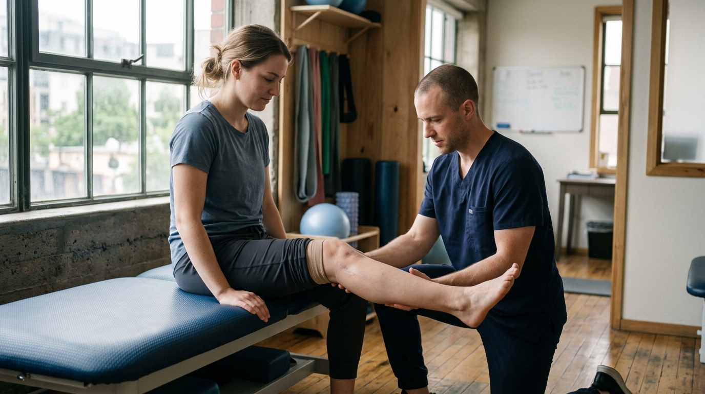
A physical therapist can distinguish between joint capsule stiffness and muscular tightness through targeted tests. When dorsiflexion is restricted with both a straight knee and a bent knee, the joint capsule or bony anatomy is often a primary contributor. If dorsiflexion improves significantly when the knee is bent, the gastrocnemius muscle is more likely the main source of restriction, as bending the knee takes slack off that two-joint muscle.

Use ReboundBody resources to understand common recovery stages, rehab terms, movement limits and strength rebuilding. Each guide is designed to make a complex comeback easier to understand.


Read practical guidance on injury recovery, rehabilitation, mobility and rebuilding strength as you work your way back to activity.
Explore Resources