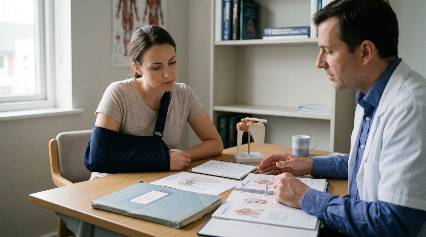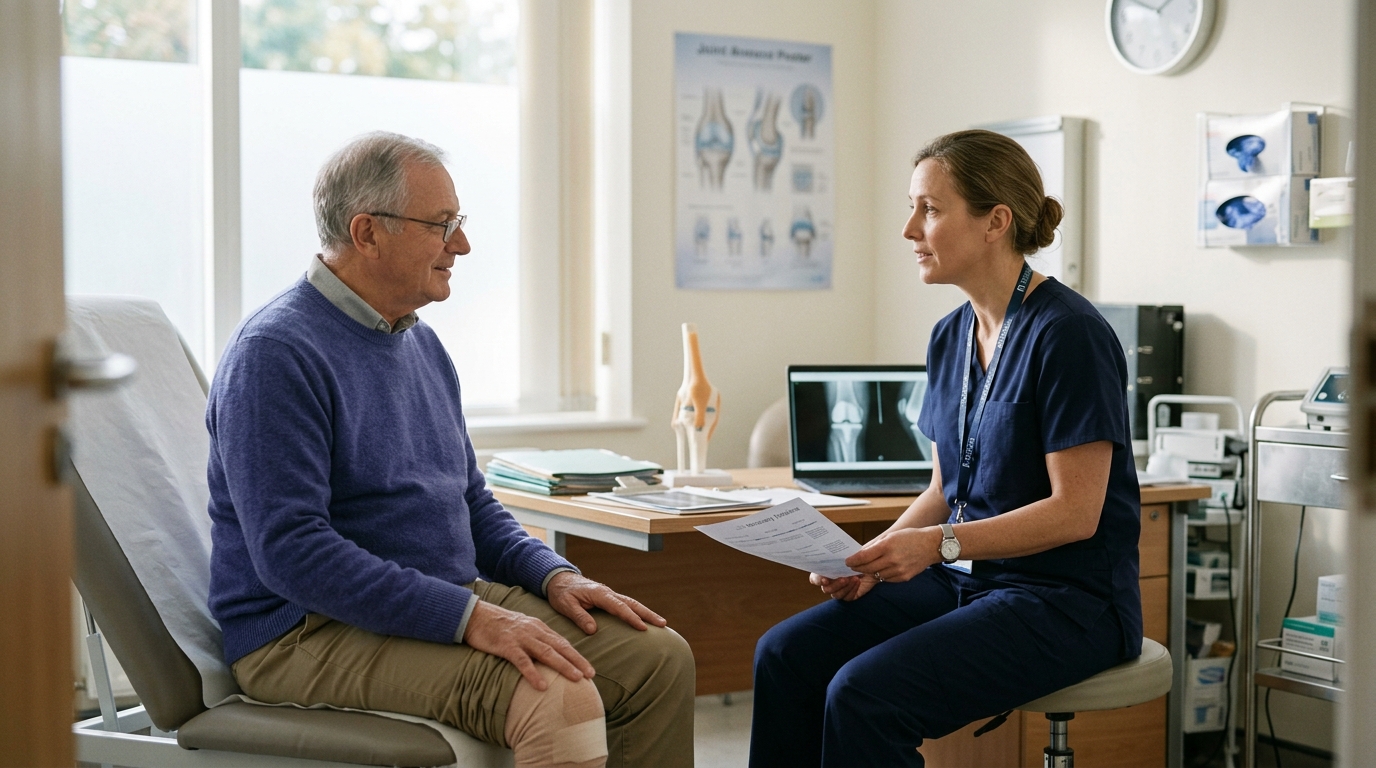

Orthopedic rehabilitation concepts are clarified in this glossary to help patients make sense of clinical terms regarding pain duration, swelling, and joint mobility.





If you have ever left an orthopedic clinic or physical therapy session wondering what "partial weight-bearing" or "passive range of motion" actually means for your daily routine, you are not alone. Searches for medical abbreviations, healing stages, and mobility restrictions are among the most common queries after an injury or surgery.
Orthopedic terminology can sound intimidating, technical, and rigid. In reality, these terms are clinical shorthand designed to describe the condition of your tissues, the movement of your joints, and the exact boundaries needed to protect healing structures.
This reference guide translates common orthopedic and rehabilitation terms into clear, plain language. It explains what these concepts mean in daily practice, outlines the evidence behind rehabilitation protocols, and provides practical questions you can take to your next appointment.
Medical language exists to give healthcare teams a precise, shared vocabulary. When a surgeon, physical therapist, and radiologist review a case, standardized words ensure that everyone understands the structural stability of a bone or ligament. For patients, however, these words can create unnecessary confusion and anxiety.
A term like "nonunion" might sound like an absolute disaster, while "callus" might bring to mind rough skin on your feet rather than normal bone healing. Misunderstanding clinical terms can lead to accidental mistakes during recovery. A person told to stay "non-weight-bearing" might assume resting their foot on the floor while sitting is acceptable, or someone told to move within "active-assisted range of motion" might use their muscles too aggressively.
A glossary provides clarity, but it does not replace an individualized care plan. Professional guidelines, such as those from the National Institute for Health and Care Excellence, emphasize that rehabilitation after traumatic injury must be tailored to the specific needs of the individual and their stage of recovery.
Terminology describes general principles, yet your specific protocol depends on the exact tissue injured, the surgical repair performed, and your overall health. Understanding the language allows you to follow clinical conversations accurately and advocate for clear explanations from your care team. You can find related educational materials across our injury recovery and healing resources.
Timeframes and symptom descriptions are often the first concepts discussed after an injury. Clinicians use specific categories to describe how long pain has lasted and how symptoms behave during movement.
Acute pain begins suddenly in connection with a specific injury, tissue damage, surgical procedure, or sudden inflammation. It serves as a protective warning signal from the nervous system to prevent further physical damage to the affected area.
In practical terms, acute pain is highest immediately following an injury or operation and gradually decreases as tissues repair. The National Library of Medicine notes that acute pain usually resolves as the underlying cause heals or is treated. When your doctor uses the word acute, they are referring to a recent problem with an expected healing progression rather than a permanent condition.
Chronic pain refers to pain that persists longer than three months or beyond the timeframe in which tissue healing would ordinarily be expected. This definition describes duration rather than the physical structural state of an injury.
Chronic pain does not automatically mean that tissue is actively tearing, failing to heal, or suffering ongoing damage. Over time, the nervous system can become sensitive, continuing to produce pain signals even after initial structural healing has occurred. Understanding this distinction helps prevent the fear that persistent discomfort always indicates ongoing structural harm.
A pain score is a numerical rating, typically from 0 to 10, used by healthcare providers to measure your subjective experience of discomfort at a specific moment. A zero represents no pain, while a ten represents the most severe pain imaginable.
Pain scores help clinicians track trends over time, evaluate whether medications are working, and check how you tolerate rehabilitation exercises. Pain is only one piece of the recovery picture. Clinical studies on bone fractures track pain alongside joint motion, functional ability, and daily quality of life because a reduction in pain does not always mean strength and mobility are fully restored.
A flare-up is a temporary, short-term increase in pain, stiffness, or swelling above your baseline recovery level. It often occurs after an increase in physical activity, a change in exercise intensity, or accidental overexertion.
Flare-ups are a common part of the rehabilitation process and do not necessarily mean you have reinjured the tissue. They generally indicate that healing structures were loaded slightly beyond their current capacity. Knowing how your team defines an expected flare-up versus a serious complication helps you respond calmly with rest and modified movement.
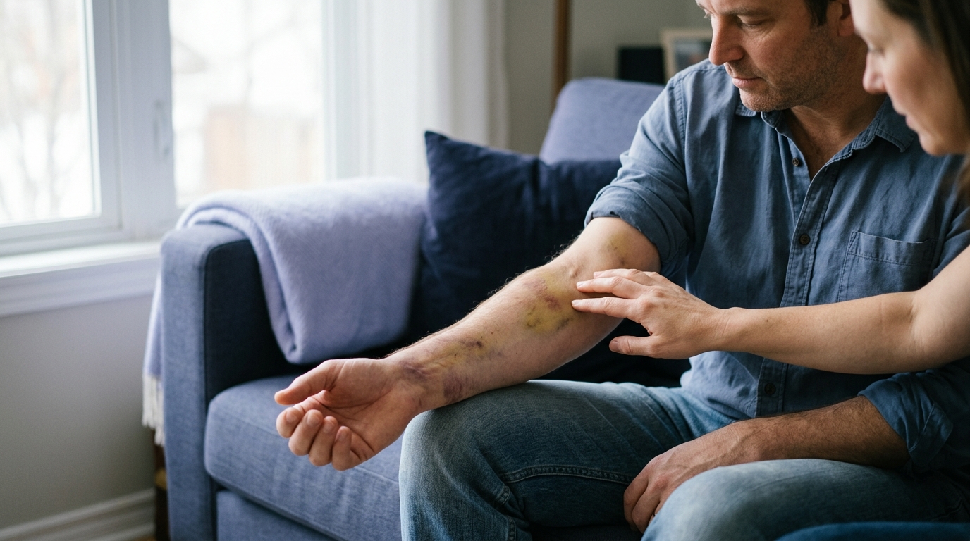
Swelling is the buildup of fluid in soft tissues or joint spaces following trauma or surgery. Bruising is visible discoloration caused by broken blood vessels beneath the skin. Tenderness refers to sensitivity or discomfort when pressure is applied directly to an area.
These three signs are normal inflammatory responses to tissue disruption. While they are common after sprains, strains, and fractures, their severity does not always correspond directly to the severity of an injury. Clinicians evaluate swelling, bruising, and tenderness together with movement and imaging to determine your overall healing stage.
Soft tissue injuries involve muscles, tendons, and ligaments. Clinicians use precise terms to identify which structure is damaged and how the injury occurred.
A sprain is a stretch or tear of a ligament, which is the tough, fibrous band of connective tissue that links bones together at a joint. Sprains commonly occur when a joint is forced beyond its normal range of motion, such as rolling an ankle or twisting a knee.
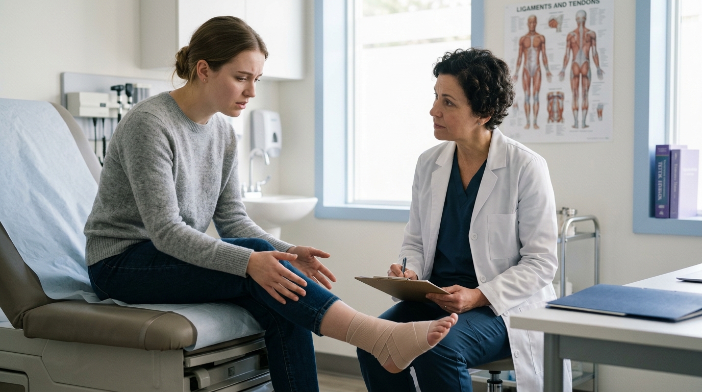
Symptoms of a sprain include local pain, rapid swelling, bruising, and joint instability. Because ligaments have a limited blood supply compared to muscles, they can take weeks or months to regain their resting tensile strength. Rehabilitation focuses on protecting the joint while restoring stability and motion.
A strain is an overstretch, partial tear, or complete rupture of a muscle or a tendon. Tendons are the strong cords of fibrous tissue that connect muscles to bones, allowing muscle contractions to move your skeleton.
Strains can happen suddenly during a rapid contraction or heavy lift, but they can also develop gradually through repetitive overuse. Common symptoms include localized muscle spasms, weakness, and cramping. Rehabilitation for a strain focuses on progressive loading to rebuild muscle fibers and tendon tolerance.
Effusion is the medical term for excess fluid that collects specifically inside a joint capsule, often referred to casually as water on the joint. Edema refers to excess fluid collecting in the soft tissues outside the joint, such as the skin and muscle layers of the lower leg.
Both conditions can limit joint movement and cause a feeling of heavy fullness. Managing effusion and edema through elevation, compression, and gentle muscle activation is often a priority in early physical therapy because excess fluid can temporarily inhibit muscle function.
Inflammation is the body's natural immune response to cellular injury, infection, or surgical intervention. It brings increased blood flow, specialized cells, and growth factors to the injured site to clear debris and initiate tissue repair.
The classic signs of inflammation are redness, heat, swelling, and localized pain. While chronic inflammation can hinder progress, early acute inflammation is a necessary first step in biological healing. Treatment strategies aim to manage excessive inflammatory symptoms without completely shutting down this vital repair process.
Restoring joint motion is a primary focus of orthopedic rehabilitation. Physical therapists use specific terms and measurements to describe how joints move and who is doing the physical work during exercise.
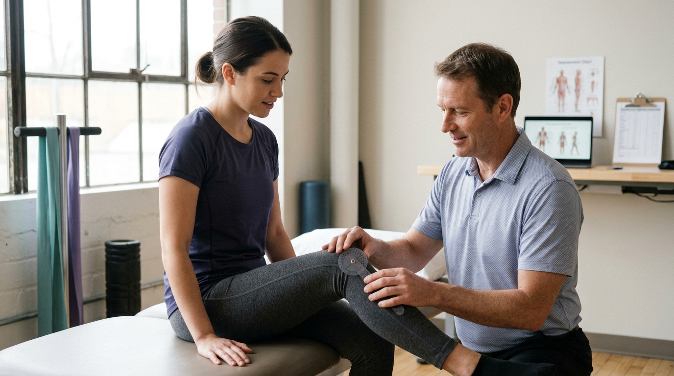
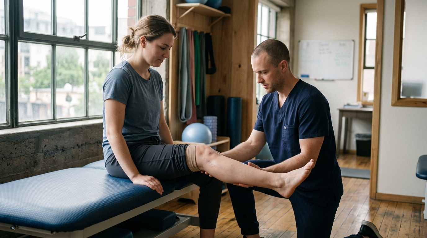
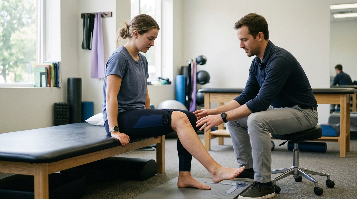
Range of motion, abbreviated as ROM, is the measurement of how far a joint can move in various directions from a neutral starting position. It is typically measured in degrees using a handheld tool called a goniometer or an inclinometer.
ROM measurements track your progress over time and compare your injured limb to your uninjured side. A specific degree measurement provides objective data, but it is only one metric. Full recovery also requires strength, coordination, and the ability to move without significant pain during daily tasks.
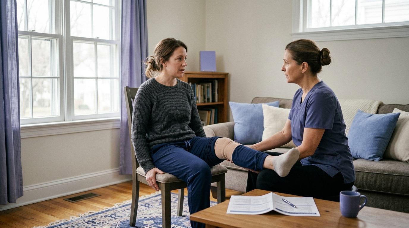
Passive range of motion, or PROM, describes joint movement produced entirely by an external force while your own muscles remain completely relaxed. The external force may be a physical therapist, a mechanical device, or your other limb.
Surgeons frequently prescribe PROM in early recovery phases when a repaired tissue, such as a sutured tendon or unstable fracture, cannot yet tolerate active muscle tension. PROM helps prevent joint capsule stiffness and maintains synovial fluid circulation without placing mechanical stress on the healing surgical site. You can read more about structured progression in our surgery and rehabilitation guides.
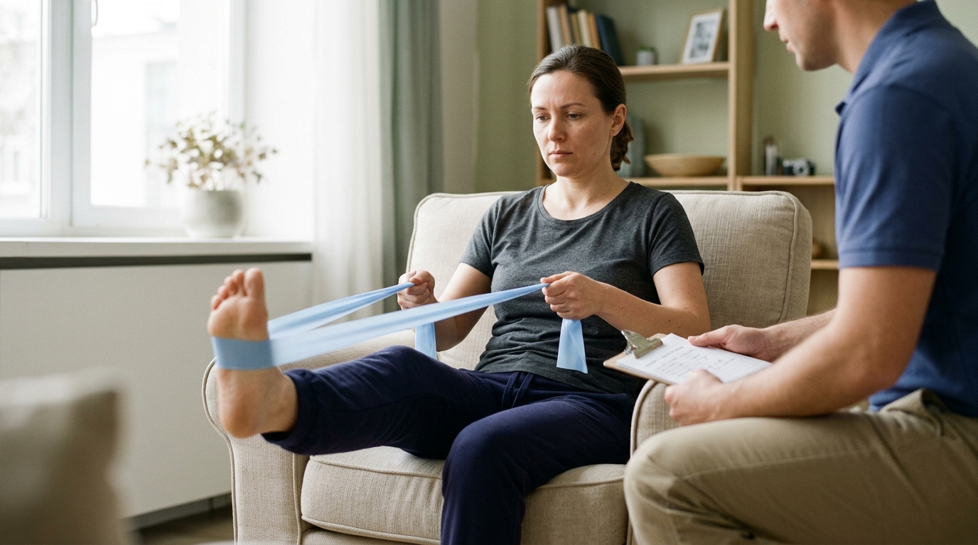
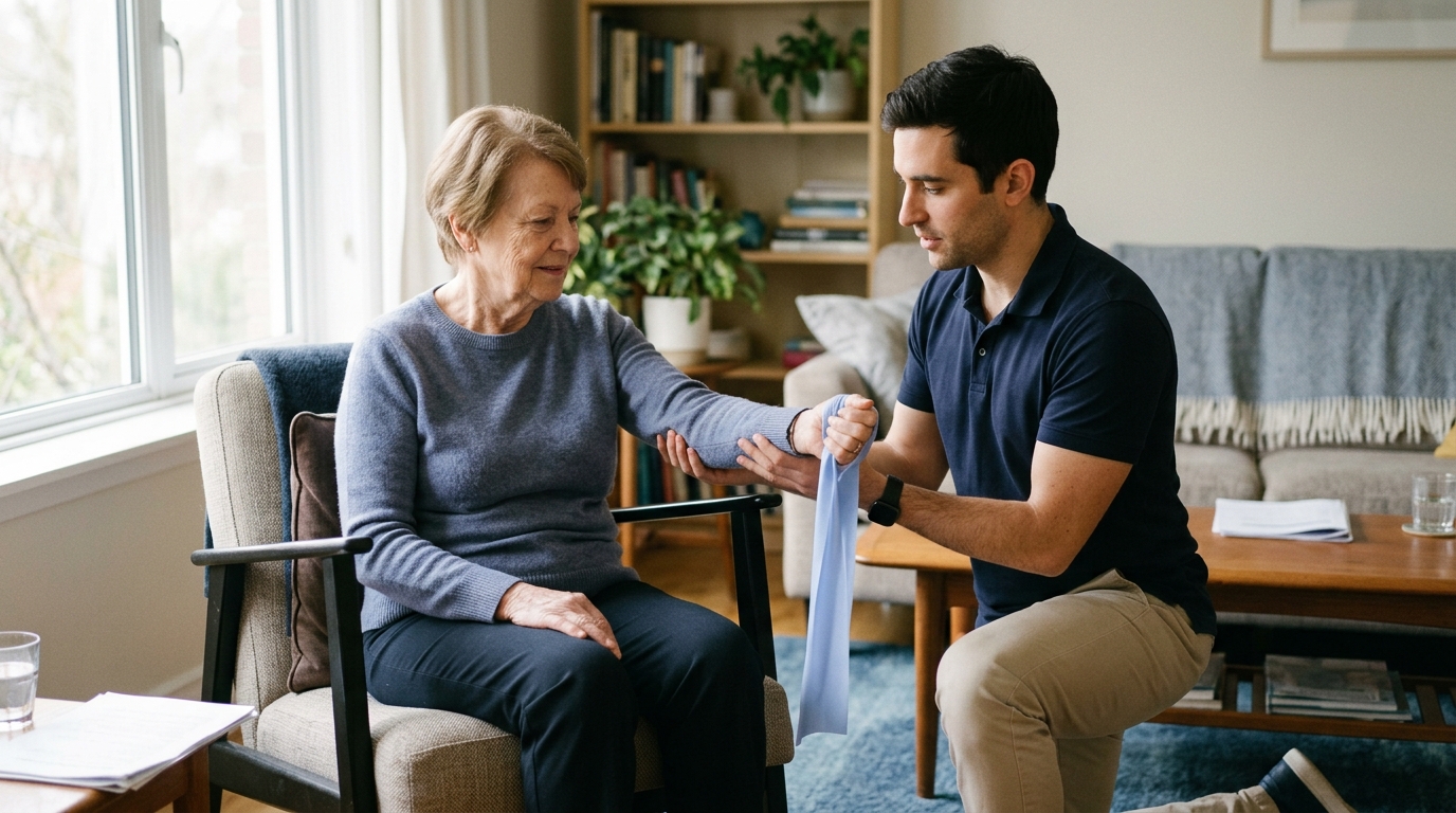
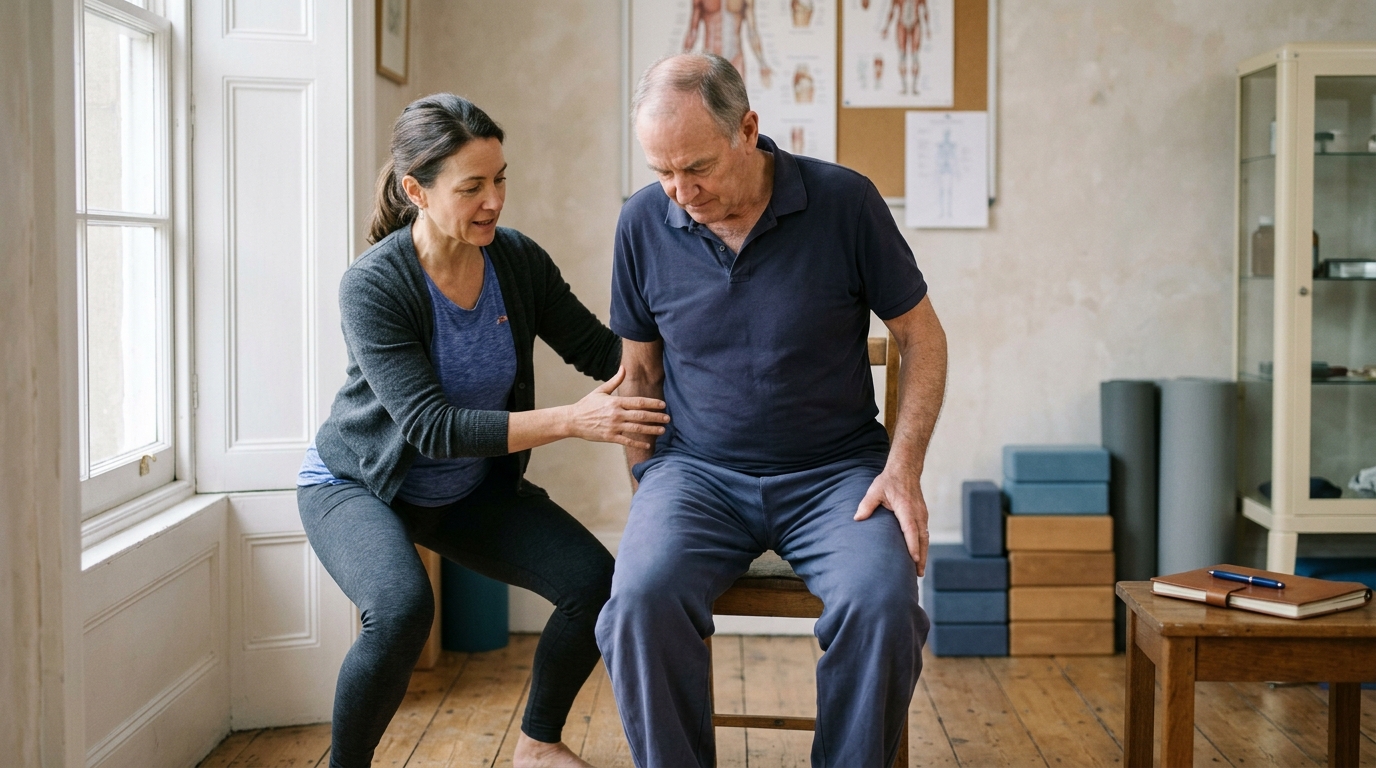
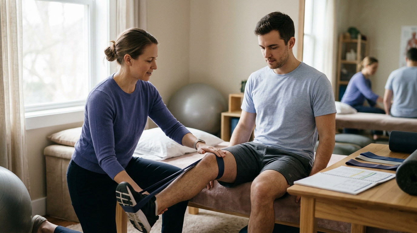
Active-assisted range of motion, or AAROM, refers to movement where you use your own muscles to move the joint as far as safely possible, with an external helper or tool providing assistance to complete the arc of motion.
Common AAROM tools include canes, pulley systems, elastic bands, and the opposite arm or leg. This stage serves as a bridge between complete muscle rest and independent movement. It allows tissues to handle light, controlled muscle contractions while protecting the joint from excessive fatigue or overload.
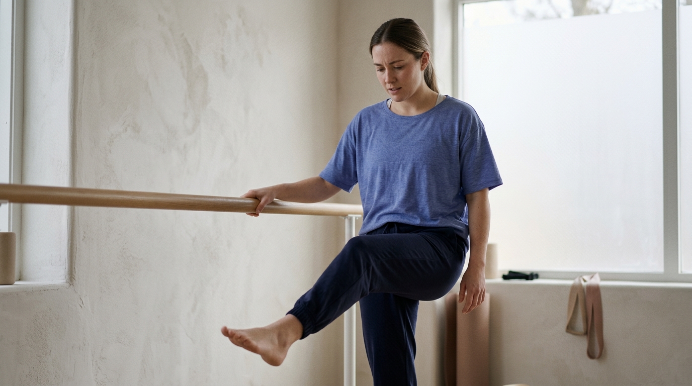
Active range of motion, or AROM, occurs when you move a joint through its available movement arc entirely using your own muscular effort, without any external physical assistance.
AROM requires both joint mobility and sufficient muscle strength to lift the weight of the limb against gravity. Transitioning to AROM is a major milestone in rehabilitation because it demonstrates that the healing tissue can safely tolerate active contraction and neuromuscular control.
Joint stiffness is a subjective feeling of tightness or an objective loss of joint mobility that limits smooth movement. It frequently occurs after periods of immobilization, such as wearing a cast, splint, or brace.
The American Academy of Orthopaedic Surgeons notes that people frequently lose muscle strength and range of motion around an injured joint. Specific, progressive mobility exercises help stretch contracted joint capsules, align collagen fibers, and restore normal flexibility.
Mobility describes a person's overall ability to move around independently within their environment. It includes changing positions in bed, standing up from a chair, navigating stairs, and walking.
Gait refers specifically to the biomechanical pattern of walking. Clinicians analyze your gait to identify compensations, such as limping or shortened stride lengths. National clinical guidance emphasizes that restoring mobility and normal gait mechanics through targeted exercise helps preserve balance, strength, and postural reflexes. For further details on functional movement, review our mobility and movement recovery resources.
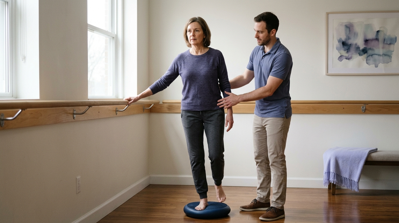
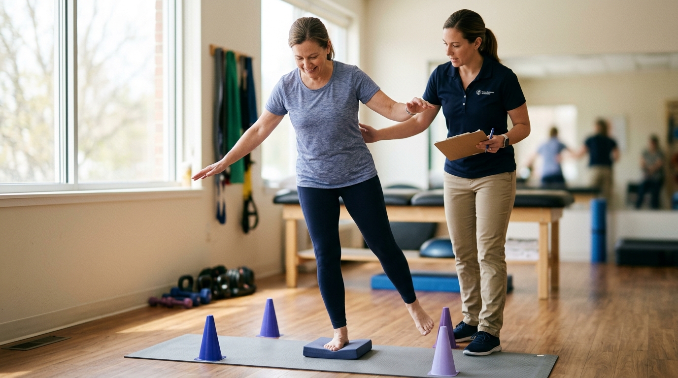
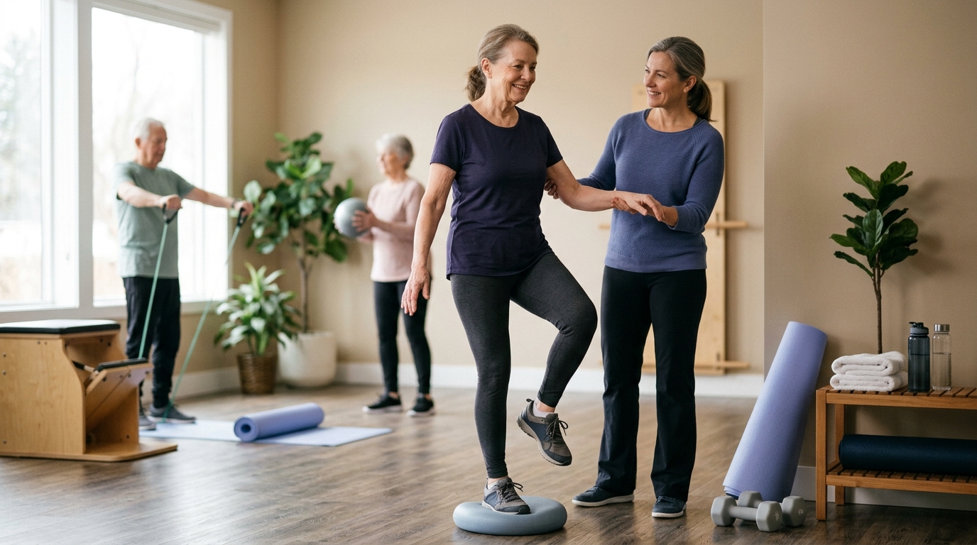
Proprioception is your body's unconscious awareness of joint position, motion, and spatial orientation. Sensory receptors located in your muscles, tendons, joint capsules, and skin continuously send feedback to the brain regarding limb position.
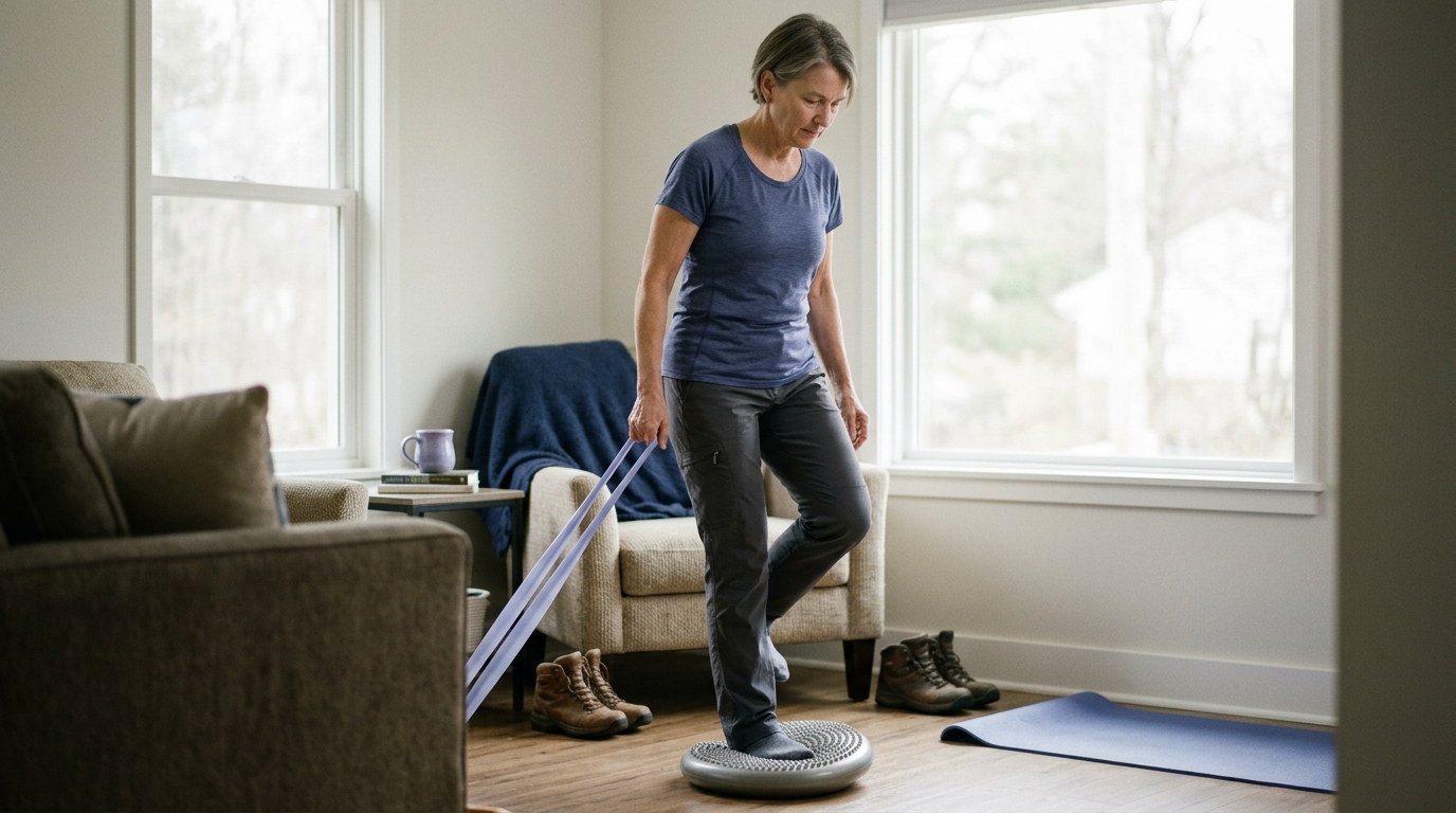
Joint injuries and surgeries disrupt these sensory receptors, which can leave a limb feeling unstable or uncoordinated. Balance training and proprioceptive exercises retrain these neurological pathways, helping you react quickly to uneven surfaces and preventing future falls or reinjury.
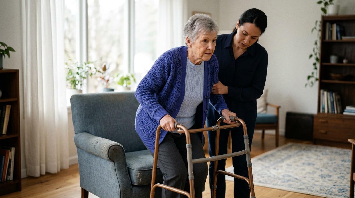
Weight-bearing orders dictate how much body weight you are permitted to place through an injured or operated limb. These instructions protect surgical hardware, bone repairs, and soft tissues while they heal. Following them precisely is essential for safe recovery.
Non-weight-bearing, commonly abbreviated as NWB, means that absolutely no body weight may be placed through the affected limb. Under clinical definitions, the foot or leg should not touch the floor during standing, walking, or transfers.
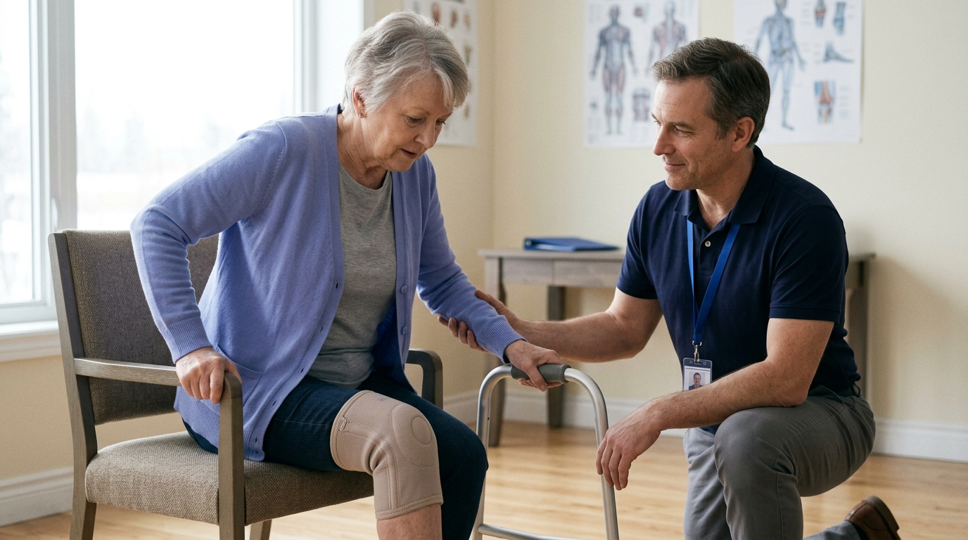
NWB requires the use of assistive devices such as axillary crutches, a walker, or a seated knee scooter. This strict restriction protects fragile bone fusions, osteotomies, or delicate cartilage restorations from mechanical forces that could displace healing structures or cause hardware failure.
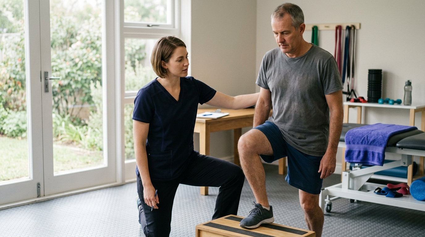
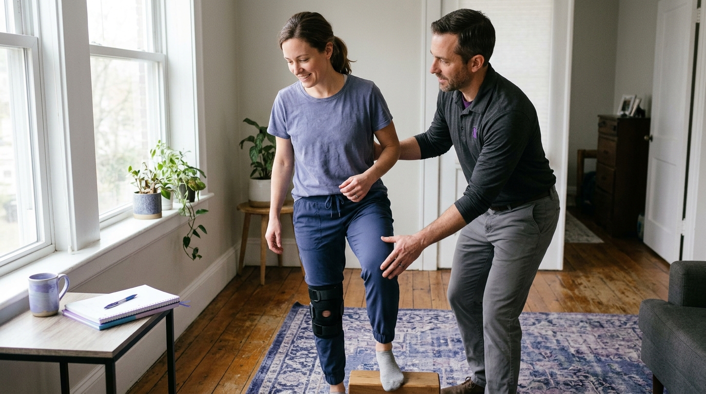
Partial weight-bearing, or PWB, permits a small, specific amount of body weight to pass through the limb. The allowed amount is usually described as a percentage of your total body weight or as a specific number of pounds.
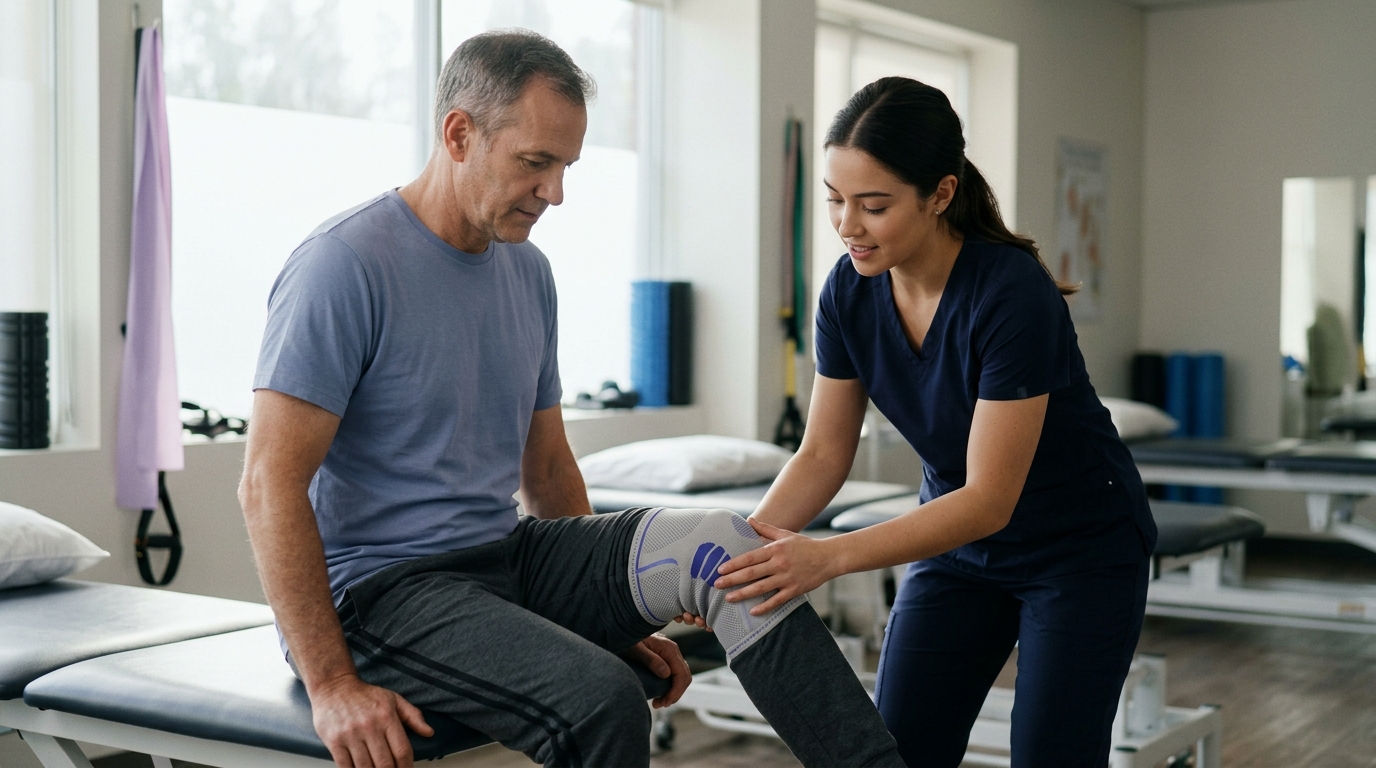
Because patients cannot accurately guess weight percentages, physical therapists often use a standard bathroom scale to help you practice what a specific load feels like. Assistive devices like crutches or walkers are required during PWB to absorb the remainder of your body weight and maintain stability.
Weight-bearing as tolerated, abbreviated as WBAT, allows you to place as much weight on the limb as you can comfortably handle without significant pain or compensatory limping.
WBAT is not a command to force full weight immediately or push through sharp pain. Instead, it allows you to progress your loading naturally based on your comfort, healing progress, and muscle control. You may still need crutches or a cane to support balance and maintain a smooth walking pattern as you build tolerance.
Full weight-bearing, or FWB, means the limb can support your entire body weight without restriction. At this stage, you no longer require assistive devices for weight reduction, though you might temporarily use a cane for balance or confidence.
Achieving FWB indicates that the bone or repaired soft tissue has developed sufficient structural integrity to handle normal standing and walking stresses. However, FWB does not mean rehabilitation is complete, as muscle strength, joint endurance, and gait symmetry still need rebuilding.
Permissive weight-bearing describes a structured rehabilitation approach where a clinician allows early loading under specific, monitored conditions. Protected weight-bearing refers to placing weight through a limb while using an external support device, such as a rigid walking boot, hinged brace, or cast.
In contrast, unprotected weight-bearing involves walking without external braces or immobilizers. Clinical research in ankle fractures highlights the importance of these distinctions. The following clinical evidence illustrates how loading protocols vary across different orthopedic injuries.
The medical understanding of weight-bearing has evolved significantly. Historically, long periods of strict immobilization were standard for most lower extremity injuries. Today, clinical research demonstrates that controlled, early loading can benefit specific conditions while posing risks to others.
In a randomized controlled trial evaluating conservative treatment for stable Weber B ankle fractures, researchers compared permissive early weight-bearing in a walking boot against non-weight-bearing immobilization in a below-knee cast. Patients in the walking boot group achieved significantly higher functional scores on the Olerud-Molander Ankle Score at six weeks and twelve weeks compared to the cast group. They also demonstrated earlier improvements in ankle range of motion. The authors concluded that early protected loading is a safe and effective strategy for this specific fracture pattern.
Another multicenter randomized trial examined weight-bearing protocols following surgical fixation of unstable ankle fractures. The study compared unprotected weight-bearing, protected weight-bearing in a brace, and non-weight-bearing protocols at six weeks post-surgery. Patients permitted to bear weight early showed superior functional ankle scores at six weeks compared to those kept non-weight-bearing, demonstrating the functional benefits of safe mechanical loading when surgical fixation is stable.
Early weight-bearing cannot be applied universally across all orthopedic procedures. A systematic review examining rehabilitation following anterior cruciate ligament (ACL) reconstruction found that accelerated weight-bearing within the first two weeks resulted in greater joint laxity and bone-tunnel widening compared to delayed weight-bearing protocols, without providing meaningful improvements in long-term functional outcomes.
These contrasting findings show why weight-bearing advice must remain specific to the injured tissue and surgical procedure. Bone fractures often benefit from controlled compressive forces that stimulate osteoblasts, whereas newly reconstructed soft-tissue grafts may stretch or loosen if loaded too aggressively before biological integration occurs. Always follow your surgeon's specific loading guidelines.
Understanding bone healing terminology helps you interpret radiology reports and follow clinical discussions about your skeletal recovery.
A fracture is a medical term for a broken bone. The break can range from a tiny, hairline crack within the bone cortex to a complex injury where the bone shatters into multiple pieces.
Regardless of whether a break is called a crack, hairline fracture, or compound break, bone tissue undergoes the same biological repair process. During recovery, surrounding muscles often weaken and joints stiffen due to disuse and immobilization. Rehabilitation exercises help restore the strength and joint motion lost during the healing phase.
In orthopedic medicine, a callus is a bridge of new, unorganized bone and cartilage tissue that forms around and between the broken ends of a fractured bone. It is completely different from the thickened skin callus that forms on your hands or feet.
Callus formation is a crucial second stage of fracture healing. The initial soft callus provides temporary structural stability, which gradually mineralizes into hard bone over several weeks. When an orthopedic surgeon sees callus formation on an X-ray, it confirms that your body is actively repairing the fracture site.
Union means that a fractured bone has successfully healed back together into a solid, continuous structure. The bone is structurally stable enough to withstand normal physiological loads without shifting or causing focal pain.
Clinicians determine union by combining clinical findings with radiographic evidence. Clinical union means the fracture site is no longer tender to touch and does not hurt during functional loading. Radiographic union means X-ray images show solid bone bridges crossing the previous fracture line.
A delayed union occurs when a broken bone takes longer than the expected medical timeframe to heal completely. The bone is still in the process of repairing itself, but the rate of progress is slower than typical averages.
Delayed union can be caused by various factors, including reduced blood supply, excessive movement at the fracture site, nicotine use, nutritional deficiencies, or underlying medical conditions. A delayed union does not mean the bone will never heal; it often requires continued protection, activity modification, or focused medical management.
A nonunion occurs when a fractured bone fails to heal and the biological repair process has completely stopped. According to the American Academy of Orthopaedic Surgeons, patients with a nonunion often experience persistent pain, tenderness, or movement at the break site long after the initial fracture pain should have resolved.
Nonunions typically require medical or surgical intervention to restart the healing process. Treatments may include surgical stabilization with metal plates and screws, bone grafting to introduce active bone cells, or specialized bone stimulator devices that use electromagnetic pulses to encourage cellular growth.
A malunion occurs when a fractured bone heals completely, but in an abnormal anatomical alignment or with a residual deformity. The bone is structurally solid, but it may be rotated, angled, shortened, or shifted from its original position.
A minor malunion may cause no functional problems and require no treatment. If a malunion significantly alters joint alignment or changes how weight passes through a limb, it can lead to premature joint wear, pain, or limited mobility, sometimes requiring corrective surgical osteotomy.
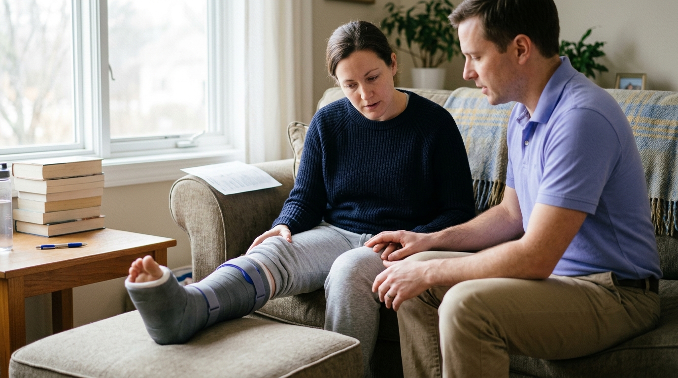
Immobilization is the practice of restricting the movement of a joint or fractured bone using an external device such as a fiberglass cast, splint, or rigid brace.
Immobilization protects broken bone ends or repaired soft tissues during the initial phases of healing, allowing fragile new blood vessels and cellular matrices to form without being torn by movement. However, prolonged immobilization can lead to muscle atrophy and joint stiffness, so modern protocols aim to minimize immobilization time as soon as tissue stability permits.
Radiographic progression refers to the visible biological changes observed across a series of medical imaging scans over time, such as X-rays, CT scans, or MRIs.
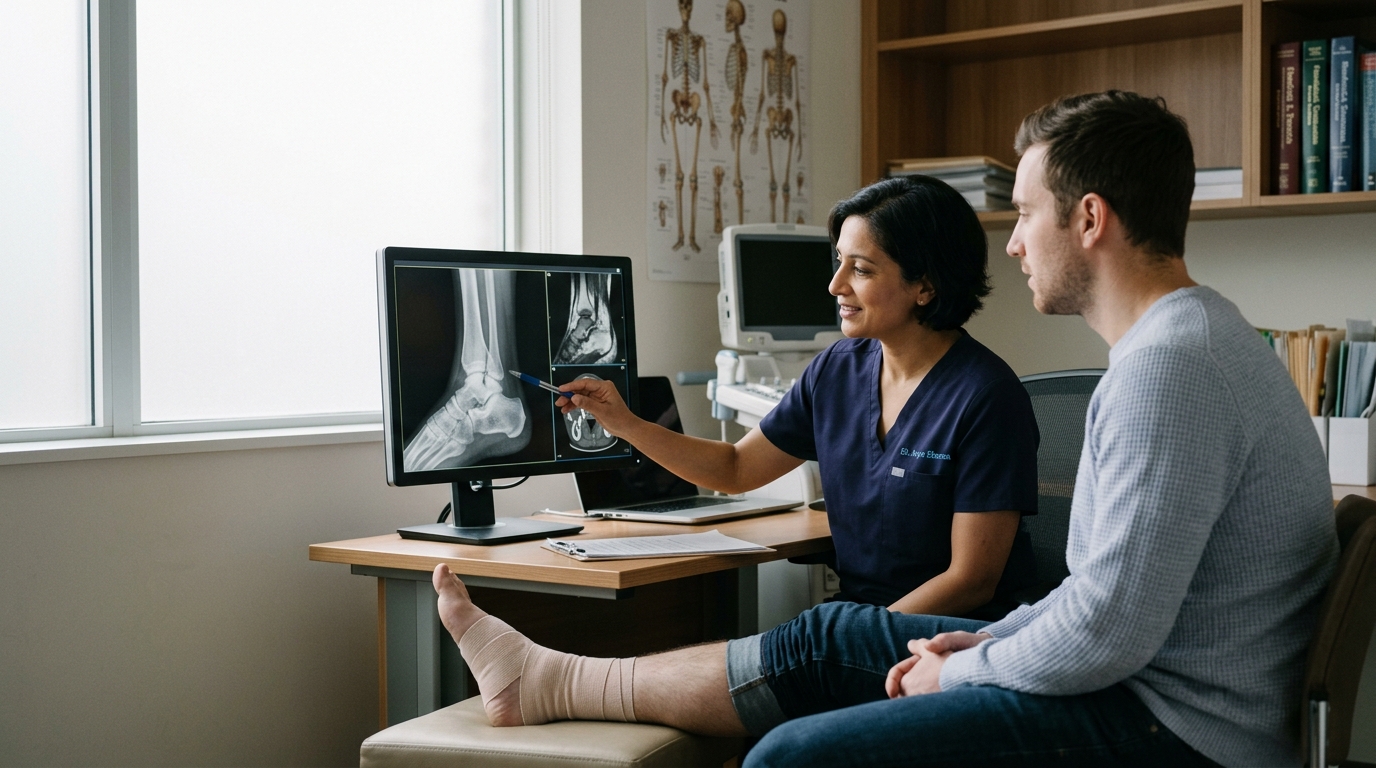
Clinicians look for specific signs of radiographic progression, including the appearance of soft callus, the mineralization of hard callus, the gradual fading of the fracture line, and the remodeling of bone along natural stress lines. Imaging results are always interpreted in combination with your physical symptoms and functional abilities.
Rehabilitation focuses on helping you regain physical capacity, independence, and the ability to participate in meaningful daily activities.
Rehabilitation is the comprehensive process of restoring physical, psychological, and functional abilities lost due to injury, illness, or surgery. The National Library of Medicine notes that physical medicine and rehabilitation helps individuals regain optimal body function and independence.
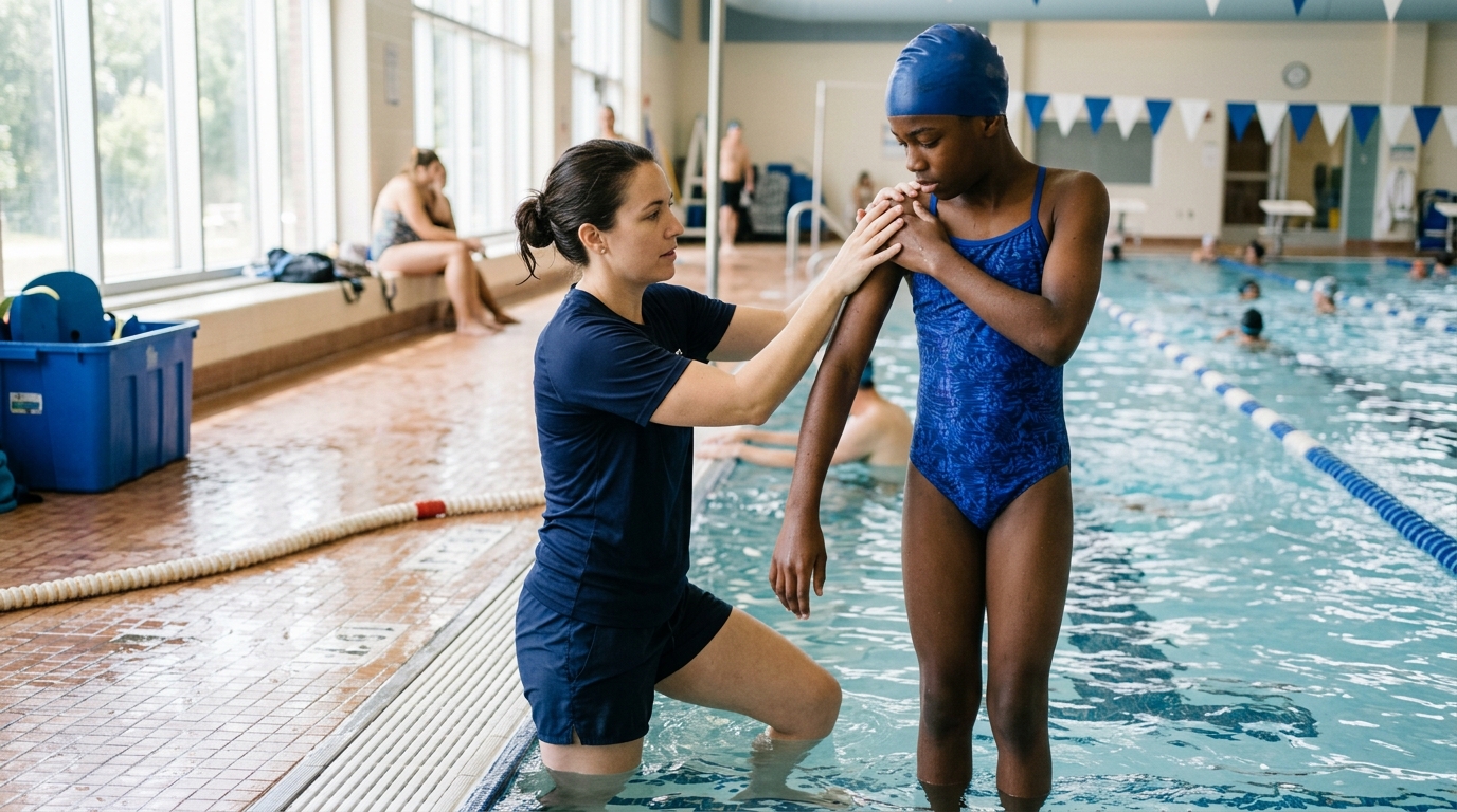
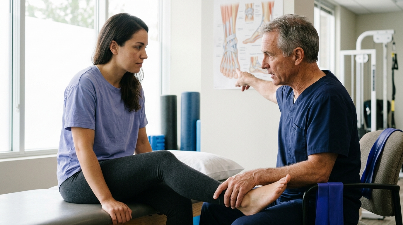
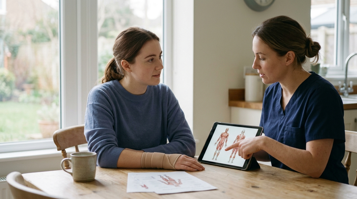
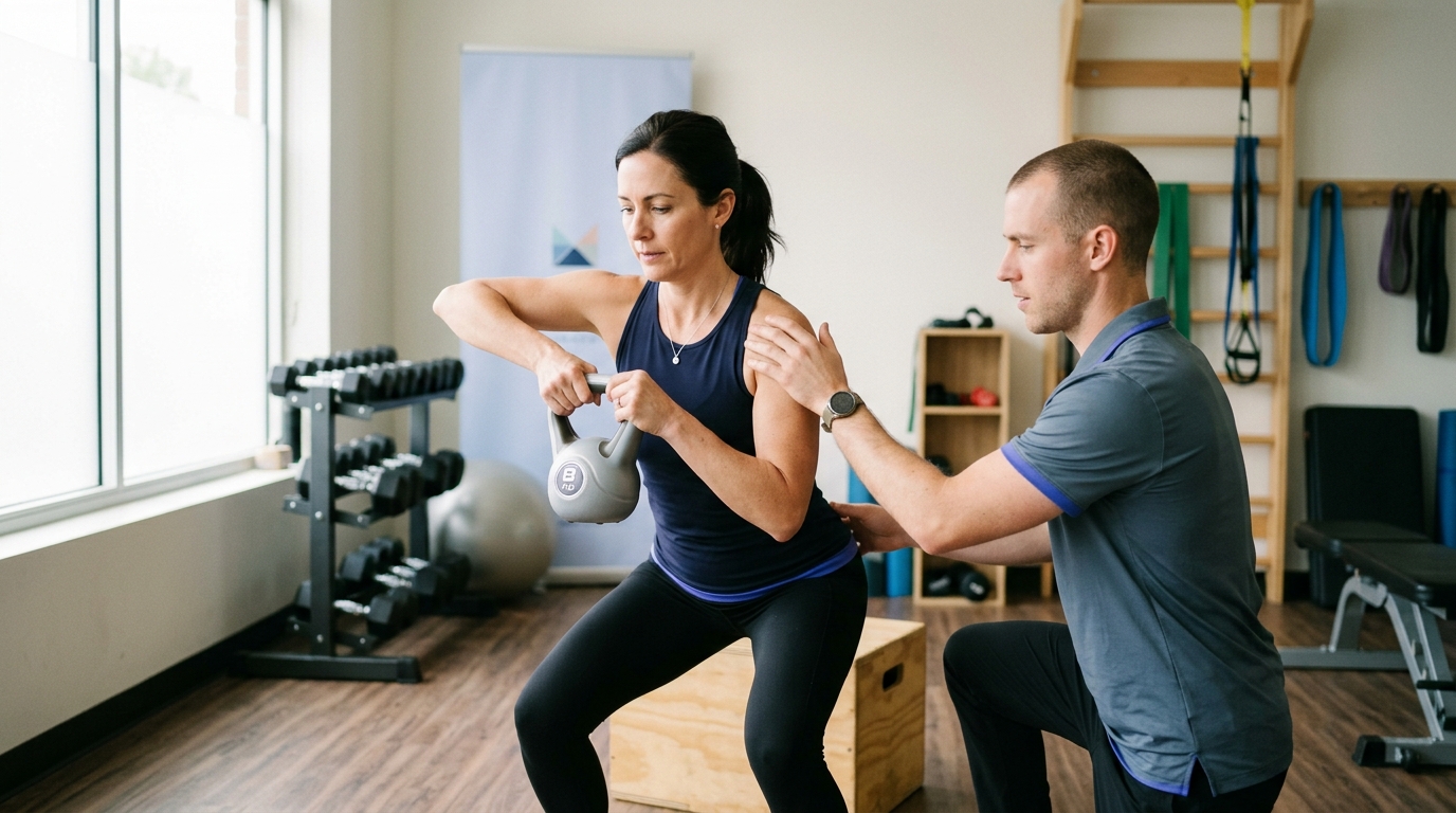
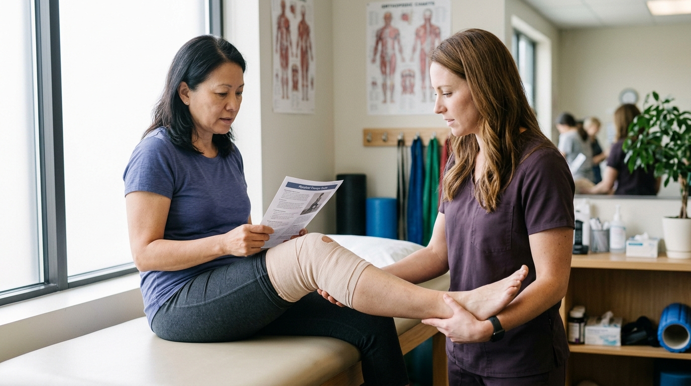
Physical therapy is a specialized healthcare discipline within rehabilitation that uses targeted exercise, manual techniques, patient education, and movement training. Physical therapists design structured recovery programs to reduce pain, improve joint kinematics, and guide safe progression back to daily life and physical recreation.
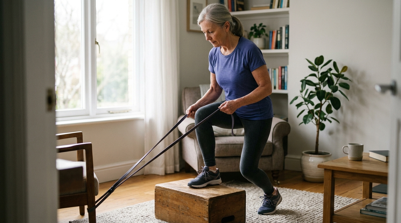
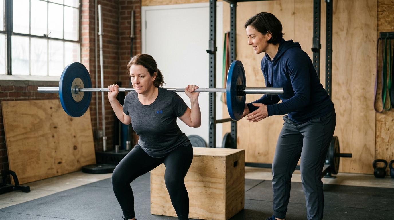
Strengthening exercises use external resistance, such as body weight, resistance bands, or free weights, to rebuild muscle mass, neural drive, and muscular endurance after periods of disuse or injury.
Flexibility refers to the ability of a muscle and its surrounding connective tissues to lengthen passively through an available range of motion. Structured rehabilitation balances strengthening with flexibility exercises to ensure that restored joint mobility is supported by strong, stable muscles. You can explore structured exercise principles in our strength and return to sport planning guides.
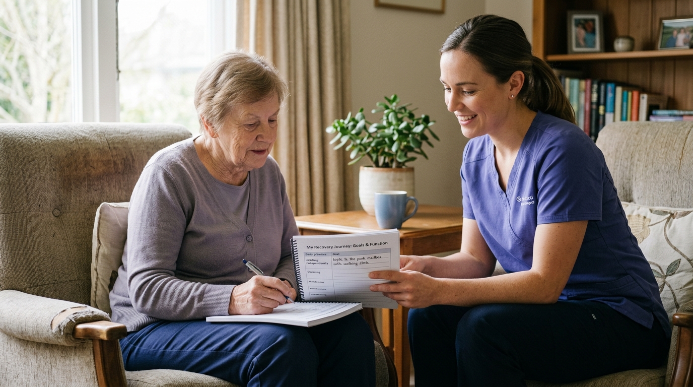
Functional recovery refers to your ability to perform meaningful real-world tasks independently and safely, rather than simply achieving a specific test score on an exam table.
Functional recovery focuses on practical milestones, such as walking down stairs without holding a rail, carrying groceries, getting up from the floor, or returning to work. A patient might have a normal X-ray and full passive motion but still require physical therapy to achieve complete functional recovery in their daily routines.
An outcome measure is a standardized test, clinical score, or measurement tool used by healthcare providers to assess your physical status and track improvement over the course of treatment.
A Patient-Reported Outcome Measure, or PROM, is a validated questionnaire completed directly by the patient without interpretation by a clinician. PROMs capture your personal perspective on your pain, physical limitations, emotional well-being, and quality of life. Common orthopedic PROMs include the Olerud-Molander Ankle Score (OMAS), the Knee Injury and Osteoarthritis Outcome Score (KOOS), and general health surveys like the RAND 36-item tool.
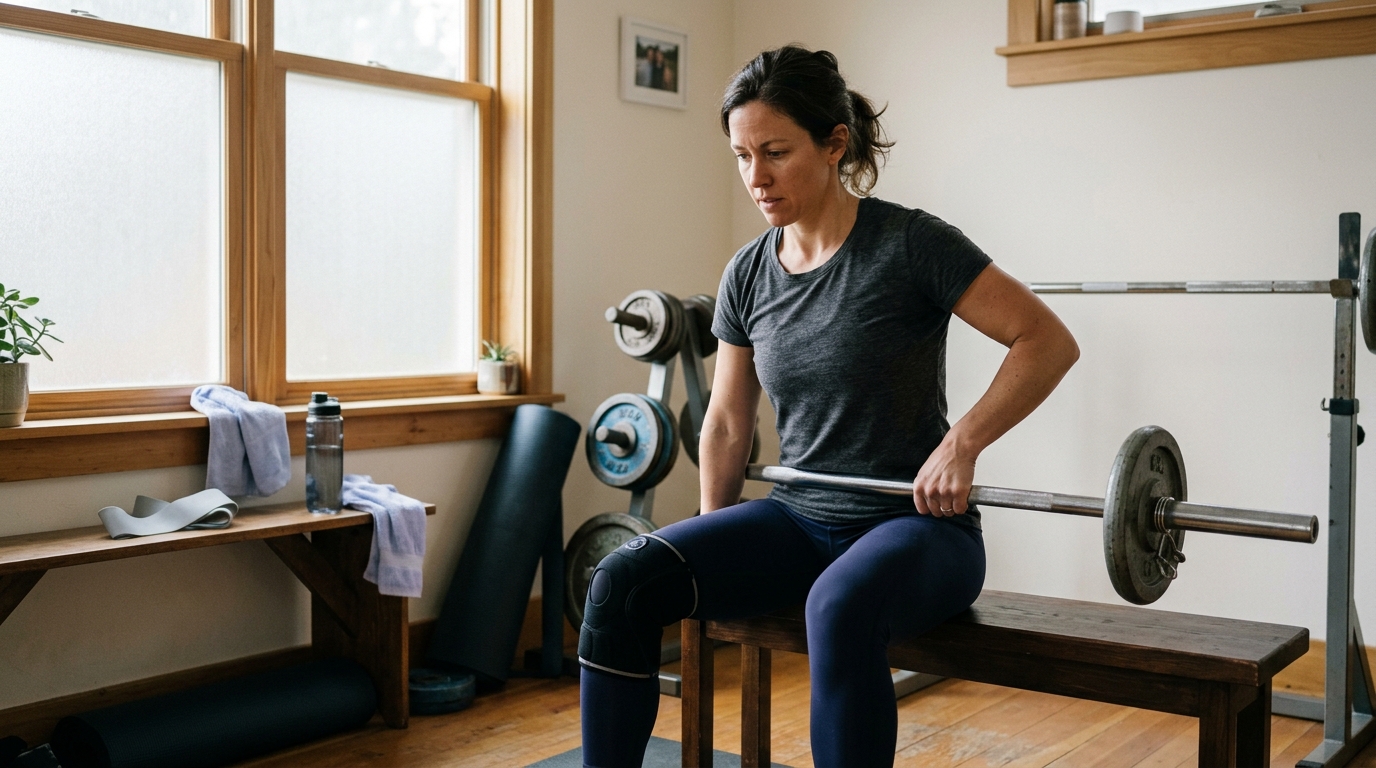
Return to activity describes the gradual, phased progression from formal clinical rehabilitation back to unrestricted recreational pursuits, occupational duties, or competitive athletic sports.
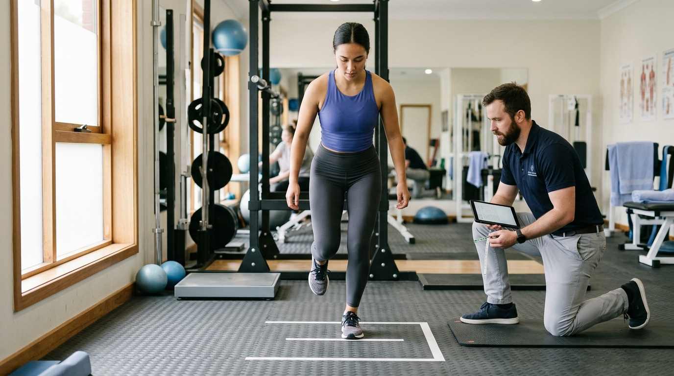
Return to activity is not a single calendar date determined on the day of surgery. It is a criteria-based progression that depends on meeting specific physical benchmarks, such as restoring equal limb strength, demonstrating symmetrical balance, passing functional hop tests, and building cardiovascular endurance.
Community health guidelines from the National Health Service emphasize that progression must be gradual. Warning signs that indicate you are progressing too quickly include sharp or increasing joint pain, swelling that returns after activity, or a noticeable loss of joint motion and strength.
Reconditioning is the process of rebuilding total-body cardiovascular fitness, muscular endurance, metabolic capacity, and neuromuscular coordination following a period of prolonged physical inactivity or bed rest.
When an injury forces you to rest a specific limb, the rest of your body also experiences physical deconditioning. Clinical guidelines from NICE recommend incorporating comprehensive reconditioning, balance training, and fitness exercises as early as safely possible to support overall health and ease the transition back to an active lifestyle.
Recovery timelines presented online or in brochures are general population estimates, not fixed schedules. Many biological and behavioral factors influence how quickly tissues heal and how smoothly rehabilitation proceeds.
Different tissues heal at vastly different rates based on their local vascular supply and cellular turnover:
Biological age and general health significantly impact recovery capacity. As we age, cellular repair mechanisms slow down, bone mineral density naturally decreases, and muscle protein synthesis becomes less efficient.
Underlying medical conditions, including diabetes, peripheral vascular disease, rheumatoid arthritis, and thyroid disorders, can prolong inflammatory phases and slow tissue healing. Lifestyle factors also play a critical role. Adequate protein intake, quality sleep, and tobacco avoidance support bone mineralization and soft-tissue repair, whereas chronic stress and poor nutrition can delay recovery milestones.
The specific surgical approach used to treat an orthopedic condition dictates early rehabilitation rules. A minimally invasive arthroscopic procedure causes less disruption to surrounding muscles and skin than a large open reconstruction, often allowing for earlier active motion.
The mechanical stability of surgical hardware also shapes your protocol. Rigid internal fixation with sturdy titanium plates and compression screws often allows for earlier protected weight-bearing, whereas delicate soft-tissue repairs, such as rotator cuff or labral repairs, require strict early protection to prevent surgical suture pullout.
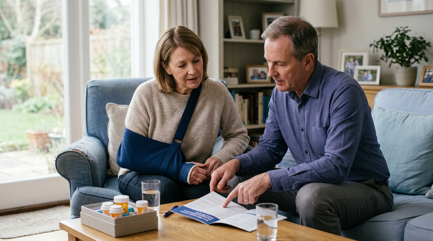
Clear communication with your orthopedic surgeon, physician, and physical therapist is essential for navigating rehabilitation safely. Use these practical questions to clarify common clinical situations.
If your discharge paperwork or clinical notes list a weight-bearing restriction, ask your clinician for precise boundaries:
If your care team prescribes range of motion exercises, make sure you understand who is doing the physical work:
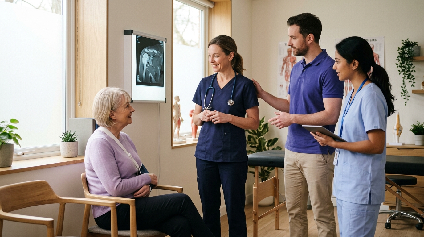
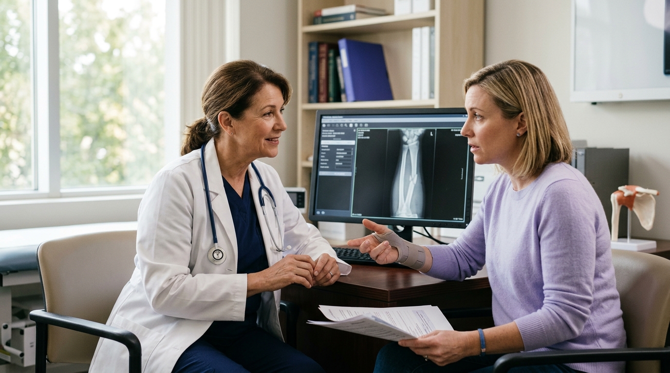
When reviewing follow-up X-rays or imaging results with your orthopedic doctor:
When preparing to transition from basic physical therapy exercises back to recreational sports, gym workouts, or running:
Navigating orthopedic recovery requires patience, active participation, and clear communication with your healthcare team. Clinical terms are not meant to confuse you; they exist to describe the precise boundaries needed to protect your body while it repairs itself.
Healing is rarely a straight line. Temporary flare-ups, joint stiffness, and periods of slower progress are normal parts of the rehabilitation process. Understanding the language of orthopedic care helps you make informed choices, follow your rehabilitation plan accurately, and work constructively with your care team.
To put this guide into practice over the coming days:

Use ReboundBody resources to understand common recovery stages, rehab terms, movement limits and strength rebuilding. Each guide is designed to make a complex comeback easier to understand.


Read practical guidance on injury recovery, rehabilitation, mobility and rebuilding strength as you work your way back to activity.
Explore Resources