
A clear clinical framework helps you reassess stalled rehabilitation by evaluating original diagnoses, tracking functional healing markers, and adjusting recovery timelines effectively.





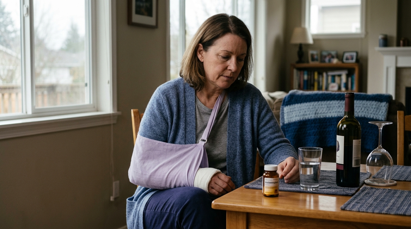
You wake up several weeks into physical therapy, and your joint still aches. You search online for why your injury stopped improving, wondering if you did something wrong or if the damage is permanent.
When physical rehabilitation slows down, plateaus, or brings sudden symptom flares, it is easy to assume the worst. A stall in progress is rarely proof of permanent failure. Instead, a slowdown is a practical prompt to review your current plan, check your functional milestones, and determine whether your recovery needs adjustment.
Recovery from an orthopedic injury or surgery is rarely a straight line. Healing involves complex biological processes, neuromuscular adaptations, and daily physical demands. A setback or a prolonged pause simply means your body and your care team need to reassess three fundamental questions: Is the working diagnosis still correct? Is the tissue healing as expected? Are outside factors influencing your symptoms or movement capacity?
Understanding what a recovery stall actually means can help you replace fear with structured action. By learning how to track objective progress, identify practical roadblocks, and talk with your medical team, you can navigate difficult phases with clarity and confidence.
A stalled recovery is a practical description of an unexpected pause or deviation in your rehabilitation course. It is not an official medical diagnosis. Clinicians do not look at a single universal calendar to decide whether an injury has failed to heal. Every tissue heals at its own rate, and every person brings unique physiological factors to the table.
Progress can deviate from expectations in several distinct ways. Understanding the specific nature of your slowdown helps you describe it accurately to your care team:
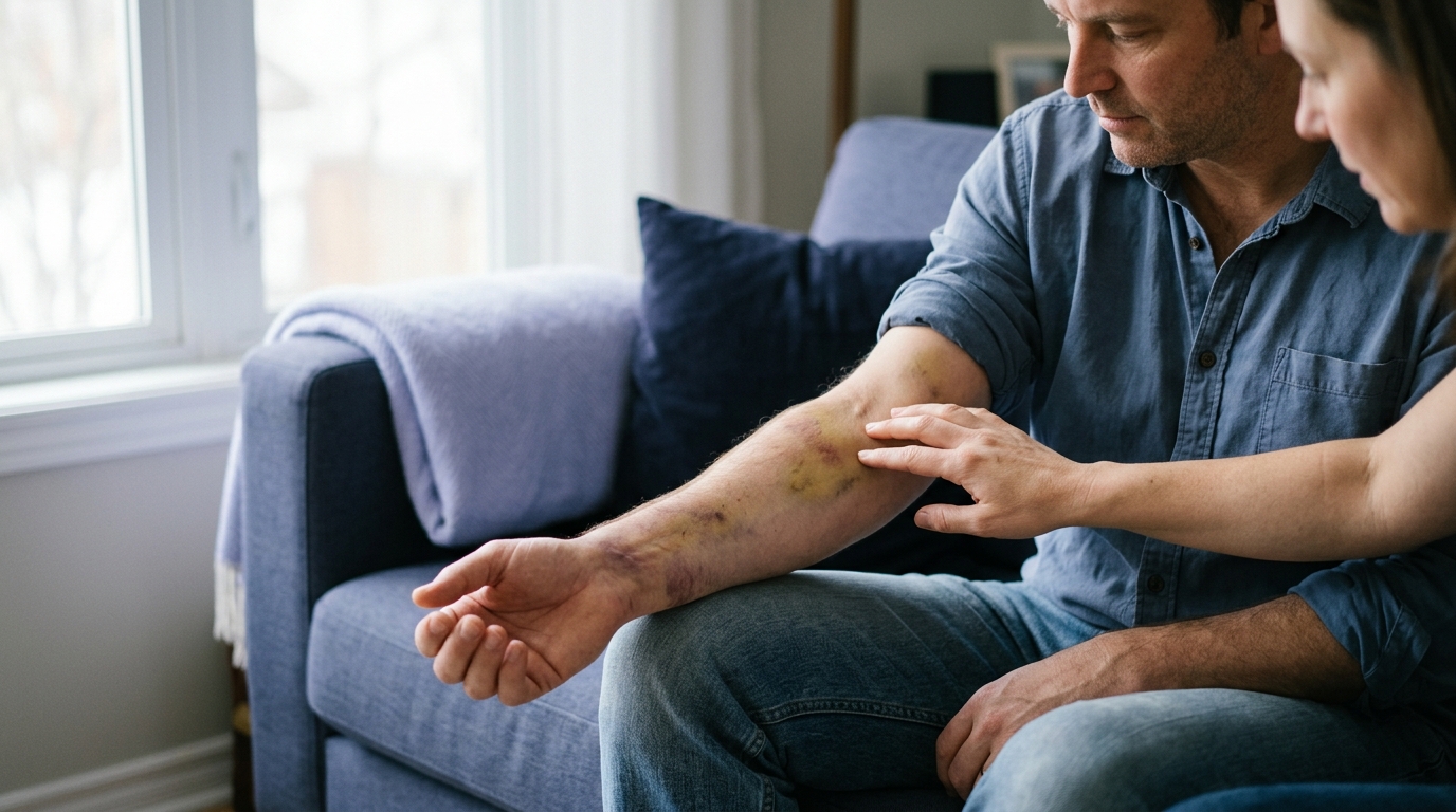
A symptom flare does not automatically mean you have reinjured the underlying tissue. Pain is a protective alarm system managed by your nervous system. That alarm can sound louder because of fatigue, increased exercise volume, poor sleep, or heightened stress, even when tissues remain structurally intact.
Similarly, persistent pain does not prove that active tissue damage is ongoing. Clinical guidelines from the Massachusetts Department of Industrial Accidents define chronic pain as pain lasting longer than three months, or pain that persists beyond the normal time expected for tissue healing. This definition describes duration rather than permanent physical damage. Having persistent discomfort does not mean your body cannot adapt, strengthen, and regain function.
To evaluate your situation clearly, explore our injury recovery and healing resources to understand typical tissue healing phases.
Medical research shows that physical recovery rarely moves along a smooth, predictable path. Healing trajectories vary widely, even among people with similar injuries who receive comparable care.
A prospective cohort study published in pain research tracked 241 adults within four weeks of acute musculoskeletal trauma. The researchers followed these individuals for a full year to observe how pain interfered with daily activities over time. The results demonstrated three clear recovery trajectories across the group:
These percentages reflect that specific study sample, rather than a universal guarantee for any single injury. However, the data highlights an essential clinical truth. A slow start does not mean long-term recovery is impossible. More than a quarter of the individuals in the study followed a delayed path, showing meaningful gains well after the early weeks of treatment.
Clinical guidelines from major health organizations emphasize that recovery milestones must be personalized. The National Institute for Health and Care Excellence (NICE) provides guidelines for rehabilitation after traumatic injury and low back pain. NICE emphasizes that rehabilitation plans should be tailored to individual risk profiles, personal goals, and baseline physical capabilities. A rigid timeline cannot account for differences in tissue vascularity, surgical techniques, or patient age.
The International Olympic Committee (IOC) published a comprehensive consensus statement on pain management in sport. The IOC panel highlighted that rehabilitation requires planned reassessments at critical time points. When pain fails to improve along the predicted timeline, or when symptoms change in distribution or quality, a structured reevaluation is warranted. Reassessment is a standard, expected component of high-level rehabilitation rather than an admission of clinical failure.
To learn more about the biological mechanisms behind tissue repair, visit our recovery science articles.
When progress stalls or symptoms flare, guessing what went wrong often creates unnecessary anxiety. A structured framework brings objectivity back to your rehabilitation. The consensus framework endorsed by the IOC offers a reliable method for evaluating a stalled recovery by asking three distinct questions.
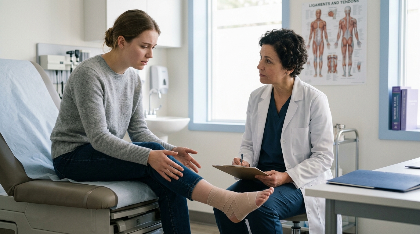
The first step in any review is confirming whether the working diagnosis accurately explains your current symptoms. Musculoskeletal structures sit close together, and pain can refer from one area to another.
A joint sprain may mask an underlying bone bruise, a tendon tear, or localized nerve irritation. In the early days following an injury, swelling and acute guarding can make precise physical examination difficult. As acute swelling subsides, the true underlying driver of your symptoms may become clearer.
NICE guidance on low back pain and sciatica emphasizes that clinicians must consider alternative diagnoses when symptoms fail to improve or when new symptoms emerge. If your joint locks, catches, gives way unexpectedly, or demonstrates localized neurological changes, your physical therapist or physician may need to perform specialized orthopedic tests or order diagnostic imaging. Confirming the structural diagnosis ensures your exercise program targets the right tissue.
The second question examines the biological repair of the injured site. Different anatomical structures heal at vastly different rates due to blood supply and cellular turnover.
Bone fractures typically form a soft callus within a few weeks, followed by progressive mineralization over several months. Tendons and ligaments have lower metabolic activity and less blood supply, requiring prolonged, carefully graded mechanical loading to realign collagen fibers. Cartilage has minimal blood flow and heals very slowly, often adapting through fluid movement rather than rapid structural regeneration.
If biological healing is delayed, your rehabilitation plan may need adjustments in load and rest. A tissue that is underloaded will not receive the mechanical signals required to lay down strong collagen. Conversely, a tissue that is consistently overloaded will remain irritated and inflamed. Finding the balance between adequate rest and progressive mechanical stress is critical for continued biological adaptation.
Pain is not a direct reflection of structural tissue damage alone. It is an integrated output of your central nervous system, shaped by biological, psychological, and environmental inputs.
When physical progress stalls, non-injury factors are often contributing to symptom sensitivity. Key non-injury contributors include:
Addressing these non-injury elements often breaks a persistent plateau without requiring aggressive medical interventions.
Relying entirely on daily pain scores to judge your recovery can be misleading. Pain naturally fluctuates based on weather, stress, fatigue, and daily activities. If you evaluate your progress solely on whether you feel zero discomfort, you may miss significant physical improvements.
NICE traumatic injury rehabilitation guidelines recommend tracking both patient-reported and clinician-reported outcome measures alongside functional goals. Massachusetts chronic pain guidelines similarly recommend tracking objective indicators such as work participation, daily tasks, joint range of motion, and muscular strength.
Meaningful recovery is about restoring your ability to live, work, and move freely. Functional milestones offer a clear, objective view of whether your physical capacity is expanding, even if baseline soreness remains steady.
Track measurable activities that matter to your daily life:
When you notice that you can walk farther, carry more weight, or stand longer before discomfort begins, your functional capacity is improving. That is meaningful progress.
Another reliable indicator of tissue capacity is symptom recovery time, or how quickly your symptoms settle back to baseline after physical effort.
In the early stages of a setback, a challenging exercise session might cause an ache that lingers for two or three days. As your tissue tolerance and neuromuscular control improve, that same exercise might only cause mild soreness that fades within two hours. Even if the peak pain rating during the activity feels similar, a shorter recovery time proves that your musculoskeletal system is becoming more resilient.
Your physical therapist uses specific clinical measurements to assess progress. You can discuss these objective markers during your regular appointments:
Tracking these objective categories alongside your daily symptoms provides a complete picture of your recovery. For structured exercise ideas that support these metrics, review our rehabilitation and mobility guidance.
When recovery slows down, individual physical, environmental, and behavioral factors are often influencing the timeline. Recognizing these factors helps you and your clinician adjust your program effectively.
Every musculoskeletal tissue operates under distinct biological rules:
Understanding the specific tissue involved prevents unrealistic expectations. Expecting a chronic tendinopathy to resolve in three weeks creates frustration, whereas recognizing that tendon remodeling takes months encourages patience and consistency.
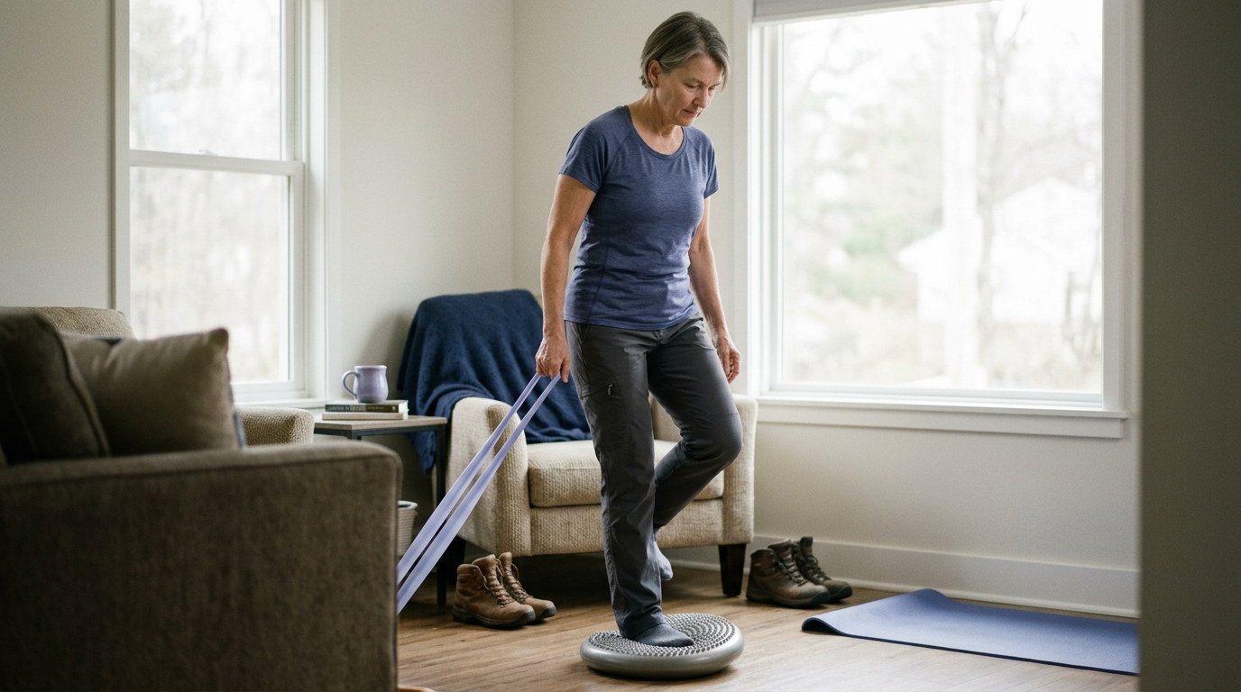
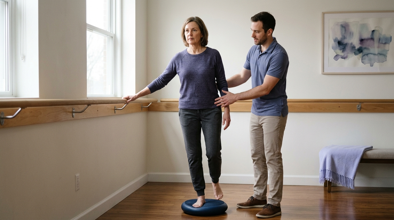
Achieving physical adaptation requires matching the exercise dose to your current tissue capacity. A recovery stall often occurs when the mechanical load is either too low or too high.
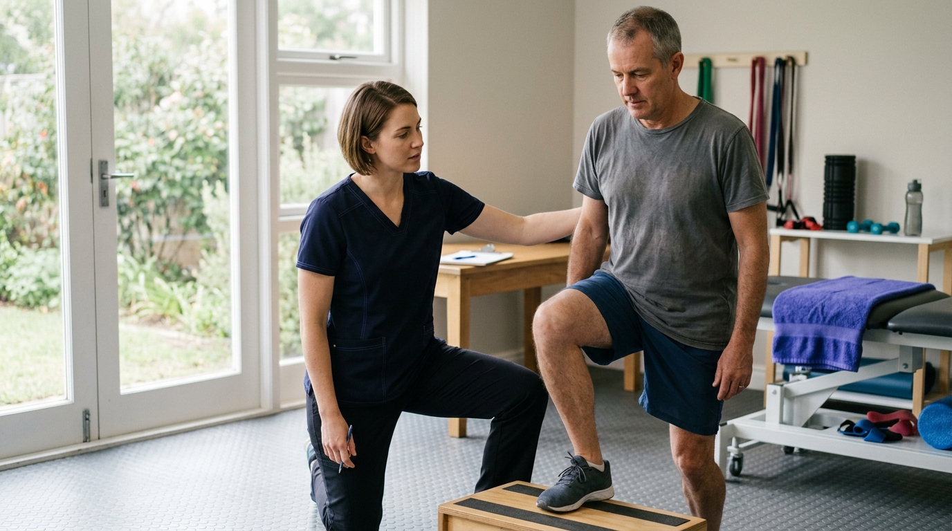
An insufficient rehabilitation dose occurs when exercises are kept too light for too long. Gentle range-of-motion movements are vital immediately after injury, but tissues require progressive resistance to build strength and capacity. If your exercises never challenge your muscles, your physical adaptation will plateau.
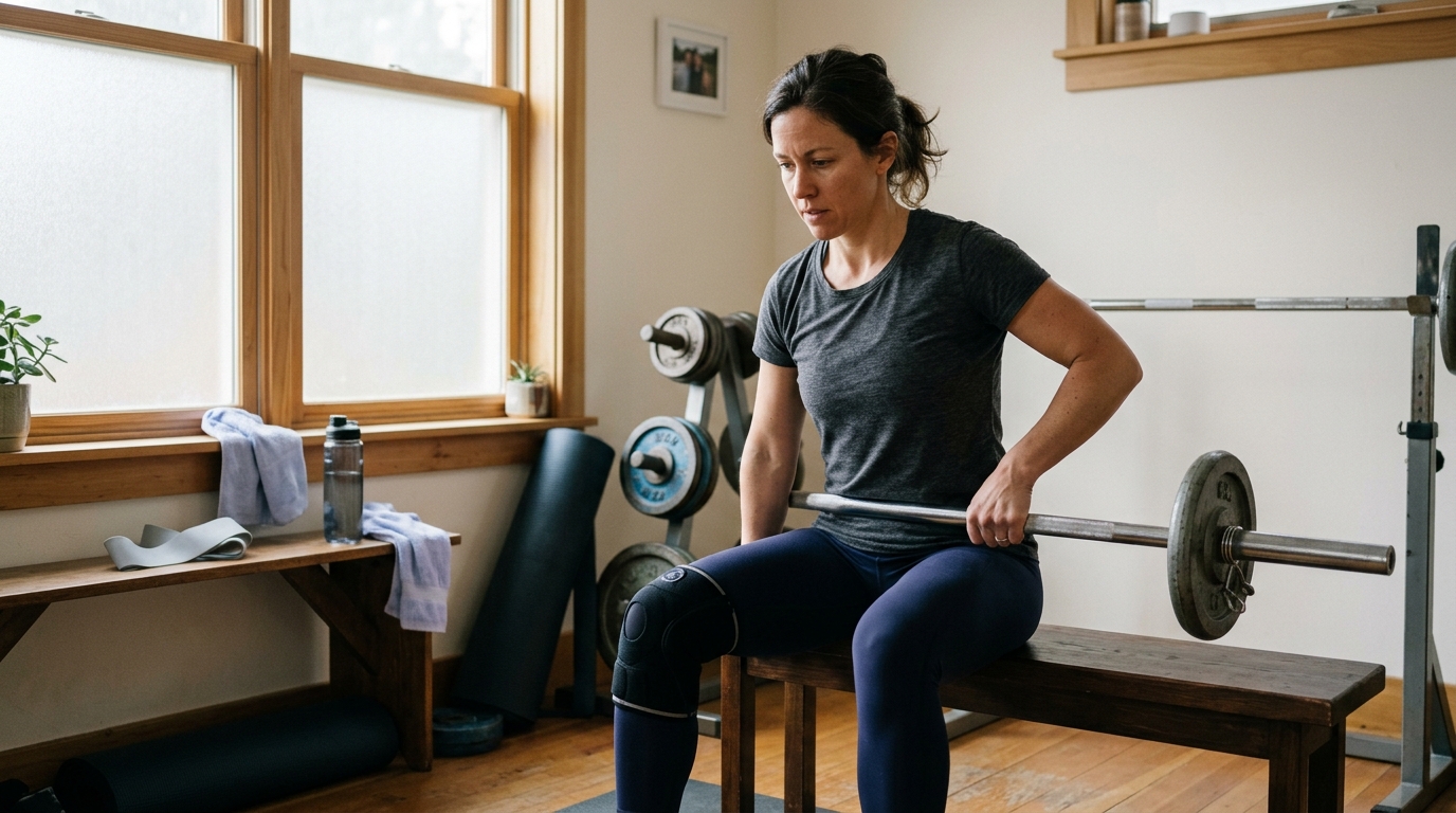
An excessive rehabilitation dose occurs when volume, intensity, or frequency increases too rapidly. Adding weight, repetitions, and daily walking distance all at once can overwhelm healing tissues. The result is recurring inflammation, stiffness, and perceived setbacks.
Age influences recovery speed, though it does not prevent successful rehabilitation. As we age, tissue elasticity decreases, microvascular circulation declines, and muscle protein synthesis slows.
An adult over 50 may require more recovery time between heavy loading sessions than a younger individual. Older adults also experience more rapid muscle loss during periods of immobilization. Rebuilding baseline strength, power, and connective tissue stiffness simply requires a dedicated, progressive approach over a longer timeframe.
Rehabilitation takes place within the context of your broader life. When outside stressors mount, recovery capacity drops.
Chronic psychological stress elevates circulating cortisol and inflammatory cytokines, which interfere with normal tissue repair. Poor sleep impairs growth hormone release, reducing the body's ability to repair cellular damage sustained during physical therapy. Inadequate dietary protein restricts the amino acids necessary to rebuild muscle tissue and synthesize collagen.
Addressing lifestyle foundations often re-energizes a rehabilitation plan that has stalled. Explore our strength and return to activity resources for guidance on safely building physical capacity.
While most recovery slowdowns require simple plan modifications, some symptom changes require immediate clinical evaluation. Distinguishing between a benign rehabilitation flare and a potential medical emergency is essential for your safety.
Emergency warning signs vary based on your injury location and general health. For example, the Centers for Disease Control and Prevention (CDC) lists specific emergency signs following a head injury or suspected concussion. These include a worsening headache that does not go away, repeated vomiting, unequal pupil sizes, slurred speech, increasing confusion, seizures, or difficulty waking up. These neurological symptoms require emergency evaluation rather than a routine therapy appointment.
For spinal injuries, red flag symptoms warranting urgent medical assessment include new loss of bowel or bladder control, numbness in the saddle area (groin and inner thighs), or progressive weakness in your legs. For peripheral joint injuries or post-surgical recovery, warning signs that warrant prompt medical contact include:
If you experience severe, rapidly worsening, or unexplained symptoms, contact your physician or local emergency services immediately. For standard musculoskeletal aches, stiffness, and mild flares, follow the structured review steps below.
Reviewing real-world recovery patterns helps illustrate how different situations can be approached productively. The following examples represent common clinical models.
A 52-year-old runner recovering from Achilles tendinopathy reports that their tendon still aches every morning. They feel discouraged and believe their recovery has stalled.
However, a review of objective data shows that four weeks ago they could only walk 15 minutes before stopping. Today, they walk 45 minutes comfortably, perform 25 single-leg heel raises, and navigate stairs without hesitation.
The care team explains that tendon pain often lags behind functional gains. The recovery plan is working because functional capacity and tissue load tolerance are steadily rising. The clinician recommends maintaining current strengthening exercises while using morning stiffness duration as a tracking metric.
Productive conversation prompt: "My morning stiffness is still present, but my walking distance has tripled. How should we adjust my loading exercises to keep building capacity while managing this background ache?"
A 44-year-old individual recovering from a lumbar disc issue experiences significant relief after four weeks of physical therapy. Feeling confident, they spend an entire weekend digging in the garden and lifting heavy bags of soil.
On Monday morning, their low back pain and muscle spasms return with significant intensity. The person panics, fearing they have re-ruptured the disc and undone all their progress.
A physical examination by their therapist reveals no new neurological deficits, normal reflexes, and intact leg strength. The flare is identified as an acute muscle spasm and tissue overload from an unaccustomed spike in activity. The care team temporarily modifies the exercise intensity for several days, introduces gentle mobility movements, and reinforces activity pacing strategies. Within a week, the flare settles.
Productive conversation prompt: "My back flared up after a heavy weekend of yard work. Are my neurological signs still stable, and what temporary modifications should we use while this settles?"
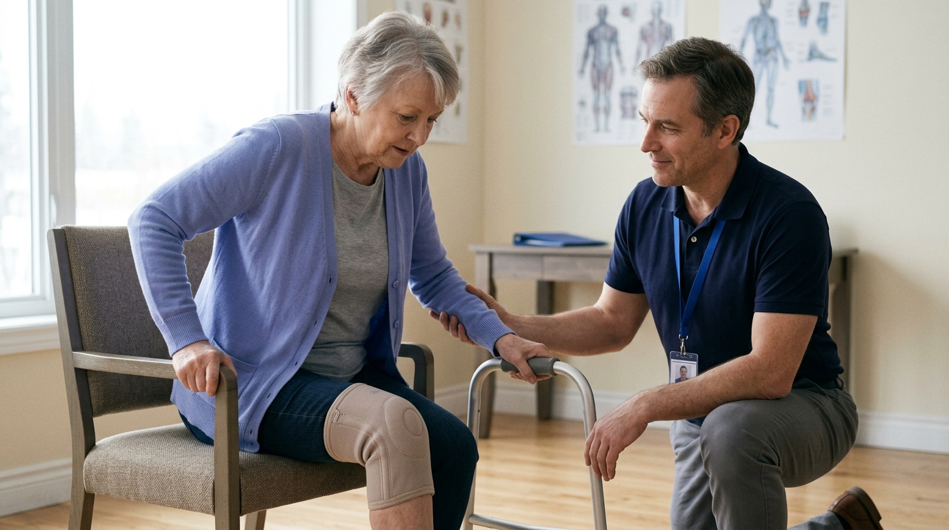
A 60-year-old individual recovering from total knee replacement achieves 105 degrees of knee flexion by week six. Over the next four weeks, their flexion remains at 105 degrees, quadriceps strength does not improve, and walking endurance remains capped at 10 minutes.
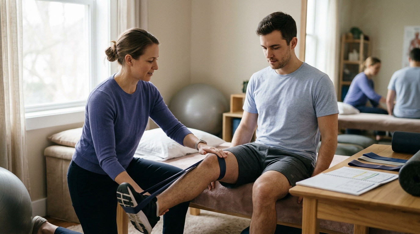
The patient and physical therapist review the current home program. They realize the patient has been doing the exact same light resistance band exercises for a month without increasing resistance or changing volume.
The therapist updates the program by introducing heavier closed-kinetic-chain exercises, adjusting manual therapy techniques, and adding stationary cycling intervals. By progressively overloading the quadriceps and stretching the joint capsule under supervision, the plateau breaks, and range of motion increases to 118 degrees over the next month.
Productive conversation prompt: "My knee flexion and walking endurance have not changed in four weeks. Is it time to progress my exercise resistance, or should we evaluate joint capsule tightness?"
A 38-year-old recreational athlete recovering from a shoulder rotator cuff strain notices that their original dull shoulder ache has changed. They now experience sharp, clicking pain in the front of the joint whenever they raise their arm overhead, accompanied by tingling down their forearm.
Because the location, quality, and triggers of the pain have fundamentally shifted, the physical therapist pauses aggressive overhead strengthening. The therapist performs a detailed reassessment, identifying secondary biceps tendon irritation and thoracic spine stiffness resulting from compensatory movement patterns.
The treatment plan is redirected to restore thoracic mobility and scapular mechanics before reloading the shoulder. The tingling resolves, and overhead movement becomes pain-free.
Productive conversation prompt: "My shoulder pain has moved to the front of the joint and feels sharp rather than dull. Can we re-evaluate my movement mechanics to see if another structure is being pinched?"
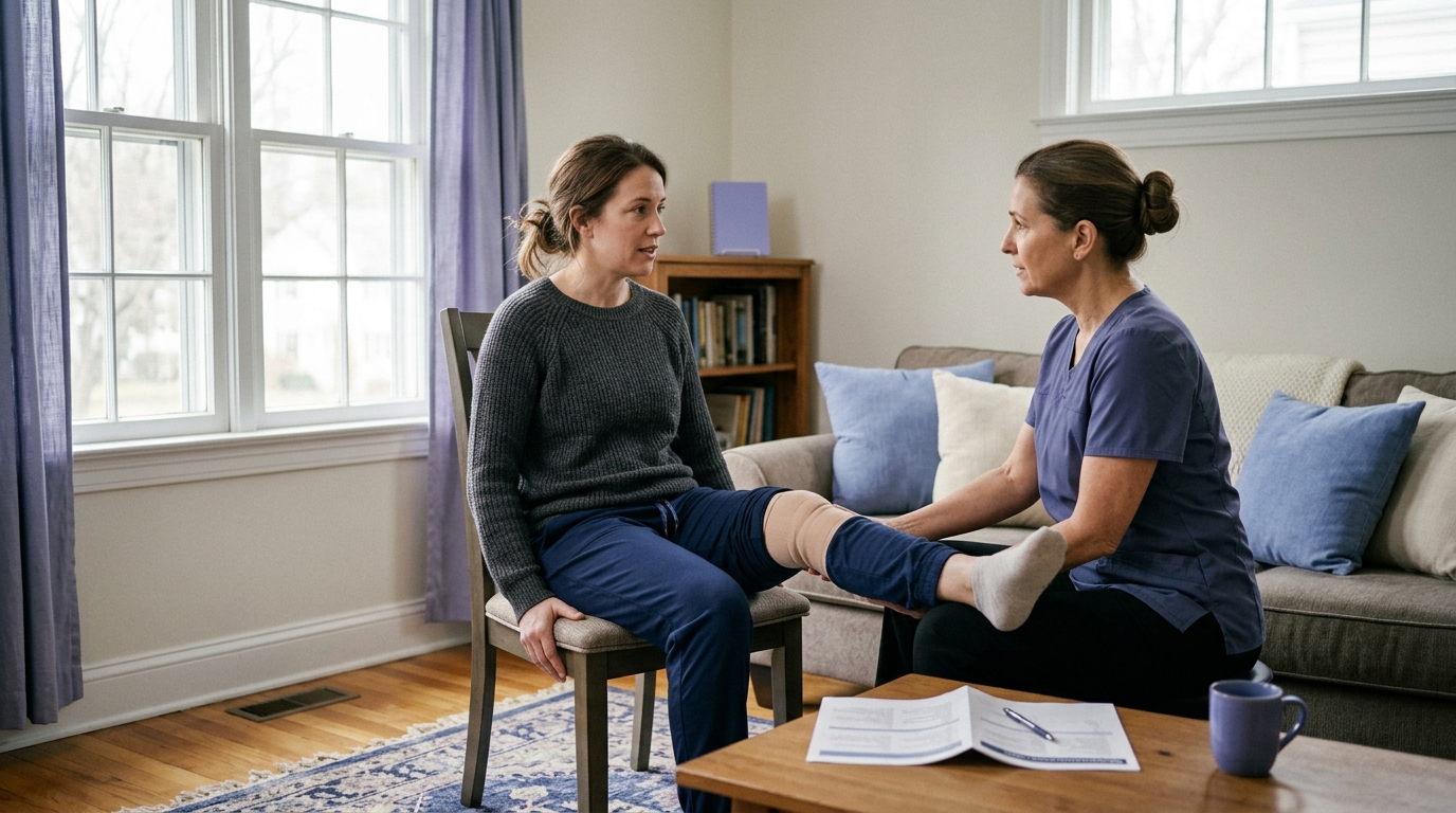
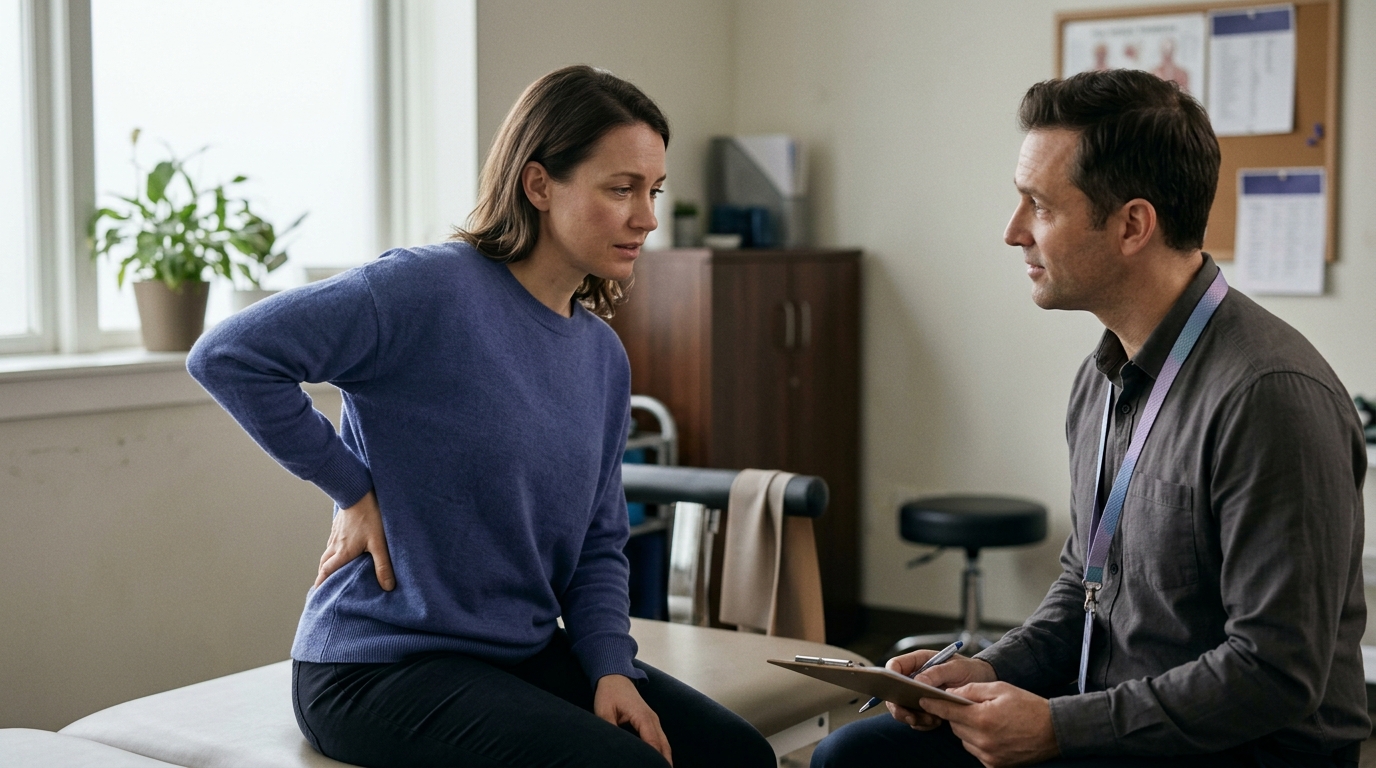
Preparing focused, objective questions transforms a standard appointment into a collaborative strategy session. Instead of simply stating that your injury hurts, use these research-backed questions to structure your discussion:
Bringing written notes regarding your symptom timeline, functional gains, and specific activity triggers helps your care team make informed clinical decisions. To learn more about our editorial approach to evidence-based recovery, read about ReboundBody.
A slowdown, plateau, or symptom flare during injury recovery is a regular clinical event, not an admission of permanent failure. Recovery trajectories vary significantly, and healing rarely follows a perfectly linear timeline.
Evaluating a stalled recovery requires a balanced, structured approach. You and your healthcare team can work through the core questions: confirm the accuracy of your diagnosis, assess biological tissue healing, and identify any physical or lifestyle factors that may be limiting your progress. By shifting your focus from short-term pain fluctuations to long-term functional milestones, you can measure real improvements in strength, endurance, and mobility.
Rehabilitation is an ongoing, collaborative process of testing, learning, and adjusting. With clear communication, objective tracking, and a willingness to refine your plan, you can navigate recovery plateaus and continue working toward your physical goals.
Use this practical checklist to review your current recovery status and prepare for your next clinical appointment:

Use ReboundBody resources to understand common recovery stages, rehab terms, movement limits and strength rebuilding. Each guide is designed to make a complex comeback easier to understand.


Read practical guidance on injury recovery, rehabilitation, mobility and rebuilding strength as you work your way back to activity.
Explore Resources